Case Report | | Peer-Reviewed

The Particularities of Pulmonary Tuberculosis in Children with Type 1 Diabetes: About 2 Cases

Received: 7 July 2024     Accepted: 9 August 2024     Published: 27 August 2024
Views:       Downloads:
Abstract

The combination of pulmonary tuberculosis and type 1 diabetes is a significant public health problem, especially in developing countries, where the incidence of both diseases is rising sharply. According to the World Health Organization (WHO), by 2022, 23% of tuberculosis cases will be in Africa. Morocco is one of the countries with a medium incidence of tuberculosis. The Ministry of Health has launched a national strategic plan for the prevention and control of tuberculosis. However, there is no national study on the prevalence of tuberculosis in diabetic children in Morocco. Several studies in the literature have investigated the specifics of this association, and have shown that there is a two-way association between tuberculosis and type 1 diabetes. Latent tuberculosis is most common in diabetic children, while active tuberculosis can give rise to severe and atypical presentations. In this study 2 cases have been reported of type 1 diabetes associated with pulmonary tuberculosis, of varying severity, in a 15-year-old child known to be diabetic and an 18-month-old infant with inaugural diabetes, in order to determine the clinical, paraclinical, therapeutic and evolutionary particularities of tuberculosis in these children.

Published in American Journal of Pediatrics (Volume 10, Issue 3)
DOI 10.11648/j.ajp.20241003.18
Page(s) 152-157
Creative Commons

This is an Open Access article, distributed under the terms of the Creative Commons Attribution 4.0 International License (http://creativecommons.org/licenses/by/4.0/), which permits unrestricted use, distribution and reproduction in any medium or format, provided the original work is properly cited.

Copyright

Copyright © The Author(s), 2024. Published by Science Publishing Group

Keywords

Diabetes Type 1, Pulmonary Tuberculosis, Mycobacterium Tuberculosis, Glycemic Imbalance

1. Introduction
Tuberculosis is an infectious opportunistic disease caused by Mycobacterium Tuberculosis (MT). It is a public health problem and a major cause of morbidity and mortality worldwide .
Type 1 diabetes (T1D) is an autoimmune disease resulting in the complete destruction of the beta cells of the islets of Langerhans. Its incidence has risen steadily over the past 2 decades. It is associated with a high risk of developing an infectious disease
The association of diabetes with tuberculosis is well established in several systematic reviews . However, the precise pathophysiological mechanisms of this association are still poorly understood . In a meta-analysis, it was reported that diabetes tripled the risk of active tuberculosis and that around 15% of tuberculosis cases worldwide are associated with diabetes.
However, studies in children are rarer than those in adults. There is a two-way association between tuberculosis and diabetes, although diabetes is a powerful risk factor for tuberculosis, tuberculosis also influences glycemic control in diabetic patients .
The prevalence of diabetes is rising sharply in developing countries where tuberculosis is also endemic This could considerably amplify the public health impact of this double burden of disease. As a result, tuberculosis and diabetes are currently among the world's top public health priorities.
2. Case Report
In this study 2 cases have been reported of children followed for T1D associated with pulmonary tuberculosis of variable occurrence and severity, followed at the pediatric endocrinology unit, Department of Childhood Diseases, Abderrahim Harouchi Mother-Child Hospital, in Casablanca, Morocco.
The first case concerns a 15-year-old child with T1D, followed for 2 and a half years with glycemic imbalance, who presented with a severe form of pulmonary tuberculosis and an aspergillosis graft. The 2nd case concerned an 18-month-old infant who presented with pulmonary tuberculosis discovered synchronously with diabetes. The diagnosis of pulmonary tuberculosis was confirmed bacteriologically in both children. The aim of this study was to analyze the clinical, radiological, evolutionary, and therapeutic features of this association.
3. Results
Case 1:
A 15-year-old male child, from a first-degree consanguineous marriage, without tuberculosis contagion, followed for T1D for 2 and a half years, on a basal bolus regimen, poorly balanced, with his mean glycated hemoglobin (HbA1c) being 11%.
He was admitted to the pediatric endocrinology unit with acute digestive symptoms of abdominal pain and vomiting, associated with a chronic productive cough dating back a month and a half, resistant to betalactamines and macrolides, complicated by the appearance of small hemoptysis 4 days before admission. All of this occurred in a context of a deteriorating general condition, with weight loss of 4kg in one month, and fever at 38°C.
On admission, the child was drowsy, with acetone breath odor, dehydrated and with sunken eyes, with no signs of respiratory distress. Skin recoloration time was less than 3 seconds. There was no mottling and the extremities were slightly cold. Respiratory rate was 38 cycles per minute.
Heart rate was 110 beats per minute. Blood pressure was 110/80 mmHg. SpO2 was 95% on room air. Temperature was 38.2°C. Pleuropulmonary auscultation revealed bilateral crepitus rales. Blood glucose level was 5.2g/dl. Urine dipstick showed 3+ of acetone and 3+ of glucose. The abdomen was soft, with no abdominal guarding or contractures.
Rehydration with 0.9% sodium chloride at a rate of 10 cc/kg/hour for two hours was started as an emergency, after performing a serum electrolytes test and renal function test.
Corrected natraemia was normal at 139 mmol/l, with corrected kalaemia at 5 mmol/l, alkaline reserves were 8 mmol/l. plasma osmolarity was 302 mOsm. Renal failure was observed, with creatinemia at 8.8 mg/l and glomerular filtration rate at 77 ml/min. Glycated hemoglobin was very high at 17%.
For frank severe cases of diabetic ketoacidosis, after two hours of rehydration with salted serum, an infusion of 3l/m2/d of 5% glucose serum was started, combined with insulin therapy using a self-pulsing syringe (SAP) at a dose of 0.05 IU/kg/h for 4 hours, then 0.1 IU/kg/h. With strict clinical, neurological, hemodynamic, and respiratory monitoring, and a serum electrolyte test at H2, H6, H12 and H24.
After the child had been stabilized and switched to subcutaneous insulin therapy, after 28 hours of SAP, a thoracic radiograph revealed an interstitial syndrome and an excavated pulmonary opacity (figure 1).
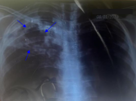
Figure 1. A thoracic radiograph revealed an interstitial syndrome and an excavated pulmonary opacity.
In view of this radiological appearance, a bacteriological workup was initiated, revealing acid-fast bacilli in the cytobacteriological examination of the sputum. The tuberculin intradermal test was negative. While the quantiFERON test was positive.
Thoracic computed tomography scan (Thoracic CT scan) showed bilateral bronchopneumopathy associated with multiple excavated lesions, with a strong suspicion of grafting of Aspergillus in the bilateral upper lobar region (figure 2).
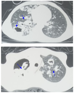
Figure 2. Thoracic CT scan showing multiple lesions excavated from the two upper lobes of the lung, the largest of which is in the right upper lobe measuring approximately 64×48 mm. Hyperdense material was seen in two of these cavities, bilaterally, measuring 24×14 mm on the right and 25×16 mm on the left. Multiple scattered micronodular and reticular infiltrates are present in both lung fields, giving the appearance of a tree-in-bud pattern. Scattered bilateral cylindrical and cystic foci of bronchiectasis associated with peribronchovascular thickening, some of which are the site of mucoid impaction. Multiple basal cervical and mediastinal adenopathies, the largest of which is subcarinal, measuring 10.5 mm in its minor axis.
In view of the strong suspicion of aspergillosis on thoracic CT, aspergillus serology was carried out, including the search for soluble aspergillus antigens (Galactomannan antigen), which was positive with an index of 3.4.
Given the severity of the clinical presentation, in addition to T1D, an associated immune deficiency was sought, but the blood count was normal and human immunodeficiency virus serology was negative. Immunoglobulins and lymphocyte subpopulations were normal.
Antituberculosis treatment was started with quadritherapy; Rifampicin (R) at a dose of 15 mg/kg/d, Isoniazid (H) at a dose of 10 mg/kg/d, Pyrazinamide (Z) at a dose of 35 mg/kg/d, and Ethambutol (E) at a dose of 20 mg/kg/d, for 2 months, combined with voriconazole at a dose of 10 mg/kg/d for 12 weeks, and ciprofloxacin at a dose of 30 mg/kg/d, then HR (Rifampicin, Isoniazid) for 4 months. Combined with insulin therapy using a basal bolus regimen with a dose of 1.4 IU/kg/d. Progression was marked by improvement in respiratory function and glycemia.
Case 2:
18-month-old male infant admitted with polyuro polydipsic syndrome for 2 weeks. History: From a non-consanguineous marriage, with good psychomotor and staturo-ponderal development. The father and older brother have been treated for pulmonary tuberculosis for 2 months.
Examination on admission revealed a drowsy infant with a Glasgow score of 13/15, dehydrated with sunken eyes and dry mucous membranes. Extremities were cold, with no mottling, and skin recoloration time greater than 3 seconds. Respiratory rate was 40 cycles per minute. Heart rate was 139 beats per minute. Blood pressure was 112/70 mmHg. SpO2 was 97% on room air. He was apyretic at 36.9°C. Capillary blood glucose was 4.82 g/dl, with ketones present on urine dip-stick. Filling with 0.9% saline at a dose of 20 cc/ kg over 10 min was performed urgently, with restoration of hemodynamic status, followed by rehydration with 0.9% saline at a dose of 10 cc/kg/hour for two hours. An H0 a serum Electrolytes test showed a normal corrected natraemia of 136 nmol/l, with normal corrected kalaemia of 3.1 nmol/l. Plasma osmolarity was 298 mOsm. Urea at 0.40 g/l, with creatinemia at 4.2 mg/l. While Alkaline reserves were < 5mmol/l. Glycated hemoglobin was 10%.
An infusion of 5% glucose solution combined with SAP insulin therapy at a dose of 0.025 IU/kg/hour for the first 4 hours, followed by 0.05 IU/kg/hour for 26 hours. Was administered with clinical and biological monitoring. During monitoring, there was normalization of consciousness and signs of dehydration. However, high blood glucose levels persisted beyond 24 hours of monitoring, with disappearance of ketone bodies. Biological balance remained correct during monitoring. The infant was then put on subcutaneous insulin therapy, according to the basal bolus regimen at a dose of 1 IU/kg/d.
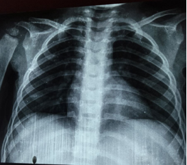
Figure 3. Front chest X-ray without abnormalities.
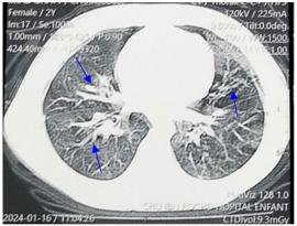
Figure 4. Thoracic CT scan showing retractile moniliform bronchiectasis of the middle lobe and left posterobasal lobes, associated with peribronchial thickening, with left posterobasal branching micronodules, pre-aortic calcified nodes, lingular and middle lobar band atelectasis.
In view of the tuberculosis contagion, a thoracic radiograph was performed, with normal results (figure 3). A bacteriological work-up was carried out to check for acid-fast bacilli in the gastric tubing fluid, with positive results. The intradermal tuberculin test was negative. However, the quantiFERON test was positive. A thoracic CT scan revealed bilateral infectious bronchopneumonia (figure 4).
Antituberculosis treatment was started with quadritherapy: Rifampicin (R) at 15 mg/kg/d, Isoniazid (H) at 10 mg/kg/d, Pyrazinamide (Z) at 35 mg/kg/d, and Ethambutol (E) at 20 mg/kg/d, for 2 months, then HR for 4 months. Regular follow-up by the pediatric endocrinologist and pediatric pulmonologist, with good clinical progress in terms of respiratory and blood sugar levels.
4. Discussion
The association of tuberculosis and T1D represents a major burden for the healthcare system, particularly in developing countries where tuberculosis is one of the most frequent causes of bacterial infection and where the prevalence of T1D is rising remarkably .
Estimates of the prevalence of tuberculosis in diabetics are heterogeneous in the literature. Pediatric studies have shown the prevalence to range from 4.2% to 29.5% . This heterogeneity between studies can be explained mainly by three factors: the quality of the studies, the geographical region, and the baseline incidence of tuberculosis .
Morocco is one of the countries with a medium incidence of tuberculosis. The incidence rate per 100,000 inhabitants has fallen from 115 in 2000 to 94 in 2021 . The Moroccan Ministry of Health has launched a national strategic plan for the prevention and control of tuberculosis (NTCP) . However, there is no national study on the prevalence of tuberculosis in diabetic child
ren in Morocco.
Indeed, diabetes is a predisposing condition for tuberculosis. People with diabetes are three times more likely to develop tuberculosis . Several meta-analyses have been carried out in this respect, showing that diabetes is associated with an increased risk of developing latent and active tuberculosis . Other studies have shown that the risk of latent tuberculosis progressing to active tuberculosis is between 5 and 15%, with an increased risk in people suffering from immunosuppressive diseases such as diabetes .
Although the precise pathophysiological mechanism remains unknown, suggested hypotheses have included depressed cellular immunity, alveolar macrophage dysfunction, low levels of interferon gamma, pulmonary microangiopathy and micronutrient deficiency . On the other hand, tuberculosis affects glycemic control in diabetic patients .
A study by Majaliwa et al found that children with unbalanced diabetes and an Glycated hemoglobin (HbA1c) of over 7% were significantly more likely to have tuberculosis than children with an HbA1c of less than 7%, which is in line with the data from 2 observations in this study where both children had unbalanced glycemia and an HbA1c of over 7%. Unfortunately, studies carried out in Africa show a high rate of poor glycemic control, thus exposing a high risk of tuberculosis .
Diabetics can present with severe forms of pulmonary tuberculosis, with atypical localizations and often extensive radiological abnormalities , which is in line with the first observation, since the child presented with severe pulmonary involvement, with multiple excavated lesions in both upper lobes, and in the right Flower, with bilateral interstitial involvement. This may explain the isolation of acid-fast bacilli in this patient's sputum cytobacteriological examinations. Diabetes has been reported to be associated with an increased likelihood of positive sputum smear results . According to Shen et al, the radiological features of tuberculosis in diabetic patients vary from study to study, and the frequency of unusual radiographic features is probably overestimated .
The QuantiFERON test is the best diagnostic test for tuberculosis after the GeneXpert . According to Shin et al and Walsh et al , diabetes does not affect QantiFERON results, unlike the tuberculin intradermal reaction, which can be affected by diabetes. However, a study by Faurholt et al showed low sensitivity of the QuantiFERON test in tuberculosis patients with T1D, due to immune dysfunctions that can affect the interferon gamma response to MK antigen. According to several studies the sensitivity of GeneXpert is better than that of microscopy and the QuantiFERON test in immunocompromised patients, particularly diabetics. In fact, the GeneXpert test shortens diagnosis times (results within 2 hours), increases tuberculosis detection rates and enables early detection of rifampicin resistance. Since 2014, the use of Gene Xpert has been adopted in Morocco's National Tuberculosis Control Program (NTCP) and replaces microscopy in the 2020 NTCP .
In the first observation in this study, the child presented with a grafting of Aspergillus suspected on CT scan in view of the graft image and confirmed by aspergillosis serology. Its occurrence is mainly conditioned by the presence of underlying respiratory pathology, and the host's immune response
Studies of pulmonary aspergillosis in children with isolated T1D are rare. The child reported in the first observation had a normal immune system, and diabetes was the only immunosuppressive factor.
However, diabetic children may present with a latent, asymptomatic form, which is the most frequent form according to several studies, notably that carried out by Hayashi et al , and Miora et al . This is in line with the second observation of this study, since the infant presented no respiratory symptoms, and the discovery was made following screening for the presence of tuberculosis contagion in the brother and father. However, according to World Health Organization (WHO) guidelines on the management of latent tuberculosis, there was no recommendation to screen patients with T1D for latent tuberculosis, due to weak evidence . Nevertheless, the WHO recommends screening and treatment of latent tuberculosis in all children under 5 years of age who have been exposed to tuberculosis through a known positive contact.
According to Casqueiro et al, patients with diabetes are more likely to develop multi-resistant tuberculosis, and treatment failure and death are more frequent in these patients . However, Gautam et al reported in a meta-analysis that diabetes does not contribute to a multi-resistant tuberculosis . Furthermore, diabetes is an independent risk factor for tuberculosis relapse .
In addition to insulin therapy, anti-bacillary treatment was started in both children with quadritherapy; rifampicin, isoniazid, pyrazinamide and ethambutol (RHZE) for 2 months, then dual therapy; rifampicin and isoniazid (RH) for 4 months, combined with voriconazole, and ciprofloxacin in the first child, regular follow-up between the pulmonologist and endocrinologist was carried out with good clinical evolution.
The increase in the number of children with diabetes, particularly in areas with a high prevalence of tuberculosis, is prompting national tuberculosis control programs to step up their efforts to focus on the treatment and follow-up of patients with diabetes and tuberculosis.
5. Conclusions
Children with type 1 diabetes have a significantly higher risk of developing tuberculosis than the general population, especially in cases of glycemic imbalance, hence the importance of good glycemic control. Studies of this association seem necessary in order to establish appropriate programs for tuberculosis control in diabetic children.
Abbreviations

T1D

Type 1 Diabetes

MT

Mycobacterium Tuberculosis

CT

Computed Tomography

SAP

Self-Pulsing Syringe

NTCP

National Tuberculosis Control Program

R

Rifampicin

H

Isoniazid

Z

Pyrazinamide

E

Ethambutol

HbA1c

Glycated Hemoglobin

WHO

World Health Organization

Author Contributions
Ilham Bouarab: Conceptualization, Resources, Investigation
Fatim Zahra Yakine: Data curation, Methodology, Supervision, Validation
Salma Tyhami: Investigation
Fatim Zahra Alaoui Inboui: Supervision, Validation
Bouchra Slaoui: Supervision, Validation
Funding
This work is not supported by any external funding.
Data Availability Statement
The data supporting the outcome of this research work has been reported in this manuscript.
Conflicts of Interest
The authors declare no conflicts of interest.
References
[1] Shen TC, Lin CL, Wei CC. Increased risk of tuberculosis in patients with type 1 diabetes mellitus: results from a population-based cohort study in Taiwan. Medicine (Baltimore). 2014 Oct; 93(16): e96.
[2] FOE-ES, Joseph Ro, KENMOE. Diabetes mellitus and tuberculosis, a systematic review and meta-analysis with sensitivity analysis for studies comparable for confounders. PLoS One, 2021, vol. 16, no 12, p. e0261246.
[3] HADJI, W., AMAR, A., ZADOUK, H.. P139 Les particularités de la tuberculose pulmonaire chez les diabètiques [The particularities of pulmonary tuberculosis in diabetics]. Diabetes & Metabolism, 2012, vol. 38, p. A64.
[4] Majaliwa ES, Muze K, Godfrey E, Latent tuberculosis in children and youth with type 1 diabetes mellitus in Dar es Salaam, Tanzania: a cross section survey. BMC Infect Dis. 2023 Oct 3 23) 23(1): 740.
[5] PLAN STRATÉGIQUE NATIONAL POUR LA PRÉVENTION ET LE CONTRÔLE DE LA TUBERCULOSE AU MAROC 2024-2030. [NATIONAL STRATEGIC PLAN FOR THE PREVENTION AND CONTROL OF TUBERCULOSIS IN MOROCCO 2024-2030 Page 15. URL
[6] Hayashi S, Chandramohan D. Risk of active tuberculosis among people with diabetes mellitus: systematic review and meta-analysis. Trop Med Int Health. 2018 Oct; 23(10): 105.1070.
[7] Casqueiro J, Alves C. Infections in patients with diabetes mellitus: A review of pathogenesis. Indian J Endocrinol Metab. 2012 Mar; 16 Suppl 1(Suppl1): S27-36.
[8] SHIN HJ, KIM, TO, OH HJ. Impact of diabetes mellitus on in determinate results of the QuantiFERON TB Gold In-Tube test: A propensity score matching analysis. PLoS One, 2017, vol. 12, no 7, p. e0181887.
[9] Walsh MC, Camerlin AJ, Miles R. The sensitivity of interferon-gamma release assays is not compromised in tuberculosis patients with diabetes. Int J Tuberc Lung Dis. 2011 Feb; 15(2): 179-84.
[10] FAURHOLT J, Daniel A, Martine G, et al. Diabetes is associated with lower tuberculosis antigen-specific interferon gamma release in Tanzanian tuberculosis patients and non-tuberculosis controls. Scandinavian journal of infectious diseases, 2014, vol. 46, no 5, p. 384-391.
[11] Hassan H, Komba Y, Etoughe F. The contribution of the Xpert MTB / RIF test in the diagnosis and treatment of tuberculosis. Rev Mali Infect Microbiol 2021, Vol 16 N°2.
[12] BAGAYOKO K, Samake EKZ, AHOU TD, et al. Accès universel aux tests de sensibilité aux antituberculeux: cas du GeneXpert à Abidjan [Universal access to anti-tuberculosis drug susceptibility testing: the case of GeneXpert in Abidjan]. Revue des Maladies Respiratoires Actualités, 2021, vol. 13, no 1, p. 232.
[13] WATANABE N, SAITO K, KIRITANI A. A case of invasive pulmonary aspergillosis diagnosed by transbronchial lung biopsy during treatment for diabetic ketoacidosis in a type 1 diabetic patient. Journal of Infection and Chemotherapy, 2020, vol. 26, no 2, p. 274-278.
[14] BLAIZE M, JABET A, PIARROUX R. Infections fongiques pulmonaires (à l’exception de la pneumocystose) [Pulmonary fungal infections (except pneumocystis)]. Revue Francophone des Laboratoires, 2024, vol. 2024, no 559, p. 51-62.
[15] Miora MA, Arielle R, Rija. Epidemiologic-clinical, biological and radiological features of pulmonary tuberculosis in patients with diabetes in Antananarivo, Madagascar. The Pan African Medical Journal, 2022, vol. 42, p. 49-49.
[16] GETAHUN, Haileyesus, MATTEELLI, Alberto, ABUBAKAR, Ibrahim, et al. Management of latent Mycobacterium tuberculosis infection: WHO guidelines for low tuberculosis burden countries. European Respiratory Journal, 2015, vol. 46, no 6, p. 1563-1576.
[17] Gautam S, Shrestha N, Mahato S. Diabetes among tuberculosis patients and its impact on tuberculosis treatment in South Asia: a systematic review and meta-analysis. Sci Rep. 2021 Jan 22; 11(1): 2113.
Cite This Article
  • APA Style

    Bouarab, I., Yakine, F. Z., Tyhami, S., Inboui, F. Z. A., Slaoui, B. (2024). The Particularities of Pulmonary Tuberculosis in Children with Type 1 Diabetes: About 2 Cases. American Journal of Pediatrics, 10(3), 152-157. https://doi.org/10.11648/j.ajp.20241003.18

    Copy | Download

    ACS Style

    Bouarab, I.; Yakine, F. Z.; Tyhami, S.; Inboui, F. Z. A.; Slaoui, B. The Particularities of Pulmonary Tuberculosis in Children with Type 1 Diabetes: About 2 Cases. Am. J. Pediatr. 2024, 10(3), 152-157. doi: 10.11648/j.ajp.20241003.18

    Copy | Download

    AMA Style

    Bouarab I, Yakine FZ, Tyhami S, Inboui FZA, Slaoui B. The Particularities of Pulmonary Tuberculosis in Children with Type 1 Diabetes: About 2 Cases. Am J Pediatr. 2024;10(3):152-157. doi: 10.11648/j.ajp.20241003.18

    Copy | Download

  • @article{10.11648/j.ajp.20241003.18,
      author = {Ilham Bouarab and Fatima Zahra Yakine and Salma Tyhami and Fatim Zahra Alaoui Inboui and Bouchra Slaoui},
      title = {The Particularities of Pulmonary Tuberculosis in Children with Type 1 Diabetes: About 2 Cases
    },
      journal = {American Journal of Pediatrics},
      volume = {10},
      number = {3},
      pages = {152-157},
      doi = {10.11648/j.ajp.20241003.18},
      url = {https://doi.org/10.11648/j.ajp.20241003.18},
      eprint = {https://article.sciencepublishinggroup.com/pdf/10.11648.j.ajp.20241003.18},
      abstract = {The combination of pulmonary tuberculosis and type 1 diabetes is a significant public health problem, especially in developing countries, where the incidence of both diseases is rising sharply. According to the World Health Organization (WHO), by 2022, 23% of tuberculosis cases will be in Africa. Morocco is one of the countries with a medium incidence of tuberculosis. The Ministry of Health has launched a national strategic plan for the prevention and control of tuberculosis. However, there is no national study on the prevalence of tuberculosis in diabetic children in Morocco. Several studies in the literature have investigated the specifics of this association, and have shown that there is a two-way association between tuberculosis and type 1 diabetes. Latent tuberculosis is most common in diabetic children, while active tuberculosis can give rise to severe and atypical presentations. In this study 2 cases have been reported of type 1 diabetes associated with pulmonary tuberculosis, of varying severity, in a 15-year-old child known to be diabetic and an 18-month-old infant with inaugural diabetes, in order to determine the clinical, paraclinical, therapeutic and evolutionary particularities of tuberculosis in these children.
    },
     year = {2024}
    }
    

    Copy | Download

  • TY  - JOUR
    T1  - The Particularities of Pulmonary Tuberculosis in Children with Type 1 Diabetes: About 2 Cases
    
    AU  - Ilham Bouarab
    AU  - Fatima Zahra Yakine
    AU  - Salma Tyhami
    AU  - Fatim Zahra Alaoui Inboui
    AU  - Bouchra Slaoui
    Y1  - 2024/08/27
    PY  - 2024
    N1  - https://doi.org/10.11648/j.ajp.20241003.18
    DO  - 10.11648/j.ajp.20241003.18
    T2  - American Journal of Pediatrics
    JF  - American Journal of Pediatrics
    JO  - American Journal of Pediatrics
    SP  - 152
    EP  - 157
    PB  - Science Publishing Group
    SN  - 2472-0909
    UR  - https://doi.org/10.11648/j.ajp.20241003.18
    AB  - The combination of pulmonary tuberculosis and type 1 diabetes is a significant public health problem, especially in developing countries, where the incidence of both diseases is rising sharply. According to the World Health Organization (WHO), by 2022, 23% of tuberculosis cases will be in Africa. Morocco is one of the countries with a medium incidence of tuberculosis. The Ministry of Health has launched a national strategic plan for the prevention and control of tuberculosis. However, there is no national study on the prevalence of tuberculosis in diabetic children in Morocco. Several studies in the literature have investigated the specifics of this association, and have shown that there is a two-way association between tuberculosis and type 1 diabetes. Latent tuberculosis is most common in diabetic children, while active tuberculosis can give rise to severe and atypical presentations. In this study 2 cases have been reported of type 1 diabetes associated with pulmonary tuberculosis, of varying severity, in a 15-year-old child known to be diabetic and an 18-month-old infant with inaugural diabetes, in order to determine the clinical, paraclinical, therapeutic and evolutionary particularities of tuberculosis in these children.
    
    VL  - 10
    IS  - 3
    ER  - 

    Copy | Download

Author Information
  • Department of Childhood Diseases, Abderrahim Harouchi Mother-Child Hospital, Casablanca, Morocco

  • Department of Childhood Diseases, Abderrahim Harouchi Mother-Child Hospital, Casablanca, Morocco

  • Department of Childhood Diseases, Abderrahim Harouchi Mother-Child Hospital, Casablanca, Morocco

  • Department of Childhood Diseases, Abderrahim Harouchi Mother-Child Hospital, Casablanca, Morocco

  • Department of Childhood Diseases, Abderrahim Harouchi Mother-Child Hospital, Casablanca, Morocco