Research Article | | Peer-Reviewed

Voltammetric Assessment of Paracetamol on a CuONPs – MWCNTs Modified Glassy Carbon Electrode

Received: 19 June 2024     Accepted: 18 July 2024     Published: 15 August 2024
Views:       Downloads:
Abstract

In this work, an electrochemical sensor using differential pulse voltammetric method for the assessment of antipyretic and analgesic drug, paracetamol was developed. The CuO nanoparticles were synthesized and characterized. A glassy carbon electrode (GCE) fabricated with the suspension of CuO nanoparticles (CuONPs) and multi-walled carbon nanotubes (MWCNTs) were used. The fabricated electrode was characterized using Potassium ferricyanide as a redox probe, which showed increase in the electro active area in the modified electrode. The modified electrode showed improved anodic peak current enhancement in phosphate buffer solution. The consequence of pH of supporting electrolyte and amount of nanoparticles suspension were investigated at a physiological pH of 7.4. Using differential pulse voltammetry, the fabricated electrode showed linear dynamic range from 9 to 160 nM of paracetamol concentration. From the calibration plot, the computed detection limit was 5.06nM and quantification limit was16.88 nM. The developed method was checked for its reproducibility and assay during a day and intraday as well and the results were good with permitted range of errors. The developed process was fruitfully applied to detect paracetamol in pharmaceutical formulations.

Published in American Journal of Physical Chemistry (Volume 13, Issue 3)
DOI 10.11648/j.ajpc.20241303.11
Page(s) 59-65
Creative Commons

This is an Open Access article, distributed under the terms of the Creative Commons Attribution 4.0 International License (http://creativecommons.org/licenses/by/4.0/), which permits unrestricted use, distribution and reproduction in any medium or format, provided the original work is properly cited.

Copyright

Copyright © The Author(s), 2024. Published by Science Publishing Group

Keywords

Paracetamol, Nanoparticles, Voltammetry, Sensor, Modified Electrode

1. Introduction
Paracetamol (PCM), is the most widely studied pharmacological and hepatotoxic drug used by billions of people globally as an antipyretic and analgesic therapeutic drug. It is believed to be safe when administered in lower doses and it relieves moderate to severe pain and is being used to treat fever. The administered PCM in healthy adults metabolizes in liver and eliminates through urine . Among the administered dosage, about 1-4% only is released as pure PCM and majority amount of it is converted into conjugated forms as acetaminophen glucuronide, about 47-60% and as acetaminophen sulphate, about 25-35%, and are excreted from the human body. At higher dosage, PCM proved to be deadly or humans and also causes disturbance of heart and mind. The extended and too much use of PCM may lead to toxic metabolite amassing and this can cause failure of kidney and liver or even death. Therefore, there is a need for development of an analytical scheme to analyze PCM in various samples.To date, there are many analytical methods are being used to analyze and quantize various drug molecules either in real samples or pharmaceutical samples. Among the methods developed, spectroscopic, chromatographic, titrimetric and electrochemical methods are being used widely . Amongst all of these methods, the electrochemical methods are being developed heavily in recent years, since these methods are cost effective, reliable, require less time for analysis, very simple with good selectivity and high sensitivity. These methods also fall in sustainable analytical methods because of no or less solvent usage. The electrodes based on carbon materials are being employed widely in electrochemical analysis, since they have broad potential window, brilliant electrical conductivity, very little background current, more surface area for reaction, chemically and electrochemically inert, highly stable and very much cost effective . High sensitivity and excellent selectivity were achieved by employing modified electrodes using various types of modifiers, especially with the nano-dimensional materials, either carbon based nano-materials or metal based nano-materials or even nano-composites . To date, PCM was one of the drug molecule which was extensively studied and detected using various modified electrodes with very low detection limits in different formulations. There was a report on paracetamol sensing using graphite flakes . MWCNTs decorated silver nanoparticles were used to sense PCM with good detection limit and recovery . There was an attempt to study electrochemical kinetics of PCM and its detection on a carbon paste electrode, eventhough this is economical, but it was bit dfficult to produce same surface area using carbon paste electrode . Simultaneous detection of PCM and caffeine was reported using bare graphite electrode, but this lacks in sensitivity . A polyglycine modified GCE was used to detect PCM in syrup with good sensitivity and selectivity. A screen printed carbon electrode was used successfully for the assessment of PCM, acetylsalicylic acid, caffeine simultaneously with good selectivity . A sensor for PCM detection was developed using guanine modified electrode, but which is lacking in selectivity and sensitivity . A nanomaterial-poly composite electrode was fabricated for PCM detection in biological fluids with excellent detection limit and sensitive . There was an attempt to recover graphene oxide from Zn-C battery waste to construct a sensor for detection of PCM . The recovery procedure was not so convincing and limit of detection reported was on higher side. PCM and ciprofloxacin were detected simultaneously on a sub-microparticle modified electrode with good linearity range . There was an attempt to develop an organometallic complex encapsulated in nanozeolite electrode . The modified electrode shown excellent electrocatalytic activity for the detection of PCM and acsorbic acid together with good linear range and detection limit. In this work, the CuONPs-MWCNTs modified electrode was fabricated and the signal of oxidation in PBS of physiological pH was significantly enhanced in presence of nanomaterials. All the parameters were optimized using DPV and the developed method was applied to detect PCM in tablet dosage forms.
2. Materials and Methods
2.1. Chemicals and Reagents
Multi Walled Carbon Nanotubes (MWCNTs; O.D × L of 7–15 nm × 0.5–10 μm), PCM and K3Fe(CN)6 and were procured from Sigma-Aldrich. From Rankem, analytical grade Methanol, NaH2PO4, Na2HPO4 and acetonitrile are procured. To prepare the phosphate buffer solution (PBS), doubly distilled water was employed. The pH of PBS was varied from 5.4 to 8.0 using various volumes of NaH2PO4, and Na2HPO4stock solution. In methanol, the stock solution of PCM was prepared. The tablets containing PCM (Paracip – 500mg, Dolo – 650mg and Pyrigestic – 500mg) were collected from a local drug store. Then the tablets arre squashed to a powder. From this, a known part was weighed accurately and used to prepare stock solution. Thus prepared solution was used to analyze PCM after proper dilution with PBS.
2.2. Instrumentation
Voltammetric runs were done on electrochemical analyzer 660E from CH Instruments. A three-electrode convention cell consisting of Platinum wire as counter electrode, a silver/silver chloride as reference electrode, and a GCE of 2 mm diameter (unmodified and modified) were used as working electrodes, respectively. An alumina powder 0.05μm was used to polish the surface of working electrode and was cleaned ultrasonically. Then to characterize the prepared CuO nanoparticles, Scanning electron microscope model SEM XL 30 ESEM with EDAX was used.
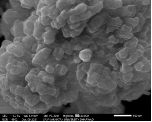
Figure 1. Scanning electron microscopic image of prepared cuo nanoparticles.
2.3. Synthesis of Nanoparticles
The CuO nanoprticles were prepared as reported elsewhere . By mixing slowly 0.5 M Na2CO3solution slowly at about 80°C with vigorous stirring to 0.5 M CuSO4solution the precursor was prepared. This results in basic copper sulphate precipitate, which was filtered, and washed to remove any ions left behind with doubly distilled water. Then it was filtered and mass was left for drying in air for 8 hours. The precursor was thermally decomposed in a muffle furnace for about 4 hours at 800°C that results in the CuO nanoparticles formation. Then the Scanning electron microscope image of the CuONPs was recorded and is as shown in Figure 1.
2.4. Analysis Method
By adding 2 mg of CuONPs and MWCNTs each in 10 mL acetonitrile, the CuONPs-MWCNTs suspension which is relatively stable was prepared using an ultrasonicator. The glassy carbon electrode was polished with the slurry of 0.05μm alumina powder on a polishing unit, and then kept in an ultrasonic bath containing mixture of methanol and water for 10 minutes, respectively. This cleaned electrode was initially coated by drop-coating 5 μL of the black suspension of CuONPs- MWCNTs and dried in open atmosphere.
The CuONPs-MWCNTs modified electrode was first activated in PBS with pH 7.4 by cyclic voltammetric runs from 0 to 0.8 V til stable voltammograms were obtained at a scan rate of 0.050 V s−1 (10 cycles). After that, electrodes are putted into another cell of PBS with pH 7.4, that contains accurate amounts of PCM. With an increment of 0.04 V, amplitude of 0.05 V, pulse width of 0.05 s, sample width of 0.0167 s and pulse period of 0.5 s, the differential pulse voltammograms were recorded from 0.0 and 0.8 V. All experiments were carried out at 25°C and in presence of dissolved oxygen.
3. Results and Discussion
3.1. Charecterization of Modified Electrode
A 1.0 mM solution of K3Fe(CN)6 in 0.1 M KCl was used to record CVs at CuONPs-MWCNT modified GCE at different scan rates. The below Randles-Sevcik equation for a reversible process was used to calculate electroactive surface area of bare and modified electrode.
Ipa= (2.69 × 10⁵)n3/2AD1/2Co ν1/2
where Ipais the anodic peak current, A is the electrode surface area, n is the number of electrons transferred, D is diffusion coefficient, ν is the scan rate and Co is the concentration of K3Fe(CN)6. In 0.1 M KCl electrolyte for 1.0 mM solution of K3Fe(CN)6, the values of n and D are 1 and 7.6 x 10⁻⁶ cm2s-1. By slope of the plot of Ip v/s ν1/2, the active surface areas obtained are 0.1462 cm2 for modified GCE and 0.03925 cm2 for bare GCE. From these values, it was concluded that active surface area has become significantly increased after fabrication.
3.2. Voltammetric Response of PCM
The DPV was recorded for 100 nM PCM with 10 μL of CuONPs-MWCNTs on GCE. Without nano particles, the oxidation peak at about 0.41 V was a bit weaker (Figure2(a)) than as compared with the peak in presence of nano particles at about 0.38 V, with significant enhancement in peak current and negative shift in peak potential (Figure 2(b)). The introduction of nano particlesonto the electrode effectively reduces the interface between the electrode and PCM. This reduction in the interface is clear from the enhancement in the peak current as indicated in (Figure 2(b)). The added nano particlesare facilitating the electro-oxidation process there by permitting the analyte to get to the surface of the electrode and take part in redox reaction more conveniently, by diminishing the interaction between surface of electrode and PCM.
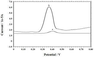
Figure 2. Differential pulse voltammogram of 100 nm pcm in 0.1 m pbs of ph 7.4 at (b) cuonps-mwcnts modified gce and (a) bare gce.
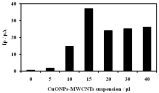
Figure 3. Plot of variation of volume of cuonps-mwcnts suspension with peak current.
3.3. Effect of Concentration of Nanoparticles
The consequence of quantity of CuONPs-MWCNTs suspension on anodic peak current was investigated. Highest oxidation peak current was obtained when 15 μl of suspension was employed for drop coating. The peak current increased significantly with increasing the amount of the suspension till 15 μl and afterwards, peak current decreased gradually and remain almost constant for higher concentrations of the suspension (Figure 3).
This was owing to the fact that, at higher quantity the film was thicker and that result in reduced conductivity which results in decreased oxidation current. Therefore, 15 μl of suspension was utilized for further studies. The effect of pH was investigated from 8.0 to 5.8 using PBS, since pH of the supporting electrolyte plays key role in the reactions which take place at the electrode. The investigation indicated that, the oxidation potential was shifted to lesser values with increase in pH of the reaction medium. There was no significant variation in peak current, but physiological pH was employed for further studies.
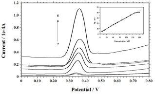
Figure 4. Differential pulse voltammograms with increasing quantities of pcm from (a) 9, (b) 20, (c) 40, (d) 60, (e) 80, (f) 120, AND (g) 160 nM AT CuONPS-MWCNTS Modified gce in pbs. inset: plot of peak current vs concentration of Pcm.
3.4. Calibration Graph
To develop a detection method using voltammetry for the estimation of PCM, DPV was used since the peaks are sharper and well defined using pulse techniques with lower background current. Using PBS as supporting electrolyte at physiological pH, voltammograms were recorded with increasing amount of PCM. The oxidation peak current roselinearly from 9.0 to 160 nM of PCM as shown in Figure 4. The calibration plot was constructed (Inset Figure 4.) which yields the equation Ip = 0.22 C + 3.58; R2 = 0.996 (C is in nM). It was also observed that, variation from linearity for the higher quantity of PCM, due to the adsorption of analyte or its product after electro-oxidation at the electrode surface.
Using the equations, 3s/m and 10s/m, the detection limit and quantification limits were computed. The obtained detection limit and quantification limit are 5.06 nM and 16.88 nM respectively. These acquired outcomes were matched up with previously reported values and were tabulated in Table 1.
3.5. Reproducibility Study
To study the reproducibility of the fabricated electrode, voltrammograms were recorded with 100 nM PCM at the fabricated electrode, which was re-fabricated each time for every several hours during a day. The relative standard deviation (RSD) of 2.13% was found that for anodic peak current for 12 number of runs. The reproducibility of results between days was analogous to that of within a day at constant temperature. However, the oxidation product was strongly adsorbing on to the electrode and hence the electrode has to be fabricated all over again after each run.
To substantiate the accuracy and precision of the above developed process, the assay of PCM was carried out during inter-day and intra-day. This was carried out at various amounts of PCM, i.e., at 10, 80 and 150 nM. Using DPV, voltammograms were recorded one time during a day (n = 8) for five successive days for the inter-day analysis and under alike state at the identical amounts of PCM (n = 8) for five times during a day to compute assay in a day.
During a day assay, the accuracy values obtained were ±1.20%, ±0.43%, and ±0.75% for low, medium and high concentrations levels with recovery values of 101.20%, 99.56% and 100.75%, respectively (Table 2). For inter-day assay, the accuracy values of ±0.60%, ±0.53% and ±0.58% were got with recovery values of 99.40%, 100.53% and 99.42% for low, medium and high quantity of PCM, respectively. The average precision values obtained werein the range of 4.67% to 10.24% (Table 2). The precision and accuracy values computed were within ±15%, and are hence within tolerable limits .
Table 1. Comparison of analytical parameters for the detection of PCM by voltammetry at various electrodes.

Electrode

Method

LDR (μM)

LOD (μM)

Ref.

ePAD

DPV

1-60

0.2

13

AgNPs@HOOC-MWCNT@SPCE

SWV

0.5-1000

0.24

14

Stv-CPE

DPV

0.6-100

0.2

15

BGE

DPV

5-150

0.2

16

Polyglycine-GCE

DPV

0.5-75

0.03

17

SPCE

SWV

---

1.2

18

Guanine-GCE

DPV

0.005-10

0.9

19

MWCNT/GO/Poly(Thr)/GCE

DPV

3-140

0.16

20

ERGO-GCE

DPV

---

0.14

21

C-HAP-GCE

DPV

0.01-1310

0.139

22

NiCoSalenA/CPE

DPV

1.71-32.5

0.51

23

SDS/CuONPs-MWCNTs/GCE

DPV

0.009-0.16

0.005

This work

ePAD - Electrochemical paper based analytical device; AgNPs – Silver nanoparticles; MWCNT – Multi walled carbon nanotubes; SPCE – Screen printed carbon electrode; Stv-Stevensite; CPE- Carbon paste electrode; BGE – Bare graphite electrode; GCE – Glassy carbon electrode; GO – Graphene oxide; ERGO – Elctrochemically reduced graphene oxide; HAP – Hydroxyapatite; NiCoSalenA - Nickel-Cobalt Salennanozeolite; PGE – Pencil graphite electrode; NiZ – Nickel ion zeolite; LSG – Laser scribed graphene; CCE- Carbon cloth electrode; SDS – Sodium dodecyl sulphate; LDR – Linear dynamic range; LOD – Limit of detection; DPV – Differential pulse voltammetry; SWV – Square wave voltammetry
Table 2. Statistics obtained for PCM assay during a day and inter-day.

Quantity (nM)

Estimated quantity (nM)

Recovery (%)

Accuracy (%)

Precision(%RSD)

Intra-day

10

10.12±0.42

101.20

±1.20

6.13

80

79.65±0.64

99.56

±0.43

9.75

150

151.13±.32

100.75

±0.75

5.32

Inter-day

10

9.94±0.21

99.40

±0.60

10.24

80

80.43±0.17

100.53

±0.53

4.67

150

149.13±0.35

99.42

±0.58

8.74

3.6. Detection in Pharmaceutical Formulations
By following the identical conditions described above, the assessment of the analyte was carried out. The stock solution was diluted with doubly distilled water so that PCM concentration fall in LDR and introduced into the cell containing accurate quantities of PBS of pH 7.4. DPV runs were recorded and standard addition method was used to compute the concentration of PCM in tablet samples. The computed results were as shown in Table 3. It was found that, PCM concentrations obtained by the present method were matching with the labeled claim.
Table 3. Results computed for PCM assay in tablet dosage forms.

Paracipa

Doloa

Pyrigestic

Labeled claim (mg)

500

650

500

Amount found (mg)b

495

653

502

Added (nM)

140

140

140

Found (nM)

139

142

141

Recovered (%)b

99.28

101.42

100.71

aName of tablets
bMean of six experiments
4. Conclusion
In the present effort, a glassy carbon electrode was fabricated with CuO nanoparticles-Multi walled carbon nanotubes for the electro-oxidation and detection of PCM in PBS. The fabricated GCE demonstrated electro-catalytic nature for the electro-oxidation of PCM, as indicated by improvement in anodic peak current. This modified electrode showed higher sensitivity and good recovery values. The PCM present in various pharmaceutical formulations were detected using the developed method productively.
Abbreviations

CuONPs

CuO Nanoparticles

GCE

Glassy Carbon Electrode

MWCNTs

Multi-walled Carbon Nanotubes

PCM

Paracetamol

PBS

Phosphate Buffer Solution

Acknowledgments
The authors acknowledge the financial support from Department of Science and Technology, New Delhi – 110016 under Level 0 of Funds for Improvement of S & T Infrastructure in Universities and Higher Educational Institutions (FIST) grant (Letter No: SR/FST/COLLEGE-/2020/914 dated 5th March 2021).
Author Contributions
Rajesh N Hegde: Conceptualization, Data curation, Investigation, Methodology, Supervision, Validation, Writing – original draft, Writing – review & editing
P Vishwanatha: Supervision, Writing – review & editing
Kiran Kamath: Formal Analysis, Investigation, Resources
Conflicts of Interest
The authors declare no conflicts of interest.
References
[1] C. D. van der Marel, B. J. Anderson, and R. A. van Lingen, Paracetamol and metabolite pharmacokinetics in infants. European Journalof Clinical Pharmacology, 59, 243-251(2003).
[2] M. R. Siddiqui, Z. A. Othman, and N. Rahman, Analytical techniques in pharmaceutical analysis: A review. Arab Journal of Chemistry, 10, S1409- S1421(2017).
[3] S. Sharma, N. Singh, and A. D. Ankalgi, Modern Trends in Analytical Techniques for Method Development and Validation of Pharmaceuticals: A Review Journal of Drug Delivery and Therapy, 11, 121-130(2021).
[4] A. Chavan, and R. Gandhimathi, Modern analytical tools for the determination of the active pharmaceutical ingredients: a review. International Journal of Pharmaceutical Science Research, 13, 61-69(2022).
[5] S. Ahmed, M. S. Islam, and B. Ullah, A Review Article on Pharmaceutical Analysis of Pharmaceutical Industry According to Pharmacopoeias. Orient Journal of Chemistry, 36, 1-10(2020).
[6] M. Majidian, G. Ozcelikay, and A. Cetinkaya, Nanomaterial-based electrochemical sensing platform for the determination of Olaparib. Electrochimica Acta, 449, 142198(2023).
[7] B. Davani, Pharmaceutical Analysis for Small Molecules, 1, 37(2017), John Wiley & Sons, Inc.
[8] S. Michalkiewicz, A. Skorupa, and M. Jakubczyk, Carbon Materials in Electroanalysis of Preservatives: A Review. Materials, 14, 7630(2021).
[9] J. Wang, Analytical Electrochemistry, 2000, 2nd edition, Wiley-VCH, New York, USA.
[10] R. N. Hegde, P. Vishwanatha, and S. T. Nandibewoor, Voltammetric Assessment of 8-Oxoguanine at a Nano-Structured Carbon Materials Based Modified Glassy Carbon Electrode. Brazilian Journal of Analytical Chemistry, 9, 84-93(2022).
[11] L. Qian, S. Durairaj, and S. Prins, Nanomaterial-based electrochemical sensors and biosensors for the detection of pharmaceutical compounds. Biosensors and Bioelectronics, 175, 112836(2021).
[12] P.A. Pushpanjali, J. G. Manjunatha, and N. Hareesha, An overview of recent developments of carbon-based sensors for the analysis of drug molecules. Journal of ElectrochemicalScience Engineering, 2021, 11, 161-177.
[13] L. C. Oliveira, D. S. Rocha, and H. A. Silva-Neto, Polyester resin and graphite flakes: turning conductive ink to a voltammetric sensor for paracetamol sensing.MicrochimicaActa, 190, 324(2023).
[14] S. Weheabby, Z. Wu, and A. Al-Hamry, Paracetamol detection in environmental and pharmaceutical samples using multi-walled carbon nanotubes decorated with silver nanoparticles. Microchemical Journal, 193, 109192(2023).
[15] M. Gharous, L. Bounab, and F. J. Pereira, Electrochemical Kinetics and Detection of Paracetamol by Stevensite-Modified Carbon Paste Electrode in Biological Fluids and Pharmaceutical Formulations. International Journal of Molecular Science, 24, 11269(2023).
[16] M. Stoytcheva, R. Zlatev, and Z. Velkova, The validity of using bare graphite electrode for the voltammetric determination of paracetamol and caffeine. International Journal of Electrochemical Science, 18, 100120(2023).
[17] N.İslamoğlu, I. E. Mülazımoğlu, and A. D. Mülazımoğlu, Sensitive and selective determination of paracetamol in antipyretic children's syrup with a polyglycine modified glassy carbon electrode.Analytical Methods, 15, 4149-4158(2023).
[18] K. Cortés, J. J. Triviño, and V. Arancibia, Simultaneous voltammetric determination of acetylsalicylic acid, caffeine and paracetamol in pharmaceutical formulations using screen-printed carbon electrode. Electroanalysis, 35, e202200484 (2023).
[19] N. Islamoglu, and A. D. Mulazımoglu, Use of guanine-modified glassy carbon electrode as an electrochemical sensor for the determination of paracetamol. Bangladesh Journal of Pharmacology, 18, 97-104(2023).
[20] G. V. Prasad, V. Vinothkumar, and S. J.Jang, Multi-walled carbon nanotube/graphene oxide/poly (threonine) composite electrode for boosting electrochemical detection of paracetamol in biological samples. Microchemical Journal, 184, 108205(2023).
[21] R. M. Silva, G. H. Sperandio, and A. D. da Silva, Electrochemically reduced graphene oxide films from Zn-C battery waste for the electrochemical determination of paracetamol and hydroquinone. Microchimica Acta, 190, 273(2023).
[22] S. Anitta, and C. Sekar, Voltammetric determination of paracetamol and ciprofloxacin in the presence of vitamin C using cuttlefish bone-derived hydroxyapatite sub-microparticles as electrode material. Results in Chemistry, 5, 100816(2023).
[23] N. Masihpour, S. K. Hassaninejad-Darzi, and A. Sarvary, Nickel-Cobalt Salen Organometallic Complexes Encapsulated in Mesoporous NaANanozeolite for Electrocatalytic Quantification of Ascorbic Acid and Paracetamol. Journal of Inorganic andOrganometalic Polymers and Materials, 33, 2661-2680(2023).
[24] E. Darezereshki, and F. Bakhtiari, A novel technique to synthesis of tenorite (CuO) nanoparticles from low concentration CuSO4 solution. Journal of Mining and Metallurgy Section B: Metallurgy, 47, 73-78(2011).
[25] Bioanalytical method validation: guidance for industry,
Cite This Article
  • APA Style

    Hegde, R., Poojary, V., Kamath, K. (2024). Voltammetric Assessment of Paracetamol on a CuONPs – MWCNTs Modified Glassy Carbon Electrode. American Journal of Physical Chemistry, 13(3), 59-65. https://doi.org/10.11648/j.ajpc.20241303.11

    Copy | Download

    ACS Style

    Hegde, R.; Poojary, V.; Kamath, K. Voltammetric Assessment of Paracetamol on a CuONPs – MWCNTs Modified Glassy Carbon Electrode. Am. J. Phys. Chem. 2024, 13(3), 59-65. doi: 10.11648/j.ajpc.20241303.11

    Copy | Download

    AMA Style

    Hegde R, Poojary V, Kamath K. Voltammetric Assessment of Paracetamol on a CuONPs – MWCNTs Modified Glassy Carbon Electrode. Am J Phys Chem. 2024;13(3):59-65. doi: 10.11648/j.ajpc.20241303.11

    Copy | Download

  • @article{10.11648/j.ajpc.20241303.11,
      author = {Rajesh Hegde and Vishwanatha Poojary and Kiran Kamath},
      title = {Voltammetric Assessment of Paracetamol on a CuONPs – MWCNTs Modified Glassy Carbon Electrode
    },
      journal = {American Journal of Physical Chemistry},
      volume = {13},
      number = {3},
      pages = {59-65},
      doi = {10.11648/j.ajpc.20241303.11},
      url = {https://doi.org/10.11648/j.ajpc.20241303.11},
      eprint = {https://article.sciencepublishinggroup.com/pdf/10.11648.j.ajpc.20241303.11},
      abstract = {In this work, an electrochemical sensor using differential pulse voltammetric method for the assessment of antipyretic and analgesic drug, paracetamol was developed. The CuO nanoparticles were synthesized and characterized. A glassy carbon electrode (GCE) fabricated with the suspension of CuO nanoparticles (CuONPs) and multi-walled carbon nanotubes (MWCNTs) were used. The fabricated electrode was characterized using Potassium ferricyanide as a redox probe, which showed increase in the electro active area in the modified electrode. The modified electrode showed improved anodic peak current enhancement in phosphate buffer solution. The consequence of pH of supporting electrolyte and amount of nanoparticles suspension were investigated at a physiological pH of 7.4. Using differential pulse voltammetry, the fabricated electrode showed linear dynamic range from 9 to 160 nM of paracetamol concentration. From the calibration plot, the computed detection limit was 5.06nM and quantification limit was16.88 nM. The developed method was checked for its reproducibility and assay during a day and intraday as well and the results were good with permitted range of errors. The developed process was fruitfully applied to detect paracetamol in pharmaceutical formulations.
    },
     year = {2024}
    }
    

    Copy | Download

  • TY  - JOUR
    T1  - Voltammetric Assessment of Paracetamol on a CuONPs – MWCNTs Modified Glassy Carbon Electrode
    
    AU  - Rajesh Hegde
    AU  - Vishwanatha Poojary
    AU  - Kiran Kamath
    Y1  - 2024/08/15
    PY  - 2024
    N1  - https://doi.org/10.11648/j.ajpc.20241303.11
    DO  - 10.11648/j.ajpc.20241303.11
    T2  - American Journal of Physical Chemistry
    JF  - American Journal of Physical Chemistry
    JO  - American Journal of Physical Chemistry
    SP  - 59
    EP  - 65
    PB  - Science Publishing Group
    SN  - 2327-2449
    UR  - https://doi.org/10.11648/j.ajpc.20241303.11
    AB  - In this work, an electrochemical sensor using differential pulse voltammetric method for the assessment of antipyretic and analgesic drug, paracetamol was developed. The CuO nanoparticles were synthesized and characterized. A glassy carbon electrode (GCE) fabricated with the suspension of CuO nanoparticles (CuONPs) and multi-walled carbon nanotubes (MWCNTs) were used. The fabricated electrode was characterized using Potassium ferricyanide as a redox probe, which showed increase in the electro active area in the modified electrode. The modified electrode showed improved anodic peak current enhancement in phosphate buffer solution. The consequence of pH of supporting electrolyte and amount of nanoparticles suspension were investigated at a physiological pH of 7.4. Using differential pulse voltammetry, the fabricated electrode showed linear dynamic range from 9 to 160 nM of paracetamol concentration. From the calibration plot, the computed detection limit was 5.06nM and quantification limit was16.88 nM. The developed method was checked for its reproducibility and assay during a day and intraday as well and the results were good with permitted range of errors. The developed process was fruitfully applied to detect paracetamol in pharmaceutical formulations.
    
    VL  - 13
    IS  - 3
    ER  - 

    Copy | Download

Author Information
  • Abstract
  • Keywords
  • Document Sections

    1. 1. Introduction
    2. 2. Materials and Methods
    3. 3. Results and Discussion
    4. 4. Conclusion
    Show Full Outline
  • Abbreviations
  • Acknowledgments
  • Author Contributions
  • Conflicts of Interest
  • References
  • Cite This Article
  • Author Information