Pellucid marginal degeneration (PMD) is a bilateral, peripheral corneal ectasia characterized by inferior corneal thinning and subsequent irregular astigmatism, often leading to poor visual outcomes with glasses or traditional contact lenses. This case report presents a 45-year-old female previously misdiagnosed with keratoconus and advised to consider corneal transplantation due to poor visual acuity and intolerance to earlier contact lens fittings. Comprehensive examination revealed PMD, and the patient was successfully fitted with Onefit scleral contact lenses. The lenses provided significant improvement in both vision and comfort, enhancing her quality of life and eliminating the need for surgical intervention. This case highlights the importance of accurate diagnosis and the efficacy of scleral lenses, particularly the Onefit design, in managing moderate to advanced PMD cases.
This is an Open Access article, distributed under the terms of the Creative Commons Attribution 4.0 International License (http://creativecommons.org/licenses/by/4.0/), which permits unrestricted use, distribution and reproduction in any medium or format, provided the original work is properly cited.
Pellucid marginal degeneration (PMD) is a bilateral, non-inflammatory, ectatic disease characterized by thinning of the peripheral cornea.PMD typically presents with an area of inferior thinning separated from the limbus by 1-2mm. The incidence or prevalence of PMD is not clearly reported as it is often misdiagnosed as keratoconus due the similarities in clinical presentation
[1]
Jinabhai, A., Radhakrishnan, H., & O’Donnell, C. (2011). Pellucid corneal marginal degeneration: A Review. Contact Lens and Anterior Eye, 34(2), 56–63.
. The Collaborative Longitudinal Evaluation of Keratoconus (CLEK) study established that those with corneal ectasias have a significantly impaired vision-related quality of life
[3]
Rathi VM, Dumpati S, Mandathara PS, Taneja MM, Sangwan VS. Scleral contact lenses in the management of pellucid marginal degeneration. Cont Lens Anterior Eye. 2016 Jun; 39(3): 217-20.
[3]
. Given the significant impact of PMD and other ectasias, an increasing number of patients and practitioners have turned to specialty contact lenses for visual rehabilitation.
PMD results in irregular astigmatism, causing aberrations that are not correctable by standard glasses or contact lenses
[2]
Zadnik K, Barr JT, Edrington TB, Everett DF, Jameson M, McMahon TT, Shin JA, Sterling JL, Wagner H, Gordon MO, Collaborative Longitudinal Evaluation of Keratoconus (CLEK) Study Group. Baseline findings in the Collaborative Longitudinal Evaluation of Keratoconus (CLEK) Study. Invest Ophthalmol Vis Sci 1998; 39: 2537-46.
[2]
. Scleral lenses are large diameter gas permeable lenses that mask optical distortion by allowing a tear lens to form between the posterior surface of the contact lens and the irregular surface of the cornea. Advantages of scleral lenses include their ability to clear the cornea and provide vision free from distortion.
The fitting of contact lenses in PMD is in general more challenging than in other corneal degenerations due to the large inferior protrusion of the cornea
[3]
Rathi VM, Dumpati S, Mandathara PS, Taneja MM, Sangwan VS. Scleral contact lenses in the management of pellucid marginal degeneration. Cont Lens Anterior Eye. 2016 Jun; 39(3): 217-20.
[3]
. Traditional rigid gas permeable (RGP) lenses can be difficult to fit in PMD as the inferior location of the apex of the cone and large area of corneal involvement cause inferior decentration of the lens
[3]
Rathi VM, Dumpati S, Mandathara PS, Taneja MM, Sangwan VS. Scleral contact lenses in the management of pellucid marginal degeneration. Cont Lens Anterior Eye. 2016 Jun; 39(3): 217-20.
[3]
. Because scleral lenses vault the cornea entirely, they can eliminate decentration challenges and are useful in the management of PMD.
2. Case Report
A 45 year female presented to our hospital for a second opinion on 26/08/2022. She reported good general health. She had no known medical allergies. She entered this process knowing that she had keratoconus which was diagnosed approximately 5 years ago. However, it had been at least 5 years since her last eye exam and old records and testing could not be located. She was a full-time glasses wearer but had noticed her vision declining, particularly while doing her daily activity work and also difficulty to work as a shopkeeper. Her new glasses wouldn’t help due to her keratoconus, she was told she might be a good candidate for a corneal transplant. She further explained that several doctors, many years ago, had attempted to correct her vision with hard contact lenses, but the vision and comfort were not acceptable and she returned to glasses wear. Her main goal was simply to avoid surgery and also make her life easier also needed to have the visual capacity to make sales on her shop.
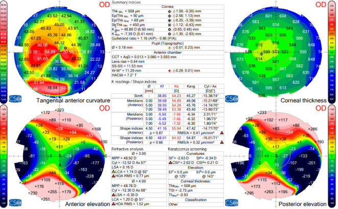
Figure 1. Corneal Topography of OD from 8th June 2022. Kmax 48.89D with classic “Crab-claw” or “Kissing Doves” topographic pattern on tangential anterior curvature.
At her initial visit, we recorded vision at 6/18 in right eye and 3/60 in left eye without correction and no improvement with glasses. Her Pupils, ocular motility, confrontation fields, and ocular alignment were normal. Intraocular pressure was obtained at 11:45 am and measured 16 mmHg in both eyes with applanation tonometry. Retinoscopy showed a characteristic scissor reflex. Manifest refraction was performed and very difficult for both patient and practitioner. Manifest Refraction OD: +2.25 – 10 X 80 and OS: +1.50 – 10 X 95; BCVA: 6/18/ BCVA: 3/60. Anterior segment evaluation showed mild blepharitis with lash scurf as well as eyelid margin telangiectasia. There was mild conjunctivochalasis in both eyes, but an otherwise clear and quiet anterior surface. Her corneas were also clear in both eyes with no scarring, striae, or Fleischer’s ring noted. Both corneas, however, had significant inferior bulging and sagging as evident with slit lamp and gross examination although no clear Munson sign was present. She was dilated with 1 drop of tropica plus in each eye (Tropic amide and Phenylephrine Ophthalmic Solution-0.8%). Internal evaluation showed grade 1+ nuclear sclerotic cataracts in both eyes. Optic nerves were healthy and well perfused with a cup to disk ratio of 0.3 in each eye. The macula in both eyes looked normal with slit lamp, and the peripheral retinas in both eyes were unremarkable. Corneal topography was ordered and performed at this visit with the MS-39 Zeus topographer.
(The Zeus MS-39 is a state-of-the-art corneal topographer and tomographer used in ophthalmology and optometry. It combines Placido-disk corneal topography with anterior segment OCT (optical coherence tomography) to deliver highly accurate and detailed imaging of the anterior eye segment.)
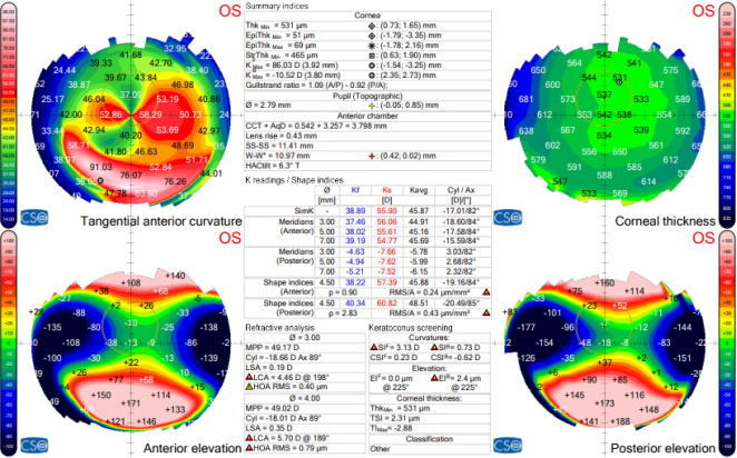
So of right eye MS-39 Zeus showed the keratometric value of K1 40.09D @ 76 and K2 55.43D @ 166 and average OD corneal astigmatism of -15.34 D @ 76 where the HVID o 11.21mm. So of left eye MS-39 Zeus showed the keratometric value of K1 38.51D @ 88 and K2 56.66D @ 178 and average OD corneal astigmatism of -18.15D @ 88 where the HVID o 11.03mm.
Figure 2. Corneal Topography of OS from 8th June 2022. Kmax 86.03D with classic “Crab-claw” or “Kissing Doves” topographic pattern on tangential anterior curvature.
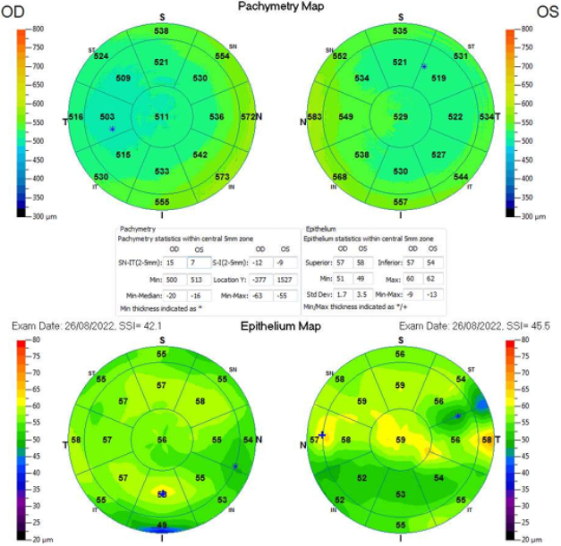
So for the corneal thickness Optovue had performed where the central corneal thickness of OD is 499 micrometers and OS is 513 micrometers and the central corneal epithelium thickness of OD is 58 micrometers and OS is 59 micrometers.
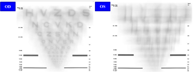
In the right eye, RMS value of lower aberrations was 8.35 micrometers and higher order aberrations was 2.65 micrometers in 6mm pupil. In the left eye, RMS value of lower aberrations was 11.79 micrometers and higher order aberrations was 1.17 micrometers in 6mm pupil.
Figure 4. Visual quality simulator for Right and Left Eye before lens fit with 6mm pupil.
4. Discussion
Pellucid marginal degeneration (PMD) is a corneal ectasia oftentimes seen as, or confused with, the more common ectasia, Keratoconus (KCN). There are unique characteristics between the two that distinguish the conditions. This is not just a distinction without a difference as proper diagnosis is important to understanding the likely course of the disease, as well as choosing the best treatment route, whether surgically or with medically necessary contact lenses
[4]
Imbornoni LM, Padmanabhan P, Belin MW, Deepa M. “Long-Term Tomographic Evaluation of Unilateral Keratoconus.” Cornea. 2017 Jul 24.
[5]
Godefrooij DA, de Wit GA, Uiterwaal CS, Imhof SM, Wisse RP. “Age-specific Incidence and Prevalence of Keratoconus: A Nationwide Registration Study.” Am J Ophthalmol. 2017 Mar; 175: 169-172.
[4, 5]
.
There are many similarities between the conditions. Both PMD and KCN cause non inflammatory breakdown of the collagen matrix of the cornea that leads to thinning and subsequent bulging of the cornea. Both conditions are usually bilateral although cases of unilaterality are not rare and both conditions are linked to allergic and vigorous eye rubbing. Additionally, both conditions have a mild male predilection but have no other ethnic, racial, socioeconomically, or geographical predisposition
[6]
Barr JT, Wilson BS, Gordon MO, et al. “Estimation of the incidence and factors predictive of corneal scarring in the Collaborative Longitudinal Evaluation of Keratoconus (CLEK) Study. Cornea 2006; 25: 16-25.
[7]
Nicholas Wolf, O. D. Pellucid Marginal Degeneration Challenges Overcome with Scleral Contact Lenses A Case Report on Avoiding a Penetrating Keratoplasty”
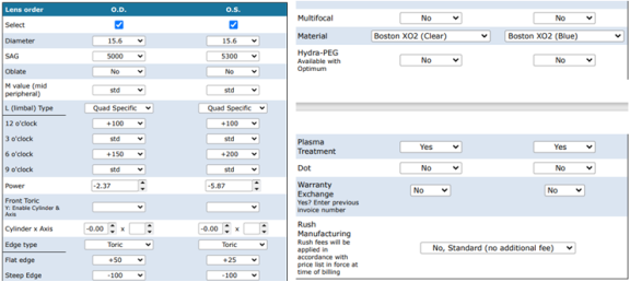
Onefit MED are intuitive and easy to use scleral lenses that allows practitioners to customize the ideal lens for a wide range of applications, when a larger diameter is needed for a healthy, highly irregular or medically indicated cornea. Designed to put the practitioner in control of the design and fit, adjustments can be made in four (4) separate zones of the lens: central, mid-peripheral, limbal and landing zone (Edge). To easily find the exact location of the mid-peripheral and limbal zones, the diagnostic lenses are etched with solid lines that can be observed at the slit lamp or OCT image. Onefit MED and Onefit MED minimize both lens thickness and fluid reservoir, maximizing oxygen transmission to the cornea and stem cells. The design serves as its own platform from which Multifocal, Oblate, Front Toric, Toric Haptic, Quadrant Specific and Controlled Peripheral Recess (NOTCHING) can be done. Onefit MED put you in the driver seat, are extremely easy to fit, save you chair time and provide the patient with exceptional visual acuity, comfort and optimal oxygen to the cornea for long-term corneal health. Onefit MED trial set consisted of 20 lenses having various combination of SAG/ POWER/ OBLATE/LIMBAL/ EDGE/ DIAMETER/ CPR/ MID and different alignment haptic. Diameter ranges from 15.6mm, 16mm and 16.4. Additionally customization is done as per requirement. Lenses in trial set consist of torric haptic. These can be further modified as per the requirement of practioner. Sag provided in the trial sets are sufficient enough to vault highly protruded cornea such as in pathological conditions like kerataglobus, corneal ectasia, PMD, Corneal Scar etc. All lenses in the trial set were a standard edge lift. Onefit MED lens was taken with the parameters of SAG OF 4900; total diameter of 15.6mm, back vertex power of -5.00 DS and standard edge lift in RE with VA 6/6p and for left eye SAG of 5000; total diameter of 15.6mm, back vertex power of -6.00 DS and standard edge lift in LE with VA 6/6p for near vision reading glasses was provided as per required of her. For finalizing the lens online calculation was done from (https://prod.blcalculator.com/blanchard/onefitmed/fittingtool/) and while fitting the lens some more customized had been done for final parameters.
Figure 6. Visual quality simulator for Right and Left Eye after lens fit with 6mm pupil.
In the right eye, RMS (Root Mean Square) value of lower aberrations was 0.00 micrometers and higher order aberrations was 0.00 micrometers in 6mm pupil. In the left eye, RMS value of lower aberrations was 0.00 micrometers and higher order aberrations was 0.00 micrometers in 6mm pupil.
5. Conclusions
PMD and KCN are often grouped together but have several important distinctions, including the location of steepest protrusion and the percentage of cornea involved. Because of these distinguishing features, PMD is notoriously more difficult to fit in contact lenses and is associated with a higher rate of graft rejection compared with KCN
[8]
Speaker MG, Arentsen JJ, Laibson PR. Long-term survival of large diameter penetrating keratoplasties for keratoconus and pellucid marginal degeneration. Acta Ophthalmol Suppl 1989; 192: 17–9.
[8]
. Pellucid marginal degeneration is an uncommon cornea ectasia that can be differentiated from keratoconus by the history, topography, and clinical presentation. This distinction is important as these two very similar conditions can have a very different clinical arc. Due to the often severe inferior corneal thinning and resultant steepening with PMD, onefit scleral contact lenses are an excellent choice. While small diameter RPGs and hybrids are an option with milder PMD, and onefit scleral lenses should be the first choice for patients with more significant disease. It was an interesting case in that her first successful contact lens experience was with a scleral contact lens at the age of 45 and while correctly diagnosed with an ectasia, PMD was the more appropriate diagnosis and better matches her clinical presentation. Her continued success, both clinically and in her day to day life, underscores that onefit scleral contact lenses are an effective and user-friendly lens modality for pellucid marginal degeneration patients.
Abbreviations
PMD
Pellucid Marginal Degeneration
CLEK
Collaborative Longitudinal Evaluation of Keratoconus Study
RGP
Rigid Gas Permeable
OD
Oculus Dexter
OS
Oculus Sinister
BCVA
Best Corrected Visual Acuity
OCT
Optical coherence tomography
K1
Keratometry1
K2
Keratomerty2
HVID
Horizontal Iris Visible Diameter
MM
Millimeter
KCN
Keratoconus
RMS
Root Mean Square
Acknowledgments
The authors did not receive any funding, grants, or other financial support from any organization for the preparation of this research. The first author is pursuing a Ph.D. in the Department of Optometry, Chandigarh University, while the second author is the co-supervisor and an Assistant Professor in the Department of Mathematics, Chandigarh University. The authors sincerely express their gratitude to the editor and the reviewers for their valuable comments and constructive suggestions, which have significantly enhanced the clarity, quality, and overall presentation of this paper.
The authors declare that there are no conflicts of interest regarding the conduct of this research, the interpretation of the results, or the preparation of this manuscript. No financial, commercial, or personal relationships were involved that could be perceived as influencing the work.
References
[1]
Jinabhai, A., Radhakrishnan, H., & O’Donnell, C. (2011). Pellucid corneal marginal degeneration: A Review. Contact Lens and Anterior Eye, 34(2), 56–63.
Zadnik K, Barr JT, Edrington TB, Everett DF, Jameson M, McMahon TT, Shin JA, Sterling JL, Wagner H, Gordon MO, Collaborative Longitudinal Evaluation of Keratoconus (CLEK) Study Group. Baseline findings in the Collaborative Longitudinal Evaluation of Keratoconus (CLEK) Study. Invest Ophthalmol Vis Sci 1998; 39: 2537-46.
[3]
Rathi VM, Dumpati S, Mandathara PS, Taneja MM, Sangwan VS. Scleral contact lenses in the management of pellucid marginal degeneration. Cont Lens Anterior Eye. 2016 Jun; 39(3): 217-20.
[4]
Imbornoni LM, Padmanabhan P, Belin MW, Deepa M. “Long-Term Tomographic Evaluation of Unilateral Keratoconus.” Cornea. 2017 Jul 24.
[5]
Godefrooij DA, de Wit GA, Uiterwaal CS, Imhof SM, Wisse RP. “Age-specific Incidence and Prevalence of Keratoconus: A Nationwide Registration Study.” Am J Ophthalmol. 2017 Mar; 175: 169-172.
[6]
Barr JT, Wilson BS, Gordon MO, et al. “Estimation of the incidence and factors predictive of corneal scarring in the Collaborative Longitudinal Evaluation of Keratoconus (CLEK) Study. Cornea 2006; 25: 16-25.
[7]
Nicholas Wolf, O. D. Pellucid Marginal Degeneration Challenges Overcome with Scleral Contact Lenses A Case Report on Avoiding a Penetrating Keratoplasty”
Speaker MG, Arentsen JJ, Laibson PR. Long-term survival of large diameter penetrating keratoplasties for keratoconus and pellucid marginal degeneration. Acta Ophthalmol Suppl 1989; 192: 17–9.
Dhankhoti, K. K., Kumari, S., Kunwar, A., Dhital, R. (2025). Pellucid Marginal Degeneration (PMD) Challenges Overcome with Onefit Scleral Contact Lenses. International Journal of Clinical and Experimental Medical Sciences, 11(5), 88-94. https://doi.org/10.11648/j.ijcems.20251105.15
@article{10.11648/j.ijcems.20251105.15,
  author = {Krishna Kumar Dhankhoti and Santoshi Kumari and Amit Kunwar and Rasmila Dhital},
  title = {Pellucid Marginal Degeneration (PMD) Challenges Overcome with Onefit Scleral Contact Lenses
},
  journal = {International Journal of Clinical and Experimental Medical Sciences},
  volume = {11},
  number = {5},
  pages = {88-94},
  doi = {10.11648/j.ijcems.20251105.15},
  url = {https://doi.org/10.11648/j.ijcems.20251105.15},
  eprint = {https://article.sciencepublishinggroup.com/pdf/10.11648.j.ijcems.20251105.15},
  abstract = {Pellucid marginal degeneration (PMD) is a bilateral, peripheral corneal ectasia characterized by inferior corneal thinning and subsequent irregular astigmatism, often leading to poor visual outcomes with glasses or traditional contact lenses. This case report presents a 45-year-old female previously misdiagnosed with keratoconus and advised to consider corneal transplantation due to poor visual acuity and intolerance to earlier contact lens fittings. Comprehensive examination revealed PMD, and the patient was successfully fitted with Onefit scleral contact lenses. The lenses provided significant improvement in both vision and comfort, enhancing her quality of life and eliminating the need for surgical intervention. This case highlights the importance of accurate diagnosis and the efficacy of scleral lenses, particularly the Onefit design, in managing moderate to advanced PMD cases.
},
 year = {2025}
}
TY  - JOUR
T1  - Pellucid Marginal Degeneration (PMD) Challenges Overcome with Onefit Scleral Contact Lenses

AU  - Krishna Kumar Dhankhoti
AU  - Santoshi Kumari
AU  - Amit Kunwar
AU  - Rasmila Dhital
Y1  - 2025/10/14
PY  - 2025
N1  - https://doi.org/10.11648/j.ijcems.20251105.15
DO  - 10.11648/j.ijcems.20251105.15
T2  - International Journal of Clinical and Experimental Medical Sciences
JF  - International Journal of Clinical and Experimental Medical Sciences
JO  - International Journal of Clinical and Experimental Medical Sciences
SP  - 88
EP  - 94
PB  - Science Publishing Group
SN  - 2469-8032
UR  - https://doi.org/10.11648/j.ijcems.20251105.15
AB  - Pellucid marginal degeneration (PMD) is a bilateral, peripheral corneal ectasia characterized by inferior corneal thinning and subsequent irregular astigmatism, often leading to poor visual outcomes with glasses or traditional contact lenses. This case report presents a 45-year-old female previously misdiagnosed with keratoconus and advised to consider corneal transplantation due to poor visual acuity and intolerance to earlier contact lens fittings. Comprehensive examination revealed PMD, and the patient was successfully fitted with Onefit scleral contact lenses. The lenses provided significant improvement in both vision and comfort, enhancing her quality of life and eliminating the need for surgical intervention. This case highlights the importance of accurate diagnosis and the efficacy of scleral lenses, particularly the Onefit design, in managing moderate to advanced PMD cases.

VL  - 11
IS  - 5
ER  -