Review Article | | Peer-Reviewed

Anomalous Coronary Arteries from the Opposite Sinus of Valsalva: A Case Series and Comprehensive Review

Received: 17 February 2026     Accepted: 2 March 2026     Published: 19 March 2026
Views:       Downloads:
Abstract

Anomalous coronary artery from the opposite sinus (ACAOS) is a rare congenital abnormality that may be encountered during routine coronary angiography. Clinical manifestations vary depending on the subtype and anatomical course of the anomaly; however, certain potentially malignant trajectories may result in myocardial ischemia, infarction, or sudden cardiac death, particularly in young individuals and athletes. Multiple imaging modalities including coronary angiography, CT angiography, and cardiac MRI are useful in the evaluation of coronary artery anomalies. Accurate delineation of the origin and course of the anomalous vessel is essential for risk stratification and therapeutic planning. Performing percutaneous coronary intervention (PCI) in patients with ACAOS can be technically challenging and often requires modifications in guide catheter selection and procedural technique. In this review, we present a series of patients with anomalous coronary arteries originating from the opposite sinus of Valsalva (ACAOS). We discuss the anatomical variations and courses of these anomalous vessels as defined by angiographic and advanced imaging findings. Particular emphasis is placed on guide catheter selection, technical challenges encountered during selective cannulation, and the strategies employed to achieve successful engagement and intervention.

Published in International Journal of Cardiovascular and Thoracic Surgery (Volume 12, Issue 2)
DOI 10.11648/j.ijcts.20261202.14
Page(s) 40-54
Creative Commons

This is an Open Access article, distributed under the terms of the Creative Commons Attribution 4.0 International License (http://creativecommons.org/licenses/by/4.0/), which permits unrestricted use, distribution and reproduction in any medium or format, provided the original work is properly cited.

Copyright

Copyright © The Author(s), 2026. Published by Science Publishing Group

Keywords

Anomalous Coronary Artery, ACAOS, Coronary CT Angiography, Percutaneous Coronary Intervention

1. Introduction
Anomalous coronary artery from the opposite sinus (ACAOS) is uncommon, and the separate origin of all three coronary arteries from the right sinus of Valsalva is exceptionally rare. The estimated incidence of coronary artery anomalies (CAAs) is 1% to 2% in the general population . The clinical manifestations are highly variable, ranging from no symptoms at all to exertional chest discomfort, breathlessness, arrhythmias, or even sudden cardiac death, depending on the anatomical path and characteristics of the anomalous vessel. The presence of ACAOS not only prolongs the procedural time but also increases the volume of contrast use and delays the revascularisation procedure. In this review, we begin by presenting a case series of patients with ACAOS, followed by a detailed discussion on the angiographic identification of anomalous coronary courses, recognition of malignant pathways, underlying mechanisms of ischemia, and the technical challenges associated with percutaneous coronary intervention (PCI).
CASE 1
A 70-year-old man, diabetic, smoker, hypertensive presented to our outpatient department with a history of effort angina NYHA class II for 1 year that has increased in intensity, frequency and duration for the last one month. The patient remained symptomatic despite optimal medical therapy. The physical examination of the cardiovascular system was unremarkable. Jugular venous pressure was normal. The patient was planned for an invasive coronary angiography with an intent to revascularize. 12-lead electrocardiogram revealed normal sinus rhythm with no significant ST-T changes. The transthoracic echocardiogram (TTE) revealed no regional wall motion abnormalities and a preserved ejection fraction. Incidentally, retroaortic anomalous coronary (RAC) sign was observed, obviating the clue for a coronary anomaly (Figure 1).
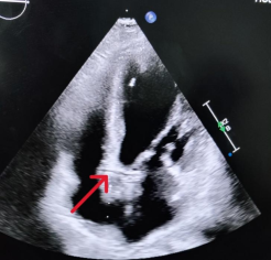
Figure 1. Transthoracic echocardiogram demonstrating the retroaortic anomalous coronary (RAC) sign in the apical four-chamber view (red arrow).
On day 2 of admission, coronary angiography and possible revascularisation was planned through right radial access. We initially encountered difficulty cannulating the left coronary artery in the left aortic sinus with a Tiger catheter (Terumo Radial TIG 5 Fr), and it was subsequently revealed that all three coronaries were arising separately from right coronary cusp. The left anterior descending (LAD) artery originated from a separate ostium located superior to the right coronary artery (RCA) origin, while the left circumflex (LCX) artery arose from an ostium positioned close to and inferior to the RCA origin. This entity is rare. The culprit lesion was identified in RCA had a discrete, critical mid-segment disease while LAD had a proximal 70% disease (Figures 2, 3).
Based on invasive coronary angiography, the LAD appeared to follow an anterior course over the right ventricle, while the LCX had a retroaortic course (Figure 2, 3). However, to confirm these findings, we proceeded with computed coronary tomography angiography (CCTA). CCTA confirmed the anomalous separate origin of LAD and LCX from right coronary sinus and delineated the course of LAD traversing through the right AV groove crossing anterior to RVOT to reach upto anterior interventricular septum and apex, thus suggesting a benign course. LCX shared an ostium with the RCA and coursed retroaortic (Figure 4). The heart chambers, aortic root and pulmonary circulation appeared morphologically normal.
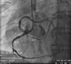
Figure 2. Coronary angiography. LAO view showing mid-discrete critical disease in RCA while LAD (originating from right aortic sinus) had a proximal 70% disease. LCX was also originating from right sinus. LAD, left anterior descending; LCX, left circumflex; RCA, right coronary artery, LAO, left anterior oblique.
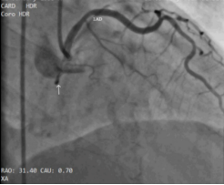
Figure 3. Coronary angiography. Injection in RAO 30 view showing a cranial anterior loop of LAD (anterior course) and LCX appearing as a dot (arrow) behind the aorta (retroaortic course).
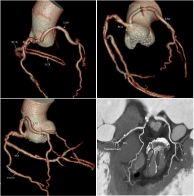
Figure 4. Reconstructed CT coronary angiography showing all three coronary arteries originating separately from the right aortic sinus of Valsalva. LAD, left anterior descending; LCX, left circumflex; RCA, right coronary artery.
Subsequently, percutaneous coronary intervention (PCI) to RCA and LAD was done successfully with a single drug eluting stent in both vessels with good angiographic result and Thrombolysis in Myocardial Infarction (TIMI) III flow (Figure 5). Initially, RCA was cannulated with 6F JR 3.5 but due to poor back up support, 6F AR 1 was taken to complete the procedure. Post-procedure, he was prescribed dual antiplatelet therapy with ticagrelor for 1 year and aspirin for life, as well as other secondary prevention medications, including high dose statin, metoprolol and ramipril. He was advised smoking cessation. The patient was discharged without any complication and was asymptomatic at one month follow up. A timeline of the diagnostic process is provided in Figure 6.
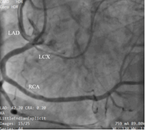
Figure 5. Percutaneous coronary intervention to left anterior descending artery and right coronary artery was done demonstrating good angiographic result with Thrombolysis in Myocardial Infarction 3 flow.
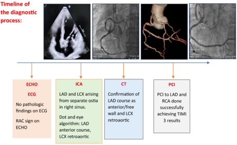
Figure 6. A timeline of the diagnostic process.
CASE 2
A 48-year-old normotensive, non-diabetic man presented with atypical chest discomfort. ECG showed nonspecific ST-T changes; echocardiography was normal. Coronary angiography revealed a normal left system and difficulty engaging the right coronary system. Subsequent manipulation revealed an anomalous RCA originating from the left coronary sinus, just above the level of the left coronary artery (LCA) (Type A) (Figure 7). All vessels had good caliber and TIMI 3 flow. CCTA confirmed a benign anatomical course of RCA. The patient was managed conservatively with medical management.
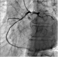
Figure 7. Coronary angiography. RCA originating from left sinus above the level of LCA.
CASE 3
A 56-year-old normotensive, non-diabetic man presented with a four-month history of exertional chest discomfort. ECG showed nonspecific ST-T changes, echocardiography revealed normal LV function. Coronary angiography with a Tiger catheter showed a normal left main and LAD but absent LCX. Cannulation of the right coronary sinus revealed LCX arising from the right sinus, sharing a common ostium with RCA. All vessels had normal caliber and TIMI 3 flow (Figure 8, 9, 10). LCX coursed retroaortic. The patient was managed conservatively with medical therapy.
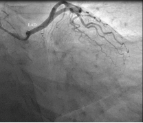
Figure 8. Coronary angiography. LAD originating from left sinus, LCX is not visualised.
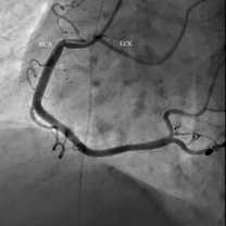
Figure 9. Coronary angiography. LCX originating from right sinus sharing same ostia with RCA.
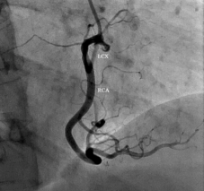
Figure 10. Coronary angiography. In the RAO 30 view, LCX coursing caudal and posterior (retroaortic course).
CASE 4
A 38-year-old normotensive man with diabetes and a family history of coronary artery disease, presented with exertional chest pain of 12 months duration. The pain was central, radiating to the left arm, and relieved by rest. On examination, the vitals were stable, and systemic examination was unremarkable. ECG showed nonspecific ST-T changes, and echocardiography revealed normal LV systolic function with no regional wall motion abnormalities. During coronary angiography, the left system could not be engaged. A Tiger catheter was subsequently used to engage the right coronary sinus, revealing that both the RCA and LCA originated from a common ostium in the right sinus. The LCA followed a caudal and anterior (septal) course, indicating a benign anatomical pattern. The left main coronary artery then emerged on the left side of the aortic root and bifurcated into the LAD and LCX, both demonstrating a normal course (Figure 11, 12).
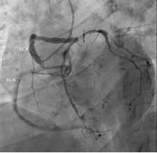
Figure 11. Coronary angiography. RCA and LCA originated from a common ostium in the right aortic sinus.
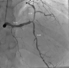
Figure 12. In the RAO 30 view, the LCA followed a caudal and anterior course, indicating a benign anatomical pattern. As per the dot and eye algorithm, the superior border of the eye is formed by LCX and inferior border by LCA, suggestive of a septal course. The left main coronary artery then emerged on the left side of the aortic root and bifurcated into the LAD and LCX, both demonstrating a normal course.
CASE 5
A 46-year-old man presented with acute extensive anterior wall myocardial infarction. Coronary angiography demonstrated a heavy thrombus burden in the proximal left anterior descending (LAD) artery, extending distally along the vessel. The left circumflex artery (LCX) was initially not visualized, raising suspicion of an anomalous origin. The right coronary artery (RCA) originated normally from the right coronary sinus and exhibited normal flow (TIMI 3). Upon further angiographic evaluation, the LCX was found to arise anomalously from the non-coronary sinus and coursed retroaortically before reaching the atrioventricular groove. The vessel had a normal calibre and maintained TIMI 3 flow, consistent with a benign retroaortic variant of anomalous LCX origin (Figure 13, 14).
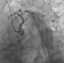
Figure 13. Coronary angiography. LAD originating from left sinus, LCX is not visualised.
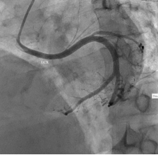
Figure 14. LCX arising from non-coronary sinus and coursed retro aortic.
2. Discussion
Coronary artery anomalies are rare with an incidence varying from 0.3% in autopsy series to 1.3% in angiographic series . These anomalies arise from disruptions in the embryological development of the heart tube. The most commonly observed variant is an anomalous left circumflex artery (LCX) originating from either the right coronary sinus or the proximal right coronary artery (RCA) . Anomalous origin of the RCA from the left coronary sinus (R-ACAOS) is particularly rare, seen in about 0.05% to 0.1% of individuals . One of the rarest CAAs is the entire LMCA arising from the right sinus of Valsalva (L-ACAOS), with a reported incidence of 0.017% to 0.15%; this entity has been described as exceedingly rare, with only isolated case reports available in the literature .
In a report of coronary anomalies from the Coronary Artery Surgery Study, Click et al. performed a detailed review of 24,959 patients and reported only three cases of all three coronary arteries arising from separate ostia in the right sinus of Valsalva .
2.1. Understanding the Anomaly
Before identifying a coronary anomaly, it is important to understand the essential features that define a coronary artery. Each coronary artery has its own definitions. LAD is a epicardial artery situated at the anterior interventricular sulcus and provides septal penetrating branches; LCX is a epicardial artery situated at the left atrioventricular sulcus and provides at least one obtuse marginal branch; RCA is a epicardial artery situated at the right atrioventricular sulcus and provides at least the acute marginal branch. This definition is essential because the coronary arteries are defined not by the origin or proximal course but by their intermediate and distal segments or their dependent microvascular bed. For example, the artery that runs in the left interventricular groove and provides septal branches is the LAD, irrespective of it is arising from the left or right aortic sinus. In addition, it is not essential that the LAD provide diagonal branches, which in turn could originate from the ramus or the obtuse marginals. When a coronary artery arises from the opposite sinus, its name, structure, and function remain unchanged—only its origin and initial course are anomalous .
By definition, a coronary artery anomaly (CAA) is a morphological abnormality present in less than 1% of the population. This criterion has been proposed as the threshold distinguishing (1) normality-including more common variations considered normal variants from (2) abnormality, which encompasses relatively infrequent and atypical anatomical variations .
Angelini proposed one of the most widely accepted classification systems for coronary artery anomalies (CAAs), categorizing them into four primary groups: (1) anomalies of origin and course, (2) intrinsic coronary artery abnormalities, (3) termination anomalies, and (4) anomalous collateral circulation .
We have provided a simplified table (Table 1) to aid in the identification of coronary artery anomalies involving individual vessels.
After arising from the opposite sinus, an anomalous coronary artery can take five courses.
1) Septal, through the proximal interventricular septum.
2) Interarterial, between the aorta and the pulmonary artery.
3) Retroaortic, posterior to the aortic root.
4) Pre pulmonic/anterior free wall, anterior to the right ventricular outflow tract.
5) Retrocardiac, behind the mitral and tricuspid valves.
Identifying the initial course of a coronary artery anomaly (CAA) using conventional angiography is often challenging because this technique offers only a two-dimensional view of the complex three-dimensional coronary anatomy. The coronary anomaly most commonly associated with increased clinical risk involves an ectopic coronary artery arising from the opposite (inappropriate) sinus of Valsalva such as the right coronary artery (RCA) originating from the left sinus, or the left main coronary artery (LMCA) from the right sinus and coursing between the aorta and pulmonary trunk .
Coronary arteries that pass anterior to the pulmonary trunk and posterior to the aorta are usually considered benign variants. The transseptal course is considered to be ‘relatively benign’ because there are reports of it being potentially fatal .
Most coronary artery anomalies do not result in signs, symptoms, or complications and usually are discovered as incidental findings at the time of catheterization.
Page et al proposed two clues that should raise suspicion of a coronary anomaly .
The ‘unperfused myocardium’ sign: a myocardial region not supplied by any visualized vessel. For example, an avascular region in the posterolateral wall of the left ventricle should suggest an anomalous origin of the LCX.
The ‘aortic root’ or Page sign: observed during ventriculography in the right anterior oblique (RAO) projection, where a vessel is seen crossing behind the aorta and pulmonary artery at the level of the aortic root.
Ishikawa and Brandt et al. (1985), followed by Serota et al. (1990), proposed a set of coronary arteriographic criteria to characterize the spatial relationship between the anomalous coronary artery and the aorta and pulmonary artery along its initial course . They introduced two methods to identify the course of anomalous coronaries: 1) Based on the orientation of the “loop” formed by the proximal portion of anomalous coronaries with the rest of the coronary tree and 2) the "dot and eye method"
The direction and configuration of the vessel loop—whether curving anteriorly, posteriorly, superior or inferiorly—can suggest its path relative to the great vessels (Figure 15). A simplified method for rapid identification of anomalous LMCA courses is illustrated in Figure 16. In summary, a cranial posterior loop indicates an interarterial course, as the vessel ascends and passes behind the aorta toward the pulmonary artery—a configuration often associated with a malignant trajectory. A cranial anterior loop suggests an anterior free-wall course, where the artery travels forward over the right ventricular outflow tract, a typically benign variant. A caudal anterior loop points to a septal course, with the vessel dipping inferiorly and running within or along the interventricular septum. Finally, a caudal posterior loop signifies a retro-aortic course, where the artery moves downward and behind the aorta before reaching its destination. These loop directions provide important visual clues during angiography and allow rapid identification of the anomalous pathway.
The "dot and eye algorithm": A dye injection in the right anterior oblique (RAO) view effectively separates the aorta and pulmonary artery when they are in their normal anatomical positions. This separation provides a clear view of the anomalous coronary artery's origin and its path in relation to the great vessels. In this method, the term "eye" describes the elliptical shape formed by the left main coronary artery (LMCA) and the early segment of the left circumflex artery (LCx), while the "dot" represents a circular radiopaque area (created by the contrast-filled coronary artery viewed directly from the end), indicating whether it lies in front of or behind the aorta.
Septal: During RAO coronary angiography, the LMCA and circumflex artery together create an elliptical configuration, with the LMCA forming the inferior limb of the ellipse and the circumflex artery forming the superior limb. The presence of septal perforators arising from the LMCA further aids in recognizing this anomaly.
Anterior/pre-pulmonic course: The anterior LMCA courses across the anterior free wall of the right ventricle, passing over the pulmonary artery, before bifurcating at the mid-septum into the LAD and circumflex arteries. On RAO coronary angiography, the LMCA forms the superior portion of the “eye,” while the circumflex artery outlines the inferior portion.
Retroaortic: The retroaortic LMCA travels posterior to the aortic root before emerging on the anterior surface of the heart, where it divides into the LAD and circumflex arteries with a normal course and length. On RAO coronary angiography, the retroaortic LMCA appears “on end” behind the aorta, visualized as a characteristic “dot,” similar to the appearance of the circumflex artery.
Interarterial: The interarterial LMCA passes between the aorta and pulmonary artery to reach its usual position on the anterior surface of the heart, where it divides into the LAD and circumflex arteries with a normal course and length. On RAO coronary angiography, the interarterial LMCA is visualized “on end,” appearing as a distinct “dot” located anterior to the aortic root.
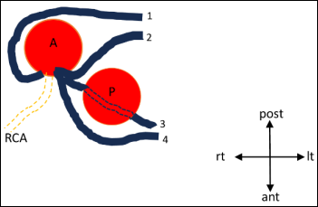
Figure 15. Diagrammatic representation of the four possible pathways for the anomalous left main coronary artery (LMCA) arising from the right coronary cusp or the right coronary artery (RCA): 1, retroaortic course; 2, interarterial course; 3, septal course; 4, anterior free wall course. A, aortic valve; P, pulmonary valve; ant = anterior; It = left; post = posterior; rt = right.
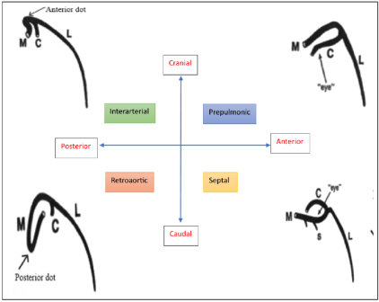
Figure 16. A simplified technique to identify the anomalous LMCA courses at a quick glance. A cranial, anterior loop indicates a prepulmonic course, forming an “eye” with the LMCA as the superior border and the LCx as the inferior border. A caudal, anterior loop suggests a septal course, forming an “eye” with the LMCA as the inferior border and the LCx as the superior border. A cranial, posterior loop corresponds to an interarterial course, visualized as a dot anterior to the aorta. A caudal, posterior loop indicates a retroaortic course, visualized as a dot posterior to the aorta. Abbreviations: M, left main coronary artery; L, left anterior descending artery; C, left circumflex artery.
The eye is therefore used to differentiate between septal and anterior course, whereas the ‘dot’ helps delineate between retroaortic and interarterial courses (Figure 17).
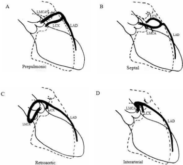
Figure 17. Diagrammatic angiographic illustration of the four possible left main coronary artery courses arising from the right coronary cusp. Illustration of the coronary angiographic images in right anterior oblique projection (redrawn from Ishikawa) showing the four possible courses of an aberrant left main coronary artery (LMCA) arising from the right coronary cusp or the right coronary artery. (LAD, left anterior descending; LCx, left circumflex).
According to Sarkar et al., the origin of the anomalous right coronary artery (ARCA) in relation to the left coronary artery (LCA) ostium and the sinotubular junction is classified into four types :
1) Type A: Origin from the aorta above the sinotubular plane
2) Type B: Origin just below the ostium of the LCA
3) Type C: Origin below the sinotubular plane, between the midline and the origin of the LCA
4) Type D: Origin along the midline of the ascending aorta
2.2. Diagnosis
Various imaging modalities—including echocardiography, invasive coronary angiography, computed tomography angiography (CCTA), and magnetic resonance angiography (MRA)—are employed to diagnose the origin and course of anomalous coronary arteries. Each technique offers unique advantages: modern CT scanners provide superior spatial resolution, while MRI avoids radiation exposure and the use of iodinated contrast agents. Together, these tools enable comprehensive evaluation of ACAOS.
Electrocardiography (ECG): ECG findings in symptomatic patients are often nonspecific and may range from ST-elevation myocardial infarction (STEMI)–like changes suggestive of ischemia to arrhythmias such as ventricular tachycardia or atrial fibrillation.
Echocardiography: Although echocardiography has limitations, signs like the retroaortic anomalous coronary (RAC) or "crossed aorta" sign and the BLEB sign may suggest the presence of an anomalous origin. RAC sign described as a binary structure above the mitral valve plane directed toward the right coronary sinus in a modified four-chamber apical view; BLEB sign seen on transesophageal echocardiography, as a round structure in mitroaortic angle in the long-axis aortic view. Frommelt et al. demonstrated that transthoracic echocardiography can reliably identify intramural courses in AOCA, enabling accurate diagnosis and guiding successful surgical management . Thus, the authors recommended using colour Doppler to assess flow direction, aiding in the identification and exclusion of coronary artery anomalies when suspected. The hallmark echocardiographic findings in Anomalous Left Coronary Artery from the Pulmonary Artery (ALCAPA) include the direct visualization of the left coronary artery (LCA) arising from the pulmonary artery (PA) and retrograde flow from the LCA into the PA, often with a dilated right coronary artery (RCA).
Coronary computed tomography angiography (CCTA): CCTA has emerged as the diagnostic modality of choice for identifying coronary anomalies, owing to its rapid acquisition time and superior spatial resolution, which enable precise delineation of both the origin and course of the coronary arteries. A study by Ghadri et al, comparing CCTA and invasive coronary angiography (ICA) found that CCTA detects a significantly higher prevalence of congenital coronary anomalies (7.9%) compared to ICA (2.1%) . CCTA’s superior ability to visualize coronary anatomy allows more precise identification of anomalies, including potentially serious ones like interarterial courses. The findings suggest that the true prevalence of coronary artery anomalies in the general population may be underestimated when relying on ICA alone. CCTA is a pivotal tool in multimodality imaging, playing a central role in the evaluation of patients with anomalous aortic origin of a coronary artery (AAOCA) and guiding clinical management decisions. The diagnosis of an intramural aortic course typically relies on the presence of key anatomical features, including an eccentricity index ≥ 2.0, a luminal diameter reduction of ≥ 50%, and an acute take-off angle of less than 45°. A 3-dimensional volume rendered CT image is a valuable tool to identify anomalous origin, ectopic courses and their relationships with the great vessels . Computational fluid dynamics simulations now enable physiological assessment through fractional flow reserve derived from CT (FFRCT).
Invasive coronary angiography (ICA): Despite the superiority of CCTA, the role of ICA remains crucial, particularly in patients with suspected atherosclerotic disease, as it allows assessment of stenoses and calcifications involving anomalous vessels. While ICA provides detailed visualization of the coronary lumen and allows for functional assessment, its limitations include difficulty in precisely delineating the three-dimensional course of anomalous vessels. In challenging cases where the coronary ostium cannot be engaged, an aortic root injection (cuspogram) can facilitate visualization. Using the dot and eye and the looping method as discussed above, we can precisely delineate the course of anomalous vessels.
Magnetic Resonance Angiography (MRA): MRA is increasingly recognized for its utility in evaluating coronary artery anomalies (CAAs), particularly in pediatric and young adult populations. Notably, it is a non-invasive modality that avoids ionizing radiation and does not require iodinated contrast agents, making it especially valuable for repeated assessments and in patients with contraindications to contrast use. While it may have limitations in spatial resolution, its non-invasive nature and advanced imaging capabilities make it a compelling choice for the assessment of coronary artery anomalies, offering a balance between diagnostic accuracy and patient safety.
Table 1. Anatomic classification of coronary artery origin anomalies.

LMCA

A. Arising from right sinus of Valsalva

(1) Interarterial

(2) Retro aortic

(3) Septal

(4) Anterior/pre-pulmonic

(5) Posterior atrioventricular groove or retrocardiac

(6) Posteroanterior interventricular groove (wraparound)

B. Absent left main trunk

C. Aberrant location in the left sinus of Valsalva (anterior or posterior)

D. Arising from the pulmonary artery

LAD

Arising from right sinus of Valsalva

Interarterial

Septal

Anterior/prepulmonic

Posteroanterior interventricular groove (wraparound)

B. Separate ostium of left anterior descending from left sinus of Valsalva

C. Arising from the pulmonary artery

LCX

A. Arising from right sinus of Valsalva

(1) Posterior atrioventricular groove

(2) Retro aortic

B. Separate ostium of left circumflex from left sinus of Valsalva

C. Rudimentary or absent left circumflex (super-dominant right coronary)

D. Arising from the pulmonary artery

RCA

A. Arising from the left sinus of Valsalva

(1) Posterior atrioventricular groove or retrocardiac

(2) Retro aortic

(3) Interarterial

(4) Septal

(5) Anterior/prepulmonic

(6) Posteroanterior interventricular groove (wraparound)

B. Posterior location in the right sinus of Valsalva

C. Absent right coronary artery (super-dominant left circumflex)

D. Arising from the pulmonary artery

2.3. Mechanism of Ischemia
The presenting symptoms of coronary artery anomalies (CAAs) can vary widely; while most cases are benign and discovered incidentally, some patients may present with ischemia or arrhythmias. The causes of ischemia can be due to 1) Atherosclerosis or ostial narrowing, 2) abnormal aortic exit or 3) coursing intramural or compression between the aortic root and pulmonary trunk. Hutchins et al. (30) proposed that the abnormal take-off angle and increased tortuosity of the proximal segment of an anomalous coronary artery may predispose it to accelerated atherosclerosis .
Additionally, other researchers have suggested that the transition zone between the intramural (bound) segment and the extramural (free) segment of the anomalous artery particularly as it courses around the aorta is especially vulnerable to lipid accumulation . Some authors propose that ischemia in coronary artery anomalies (CAAs) may result from vasospasm induced by endothelial injury along the anomalous course of the artery . Others suggest that intussusception of the proximal segment of the anomalous artery into the aortic wall may contribute to compromised blood flow .
The most common course of the proximal anomalous LAD is anterior to pulmonary trunk and anomalous LCX posterior to aorta. Both retroaortic and anterior course are considered as benign CAA. An interarterial course however, is associated with 30% risk of sudden cardiac death. Approximately half of the patients with this anomaly experience fatal events before 20 years of age . The exact reason is still debated. One hypothesis suggests that exercise causes expansion of the aortic root and pulmonary trunk, which may increase the angulation of the anomalous coronary artery, thereby narrowing its lumen. Another theory proposes that the vessel follows an aberrant course within the aortic wall, is often hypoplastic, and subjected to lateral compression along its entire proximal intramural segment .
An analysis from the CASS study demonstrated that, with the exception of the anomalous circumflex coronary artery, anomalous coronary arteries do not carry an increased risk of atherosclerosis compared to non-anomalous arteries in age- and gender-matched control patients . The elevated atherosclerotic risk in anomalous LCx may be attributed to increased mechanical stress exerted on the arterial wall by the expanding aorta. Although typically hemodynamically insignificant, an anomalous left circumflex artery (LCx) with a retroaortic course can present significant challenges during aortic valve replacement. In surgical aortic valve replacement (SAVR), there is a risk of iatrogenic occlusion due to inadvertent suture placement . With the advent of transcatheter aortic valve implantation (TAVI), concerns have emerged regarding potential extrinsic compression of the anomalous LCx by the prosthetic valve, particularly in cases where the anomalous vessel has a close anatomical relationship with the aortic annulus. To mitigate this risk, balloon aortic valvuloplasty with simultaneous coronary angiography can be performed pre-procedurally to assess the likelihood of coronary compression . During TAVI, placement of a coronary guidewire and a prophylactic unexpanded stent in the anomalous vessel is advised when there is a high risk of compression. Despite these considerations, TAVI can typically proceed as per standard practice in patients with AAOCA, provided appropriate precautions are taken.
Coronary artery anomalies can be broadly classified into relatively benign and potentially serious forms (Table 2). Benign variants are usually incidental findings with minimal hemodynamic consequence, whereas potentially serious anomalies—particularly those with an interarterial course or arising from the pulmonary artery—may be associated with myocardial ischemia, arrhythmias, or sudden cardiac death. This classification is supported by the large angiographic study by Yamanaka and Hobbs, which analyzed over 120,000 patients and highlighted the clinical relevance of specific anomalies .
Table 2. Classification of Coronary Artery Anomalies Based on Clinical Significance.

Relatively benign anomalies

Potentially serious anomalies

1) Separate origin of left anterior descending and left circumflex from the left sinus of Valsalva

1) Ectopic coronary origin from the pulmonary artery

2) Ectopic origin of the circumflex from the right sinus of Valsalva

2) Ectopic coronary origin from the opposite aortic sinus

3) Ectopic coronary origin from the posterior sinus of Valsalva

3) Single coronary artery

4) Ectopic coronary origin from the ascending aorta

4) Large coronary artery fistulae

5) Absent circumflex

5) Interarterial course

6) Intercoronary communication

7) Small size coronary artery fistulae

Adapted from Yamanaka
2.4. Role of Imaging
Intravascular ultrasound (IVUS) plays a crucial role in the evaluation and management of anomalous coronary arteries, particularly those originating from the opposite sinus of Valsalva (ACAOS). IVUS can detect slit-like orifice, acute take-off angles, intramural course, and proximal narrowing, which are associated with adverse events.
A case series by Angelini et al. highlights that symptomatic anomalous left coronary artery origination from the opposite sinus typically involves intussusception, which is accompanied by coronary segmental hypoplasia and lateral luminal compression (resulting in an ovaloid cross-section) as the pathophysiologic mechanism of ischemia .
Formato et al. used intravascular ultrasound to study anomalous aortic origin of coronary arteries (AAOCA) with intramural segments and found that these segments exhibit dynamic compression during systole, with narrowing, increased ellipticity, and resistance, unlike non-intramural sections. These findings highlight a potential mechanism for ischemia and support IVUS as a tool to assess severity .
Additional signs suggestive of an intramural course include the absence of perivascular tissue density, owing to the lack of adventitia, and segmental loss of the normal three-layered architecture of the vessel wall. Advanced IVUS systems enhance visualization of the spatial relationship between the ectopic coronary artery and the great vessels. This is crucial in interarterial variants, where the anomalous artery navigates a constrained anatomical corridor between the aorta and pulmonary artery—typically narrower than the physiological coronary caliber. As the vessel traverses posterior to the pulmonary structures, the luminal morphology evolves: it initially assumes an oval shape (eccentricity index < 2.0), progressing to an elliptical deformation (eccentricity index ≥ 2.0) in cases of intramural aortic passage. The coronary ostium may also appear slit-like, a key morphological indicator for anomalous coronaries .
2.5. Role of Physiologic Testing
Ischemia in ACAOS arises from two distinct but interrelated mechanisms:
1) Fixed Component: This involves static anatomical abnormalities such as slit-like ostia or proximal luminal narrowing—features common to atherosclerotic stenoses. These lesions tend to reduce coronary flow irrespective of hemodynamic changes.
2) Dynamic Component: This stems from the intramural segment of the anomalous artery, which is compressed between the aortic and pulmonary walls during exercise or stress. Increasing heart rate, stroke volume, and aortic wall tension exacerbate systolic compression, resulting in variable luminal narrowing not present at rest.
Adenosine-induced hyperemia, typically administered intravenously, causes microvascular dilation and is used to evaluate the fixed stenotic component of a lesion. In cases where stenosis exceeds 50%, fractional flow reserve (FFR) values generally range between 0.80 and 0.90, but rarely fall below the ischemic threshold of <0.80.
Dobutamine stress testing—often enhanced with atropine and saline infusion—more effectively uncovers dynamic compression of intramural segments by mimicking exercise-like conditions. This approach can reveal ischemia that may not be detected with adenosine alone.
Intravascular ultrasound (IVUS) provides direct visualization of dynamic compression both at rest and during stress, serving as a useful complement to pressure-based assessments.
Quantitative Flow Ratio (QFR), a non-invasive technique derived from angiographic imaging and computational flow modeling, has shown promise. Recent studies suggest that in patients with interarterial ACAOS, non-significant QFR values are associated with favorable five-year outcomes.
Emerging techniques like fluid-structure interaction (FSI) simulations, which integrate CT and catheterization data, are being increasingly used in pediatric populations. These simulations aim to non-invasively predict stress-induced FFR and have shown good concordance with invasive FFR measurements .
3. Management
Most coronary artery anomalies (CAAs) are discovered incidentally during coronary angiography performed in patients with ischemic heart disease or in those undergoing preoperative evaluation before valve replacement surgery. Current guidelines lack specific strategies for rare cases, making each reported instance clinically valuable.
The management of patients with coronary artery anomalies (CAAs) remains complex and highly individualized due to the lack of uniform guidelines and the wide anatomical variability of these anomalies. Initial assessment typically involves coronary angiography and coronary CT angiography (CCTA), with CCTA often preferred as it provides a detailed three-dimensional view of the anomalous artery’s course in relation to the aorta and pulmonary artery. High-risk anatomical features—such as an interarterial course, slit-like ostium, or long intramural segments—are associated with sudden cardiac death (SCD), especially during exertion. Therefore, restriction from competitive sports is often the first recommendation for patients with CAAs, particularly those arising from the opposite sinus. Symptomatic patients or those with high-risk features typically undergo surgical repair, including unroofing, reimplantation, or coronary artery bypass grafting (CABG), depending on the anatomy and presence of atherosclerosis. CABG is often favoured in older patients or those with coexisting coronary disease, while unroofing is suitable for long intramural courses.
Percutaneous coronary intervention (PCI) plays a limited but evolving role in select adult patients, especially those with proximal stenosis or short intramural courses. However, PCI is technically challenging due to the abnormal take-off angles, varying ostial locations, and need for careful catheter selection. Appropriate selection of the guide catheter is crucial for successful PCI in anomalous coronary arteries. Achieving selective engagement in these locations might prove challenging, often necessitating the use of non-standard diagnostic catheters. A short summary of the preferred guide catheters in anomalous coronaries is illustrated in Table 3. The catheter must be tailored to the lesion’s morphology to provide adequate support, which facilitates guidewire and balloon crossing and significantly reduces procedure time. Management strategies vary across institutions and are influenced by patient age, anatomical risk factors, clinical symptoms, and stress test results. International guidelines such as those from the ESC, AHA/ACC, and AATS provide frameworks but leave room for clinical judgment. The decision should be tailored for the individual based on the type of the anomaly.
Table 3. Preferred guide catheters in CAA .

Artery origin

Preferred guide catheters

RCA

A) RCA ostium adjacent to left main coronary artery ostium

Leya catheter, left coronary, Amplatz 45, 90 R-ACAOS Launcher, Ikari

B) RCA ostium inferior to left main coronary artery ostium

AL, AR

C) RCA ostium toward the commissure of the right and left cusps

AL, AR

D) Ostium above the sinotubular plane

JR, MP, Hockey stick

E) Ostium from right cusp superior and toward the left cusp

AL, AR, Sherpa NX balanced, 3DRC

F) Usual origin

JR

G) Shephard crook

AL

LMCA/LAD

A) Posterior origin of the left main from the left cusp or non-coronary cusp

EBU, CLS

B) Anomalous left main coronary artery from the right cusp

JR4, Hockey stick

LCX

LCX arising from right sinus

AL, AR, JR 4, Hockey stick

4. Conclusion
Anomalous coronary arteries from the opposite sinus of Valsalva are rare and encompass a wide spectrum of clinical risk. While many variants are benign and incidentally detected, interarterial or intramural courses may predispose to ischemia and sudden cardiac death.
Accurate diagnosis relies on careful angiographic assessment supported by multimodality imaging. Management should be individualized based on symptoms, anatomical risk features, and physiologic assessment. In selected patients, percutaneous coronary intervention is feasible but requires tailored catheter selection and technical expertise.
Abbreviations

RAC

Retroaortic Anomalous Coronary

RCA

Right Coronary Artery

LCX

Left Circumflex (Artery)

LAO

Left Anterior Oblique

RAO

Right Anterior Oblique

CCTA

Coronary Computed Tomography Angiography

RVOT

Right Ventricular Outflow Tract

AV

Atrioventricular

TIMI

Thrombolysis in Myocardial Infarction

LCA/LMCA

Left Coronary Artery/Left Main Coronary Artery

LV

Left Ventricle

R-ACAOS

Right Anomalous Coronary Artery from the Opposite Sinus

L-ACAOS

Left Anomalous Coronary Artery from the Opposite Sinus

MRA

Magnetic Resonance Angiography

ALCAPA

Anomalous Left Coronary Artery from the Pulmonary Artery

AAOCA

Anomalous Aortic Origin of a Coronary Artery

ICA

Invasive Coronary Angiography

FFRCT

Fractional Flow Reserve Derived from Computed Tomography

IVUS

Intravascular Ultrasound

FFR

Fractional Flow Reserve

QFR

Quantitative Flow Ratio

FSI

Fluid-Structure Interaction

SCD

Sudden Cardiac Death

CABG

Coronary Artery Bypass Grafting

ESC

European Society of Cardiology

AHA

American Heart Association

ACC

American College of Cardiology

AATS

American Association for Thoracic Surgery

Author Contributions
Sandeepan Saha: Conceptualization, Data curation, Writing – original draft, Writing – review & editing, Investigation, Methodology, Resources
Ashish Jha: Conceptualization, Data curation, Writing – original draft, Supervision, Validation, Visualization, Formal analysis
Bhuwan Chandra Tiwari: Conceptualization, Data curation, Writing – original draft, Supervision, Validation, Visualization
Abhijit Bharali: Writing – original draft, Supervision, Validation, Visualization, Formal analysis
Mithilesh Yadaw: Writing – original draft, Supervision, Validation, Visualization, Formal analysis
Conflicts of Interest
The authors declare no conflicts of interest.
References
[1] Beique F, De Tran QH, Ma F, Rudski L, Daves S, Angelini P. Anomalous right coronary artery originating from the left sinus of Valsalva. J Cardiothorac Vasc Anesth 2004; 18(6): 788-98.
[2] Click RL, Holmes DR, Vlietstra RE, Kosinski AS, Kronmal RA. Anomalous coronary arteries: location, degree of atherosclerosis and effect on survival—a report from the Coronary Artery Surgery Study. Journal of the American College of Cardiology. 1989 Mar 1; 13(3): 531-7.
[3] Yamanaka O, Hobbs RE. Coronary artery anomalies in 126,595 patients undergoing coronary arteriography. Catheterization and cardiovascular diagnosis. 1990 Sep; 21(1): 28-40.
[4] Alexander RW, Griffith GC. Anomalies of the coronary arteries and their clinical significance. Circulation. 1956 Nov; 14(5): 800–805.
[5] Angelini P, Villason S, Chan AV, Diez JG. Coronary artery anomalies. Baltimore: Lippincott Williams & Wilkins; 1999.
[6] Angelini P. Coronary artery anomalies—current clinical issues: definitions, classification, incidence, clinical relevance, and treatment guidelines. Texas Heart Institute Journal. 2002; 29(4): 271.
[7] Angelini P, Villason S, Chan Jr AV, Diez JG. Normal and anomalous coronary arteries in humans. Part 1: historical background.
[8] Angelini P. Congenital heart disease for the adult cardiologist. Circ. 2007; 115: 1296-305.
[9] Angelini P, Velasco JA, Flamm S. Coronary anomalies: incidence, pathophysiology, and clinical relevance. Circulation. 2002 May 21; 105(20): 2449-54.
[10] Patel S. Normal and anomalous anatomy of the coronary arteries. InSeminars in roentgenology 2008 Apr (Vol. 43, No. 2, pp. 100-112).
[11] PAGE JR HL, ENGEL HJ, CAMPBELL WB, THOMAS JR CS. Anomalous origin of the left circumflex coronary artery: recognition, angiographic demonstration and clinical significance. Circulation. 1974 Oct; 50(4): 768-73.
[12] Ishikawa T, Brandt PW. Anomalous origin of the left main coronary artery from the right anterior aortic sinus: angiographic definition of anomalous course. The American journal of cardiology. 1985 Mar 1; 55(6): 770-6.
[13] Serota H, Barth III CW, Seuc CA, Vandormael M, Aguirre F, Kern MJ. Rapid identification of the course of anomalous coronary arteries in adults: the “dot and eye” method. The American journal of cardiology. 1990 Apr 1; 65(13): 891-8.
[14] Sarkar K, Sharma SK, Kini AS. Catheter selection for coronary angiography and intervention in anomalous right coronary arteries. Journal of interventional cardiology. 2009 Jun; 22(3): 234-9.
[15] Frommelt PC, Frommelt MA, Tweddell JS, Jaquiss RD. Prospective echocardiographic diagnosis and surgical repair of anomalous origin of a coronary artery from the opposite sinus with an interarterial course. Journal of the American College of Cardiology. 2003 Jul 2; 42(1): 148-54.
[16] Ghadri JR, Kazakauskaite E, Braunschweig S, et al. Congenital coronary anomalies detected by coronary computed tomography compared to invasive coronary angiography. BMC Cardiovasc Disord 2014; 14: 81.
[17] Bigler MR, Kadner A, Räber L, Ashraf A, Windecker S, Siepe M, Padalino MA, Gräni C. Therapeutic management of anomalous coronary arteries originating from the opposite sinus of Valsalva: current evidence, proposed approach, and the unknowing. Journal of the American Heart Association. 2022 Oct 18; 11(20): e027098.
[18] Gentile F, Castiglione V, De Caterina R. Coronary artery anomalies. Circulation. 2021 Sep 21; 144(12): 983-96.
[19] Molossi S, Doan T, Sachdeva S. Anomalous coronary arteries: a state-of-the-art approach. Cardiac Electrophysiology Clinics. 2024 Mar 1; 16(1): 51-69.
[20] Hutchins GM, Miner MM, Boitnott JK. Vessel caliber and branch-angle of human coronary artery branch-points. Circulation research. 1976 Jun; 38(6): 572-6.
[21] Liu LB. Richardson T, Taylor CB. Atherosclerotic occlusions in anomalous left circumflex coronary-arteries: a report of 2 unusual cases and a review of pertinent literature. Par Arter lY75; 2: 55-9.
[22] Basso C, Corrado D, Thiene G. Congenital coronary anomalies as an important cause of sudden death in the young. Cardiol Rev. 2001; 9: 312-7.
[23] Angelini P, Velasco JA, Ott D, Khoshnevis GR. Anomalous coronary artery arising from the opposite sinus: descriptive features and pathophysiologic mechanisms, as documented by intravascular ultrasonography. J Invasive Cardiol. 2003; 15: 507-14.
[24] Kim D, Jeong MH, Lee KH, Lee MG, Park KH, Sim DS, Yoon NS, Yoon HJ, Kim KH, Hong YJ, Kim JH. Successful primary percutaneous coronary intervention in a patient with acute myocardial infarction and single coronary artery ostium. Korean Circulation Journal. 2012 Apr 26; 42(4): 284.
[25] Young PM, Gerber TC, Williamson EE, et al. Cardiac imaging: part 2, normal, variant, and anomalous configurations of the coronary vasculature. AJR Am J Roentgenol 2011; 197: 816–26.
[26] Arnáiz García ME, González-Santos JM, Pérez-Losada ME, López-Rodríguez J, Arnáiz J. Unexpected entrapment during surgery of anomalous circumflex coronary artery arising from right coronary artery. Turk Kardiyol Dern Ars. 2019 Apr 1; 47(3): 239-42.
[27] Tabachnick D, Obokhae B, Harrington K, Brown DL. Assessing the risk of an anomalous circumflex artery using balloon aortic valvuloplasty prior to transcatheter aortic valve replacement. Catheterization and Cardiovascular Interventions. 2020 Aug; 96(2): 497-9.
[28] Formato GM, Agnifili ML, Arzuffi L, Rosato A, Ceserani V, Zuniga Olaya KG, Secchi F, Deamici M, Conti M, Auricchio F, Bedogni F. Morphological changes of anomalous coronary arteries from the aorta during the cardiac cycle assessed by IVUS in resting conditions. Circulation: Cardiovascular Interventions. 2023 Jul; 16(7): e012636.
[29] Fretay XHD, Boudvillain O, Koutsoukis A, Degrell P, Dupouy P, Aubry P. Catheterization Techniques for Anomalous Aortic Origin of Coronary Arteries. Catheter Cardiovasc Interv. 2025 Mar; 105(4): 825-837.
[30] Ben-Dor I, Weissman G, Rogers T, Slack M, Pichard A, Ben-Dor N, Hashim H, Bernardo N, Satler LF, Waksman R. Catheter selection and angiographic views for anomalous coronary arteries: a practical guide. Cardiovascular Interventions. 2021 May 10; 14(9): 995-1008.
Cite This Article
  • APA Style

    Saha, S., Jha, A., Tiwari, B. C., Bharali, A., Yadaw, M. (2026). Anomalous Coronary Arteries from the Opposite Sinus of Valsalva: A Case Series and Comprehensive Review. International Journal of Cardiovascular and Thoracic Surgery, 12(2), 40-54. https://doi.org/10.11648/j.ijcts.20261202.14

    Copy | Download

    ACS Style

    Saha, S.; Jha, A.; Tiwari, B. C.; Bharali, A.; Yadaw, M. Anomalous Coronary Arteries from the Opposite Sinus of Valsalva: A Case Series and Comprehensive Review. Int. J. Cardiovasc. Thorac. Surg. 2026, 12(2), 40-54. doi: 10.11648/j.ijcts.20261202.14

    Copy | Download

    AMA Style

    Saha S, Jha A, Tiwari BC, Bharali A, Yadaw M. Anomalous Coronary Arteries from the Opposite Sinus of Valsalva: A Case Series and Comprehensive Review. Int J Cardiovasc Thorac Surg. 2026;12(2):40-54. doi: 10.11648/j.ijcts.20261202.14

    Copy | Download

  • @article{10.11648/j.ijcts.20261202.14,
      author = {Sandeepan Saha and Ashish Jha and Bhuwan Chandra Tiwari and Abhijit Bharali and Mithilesh Yadaw},
      title = {Anomalous Coronary Arteries from the Opposite Sinus of Valsalva: A Case Series and Comprehensive Review},
      journal = {International Journal of Cardiovascular and Thoracic Surgery},
      volume = {12},
      number = {2},
      pages = {40-54},
      doi = {10.11648/j.ijcts.20261202.14},
      url = {https://doi.org/10.11648/j.ijcts.20261202.14},
      eprint = {https://article.sciencepublishinggroup.com/pdf/10.11648.j.ijcts.20261202.14},
      abstract = {Anomalous coronary artery from the opposite sinus (ACAOS) is a rare congenital abnormality that may be encountered during routine coronary angiography. Clinical manifestations vary depending on the subtype and anatomical course of the anomaly; however, certain potentially malignant trajectories may result in myocardial ischemia, infarction, or sudden cardiac death, particularly in young individuals and athletes. Multiple imaging modalities including coronary angiography, CT angiography, and cardiac MRI are useful in the evaluation of coronary artery anomalies. Accurate delineation of the origin and course of the anomalous vessel is essential for risk stratification and therapeutic planning. Performing percutaneous coronary intervention (PCI) in patients with ACAOS can be technically challenging and often requires modifications in guide catheter selection and procedural technique. In this review, we present a series of patients with anomalous coronary arteries originating from the opposite sinus of Valsalva (ACAOS). We discuss the anatomical variations and courses of these anomalous vessels as defined by angiographic and advanced imaging findings. Particular emphasis is placed on guide catheter selection, technical challenges encountered during selective cannulation, and the strategies employed to achieve successful engagement and intervention.},
     year = {2026}
    }
    

    Copy | Download

  • TY  - JOUR
    T1  - Anomalous Coronary Arteries from the Opposite Sinus of Valsalva: A Case Series and Comprehensive Review
    AU  - Sandeepan Saha
    AU  - Ashish Jha
    AU  - Bhuwan Chandra Tiwari
    AU  - Abhijit Bharali
    AU  - Mithilesh Yadaw
    Y1  - 2026/03/19
    PY  - 2026
    N1  - https://doi.org/10.11648/j.ijcts.20261202.14
    DO  - 10.11648/j.ijcts.20261202.14
    T2  - International Journal of Cardiovascular and Thoracic Surgery
    JF  - International Journal of Cardiovascular and Thoracic Surgery
    JO  - International Journal of Cardiovascular and Thoracic Surgery
    SP  - 40
    EP  - 54
    PB  - Science Publishing Group
    SN  - 2575-4882
    UR  - https://doi.org/10.11648/j.ijcts.20261202.14
    AB  - Anomalous coronary artery from the opposite sinus (ACAOS) is a rare congenital abnormality that may be encountered during routine coronary angiography. Clinical manifestations vary depending on the subtype and anatomical course of the anomaly; however, certain potentially malignant trajectories may result in myocardial ischemia, infarction, or sudden cardiac death, particularly in young individuals and athletes. Multiple imaging modalities including coronary angiography, CT angiography, and cardiac MRI are useful in the evaluation of coronary artery anomalies. Accurate delineation of the origin and course of the anomalous vessel is essential for risk stratification and therapeutic planning. Performing percutaneous coronary intervention (PCI) in patients with ACAOS can be technically challenging and often requires modifications in guide catheter selection and procedural technique. In this review, we present a series of patients with anomalous coronary arteries originating from the opposite sinus of Valsalva (ACAOS). We discuss the anatomical variations and courses of these anomalous vessels as defined by angiographic and advanced imaging findings. Particular emphasis is placed on guide catheter selection, technical challenges encountered during selective cannulation, and the strategies employed to achieve successful engagement and intervention.
    VL  - 12
    IS  - 2
    ER  - 

    Copy | Download

Author Information