Case Report | | Peer-Reviewed

Seizures and Guillain-Barré Syndrome Leading to the Discovery of Systemic Lupus: Case of an Adolescent in Lomé and Review of the Literature

Received: 7 May 2026     Accepted: 22 May 2026     Published: 17 July 2026
Views:       Downloads:
Abstract

Background: Systemic lupus erythematosus (SLE) is a chronic idiopathic autoimmune disease. Guillain-Barré syndrome (GBS) is an acute monophasic immune-mediated polyradiculoneuropathy. Case presentation: We report the case of a 16-year-old patient, who presented in November 2023 a flu-like syndrome with diarrhea and abdominal pain. Then he had visual hallucinations and psychosis, followed by generalized tonic-clonic seizures. Urine toxicology assay was positive, a syndrome of hepatic cytolysis and cholestasis were noted. Toxic encephalopathy and hepatopathy were mentioned and he was transferred to a psychiatric ward where he persisted with an infectious syndrome, hallucinations, delirium and then flaccid tetraplegia of ascending evolution with dyspnea and respiratory distress. In January 2024, there was tetraplegia, abolished deep tendon reflexes (DTR) in all 4 limbs, hypoesthesia in all 4 limbs. Cerebrospinal fluid examination found albumin -cytological dissociation. Magnetic resonance imaging of the spinal cord was normal. He received 500 mg of intravenous methylprednisolone for 3 days then a per os relay with prednisolone 50 mg in addition to azathioprine (Aza) 50 mg x2/day and functional rehabilitation. The electroencephalogram revealed signs of non-structural generalised epilepsy in March 2024. The electroneuromyogram found a neurophysiological pattern of acute polyradiculoneuropathy in the recovery phase. In the evolution, an antinuclear antibody assay (indirect immunofluorescence) came back positive at a titer of 1/160, with a speckled appearance. On the basis of the American College of Rhumatology (ACR) with a score of 19, the diagnosis of SLE was retained and background treatment with Aza 50 mg/day continued. At the end of May 2025, there persisted a predominantly distal tetraparesis, an abolition of DTR. Conclusion: Although rare, GBS can be the mode of revelation of SLE. This SLE-GBS association is often serious and can be life-threatening.

Published in American Journal of Psychiatry and Neuroscience (Volume 14, Issue 3)
DOI 10.11648/j.ajpn.20261403.11
Page(s) 47-56
Creative Commons

This is an Open Access article, distributed under the terms of the Creative Commons Attribution 4.0 International License (http://creativecommons.org/licenses/by/4.0/), which permits unrestricted use, distribution and reproduction in any medium or format, provided the original work is properly cited.

Copyright

Copyright © The Author(s), 2026. Published by Science Publishing Group

Keywords

Guillain-Barré Syndrome, Epilepsy, Systemic Lupus, Togo

1. Introduction
Systemic lupus erythematosus (SLE) is a chronic idiopathic autoimmune disease that affects various organs, including the central nervous system (CNS) and peripheral nervous system (PNS) . Guillain-Barré syndrome (GBS) is defined as an acute monophasic immune-mediated polyradiculoneuropathy, usually post-infectious, and is the leading cause of acquired neuromuscular paralysis worldwide . The diagnosis of GBS is made based on clinical features, supported by laboratory findings and electrophysiology. Intravenous immunoglobulin (IV Ig) and plasma exchange remain the main therapeutic modalities, regardless of the electrophysiological subtype.
The presentation of severe GBS as the inaugural manifestation of SLE remains exceptional . Reported cases of GBS associated with SLE worldwide are essentially clinical cases. Cases have been reported in the Maghreb but few in sub-Saharan Africa . In Togo, studies on Guillain-Barré syndrome exist . We report the case of a 17-year-old adolescent in whom convulsive seizures followed by severe GBS revealed lupus disease. To our knowledge, this is the first case of this kind reported in Togo.
2. Methods
The study was approved by our local ethics committee and oral informed consent was obtained from the patient for the publication of this case report.
3. Case Presentation
In December 2023, a 16-year-old right-handed student and football player was hospitalized for initial generalized tonico-clonic seizures.
His personal history is poor: there is no notion of sickle cell disease, asthma, no cardio- cerebrovascular risk factors, no surgical history. There is no report of alcohol, tobacco or drug intoxication but there is an almost daily consumption of energy drinks for 3 years. His family history includes epilepsy and high blood pressure on the maternal side, a father who die in a road accident in June 2024, he is the 2nd of a family of 4 children of which the youngest is epileptic with a notion of long-term biological inflammatory syndrome. There is no consanguinity of the parents.
This adolescent presented with flu-like symptoms in November 2023, followed by diarrhea, asthenia, and anorexia. After unsuccessful self-medication and the onset of abdominal pain, he consulted a first health center in early December 2023. His weight was 53 kg for a height of 1.60 m (body mass index 20.7), blood pressure 105/60 mmHg, and a fever of 38°C. An infectious pathology is suspected and a blood count was performed, which came back normal, and a search for plasmodium falciparum trophozoites in a peripheral blood smear (PBS) was negative. The renal function tests came back normal, and the liver function tests were disturbed with hepatic cytolysis syndrome. Abdominal ultrasound revealed significant diffuse intestinal aero-stercoral stasis, hepato-bilio-pancreatic and spleno-renal morpho-structure without abnormalities. He received antipyretic and antimalarial treatment with artesunate. On day 4 of hospitalization, he presented visual hallucinations, incoherent speech with psychomotor agitation which prompted a first referral. The parents consulted another health center. The agitation persisted requiring the introduction of injectable promethazine 50 mg x2/day. He manifested tonic -clonic convulsive seizures during the hospitalization with lateral biting of the tongue (first episode of this kind). The post-critical clinical examination revealed an alteration of the general condition, an examination of the digestive system and other systems without particularity. The brain CT returned normal. The urine toxicology assay performed came back positive for (ketamine, methylenedioxymethemphetamine, benzodiazepine), negative for (cocaine, cannabis, morphine, amphetamine, methamphetamine). The blood biology found an inflammatory syndrome with an erythrocyte sedimentation rate (ESR) at 40 mm, lymphopenia, a positive C - reactive protein, a negative PBS, a normal ionogram, a normal renal assessment, a syndrome of hepatic cytolysis and cholestasis. The hepatitis B, C, human immunodeficiency virus (HIV1 and HIV2) serologies were negative. He was put on clobazam 10 mg x2/day. Given the syndrome of hepatic cytolysis and cholestasis and the toxicology assessment appearing doubtful, encephalopathy associated with toxic hepatopathy was considered and he was referred to a psychiatric department for withdrawal.
During his stay in psychiatry, he persisted with an infectious syndrome at 38-39°C, visual hallucinations, and megalomaniacal delusions. The mother's additional interview later reported that during this hospitalization, he manifested paresthesias of the type of tingling of ascending evolution then a week later, a tetraparesis of ascending evolution which lasted a week complicated by dysphagia, tachycardia with elevated blood pressure figures. A second brain scan performed came back normal, the biology found an ESR at 50 mm, a renal assessment, CRP negative, dosage of antistreptolysins (ASLO) negative, PBS negative. During hospitalization, treatment included diazepam 10 mg every 12 hours, metamizole 500 mg intravenous (IV) every 8 hours, enoxaparin 0.4 mg/day, omeprazole 40 mg/day, promethazine 50 mg/every 12 hours, vitamin B1 500 mg/day, vitamin B6 250 mg/day, gentamycin 160 mg for 5 days, ceftriaxone sulbatam IV 2g/day. The diagnosis was an infectious syndrome with elevated blood pressure complicating acute intoxication on a chronic background with ketamine and methylenedioxymethemphetamine. Given the unfavorable evolution, transfer to intensive care was indicated.
They consulted a third health center in early January 2024. During his non-medical transport, he developed dyspnea. He arrived there with respiratory distress that lasted 48 hours and required oxygen therapy. On examination, there was flaccid tetraparesis with a motor strength of 0/5 according to the Medical Research Council (MRC) Muscle Scale, abolished deep tendon reflexes (DTR) in all 4 limbs, tactile and pain hypoesthesia in all 4 limbs, regular heart sounds. The electrocardiogram of January 2024 noted a regular sinus tachycardia at 118 bpm. The pulmonary CT angiography returned normal. Magnetic resonance imaging of the spinal cord was normal. The kinetics of the liver assessment showed a cytolysis and cholestasis syndrome, an ESR at 65 mm, negative PBS. The neurological opinion concluded with Guillain-Barré syndrome, an electroneuromyogram (ENMG) was indicated as well as a lumbar puncture which was not honored. A bolus of 500 mg of intravenous methylprednisolone was performed for 3 days.
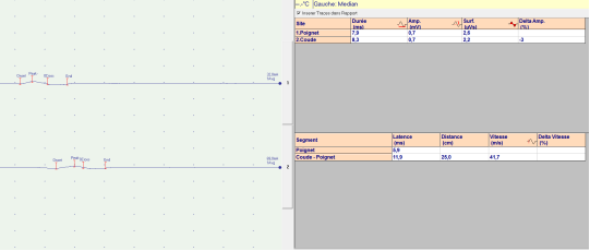
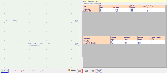
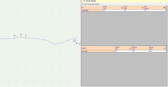
At the end of January 2024, they consulted a fourth health center where the diagnosis of Guillain-Barré syndrome was also mentioned. Biology found an ESR at 85 mm, a liver function test still disturbed. Lumbar puncture found hyperproteinorachia at 1 g/l, normal glycorachia, and an absence of cells. He benefited from short-term corticosteroid therapy 500 mg of IV methylprednisolone for 3 days then a per os relay with prednisolone 50 mg in addition to azathioprine 50 mg x2/day and functional rehabilitation. The therapies were stopped by the patient after 1 month. The patient was lost to follow-up and seen again in March 2024. He reported paresthesias such as tingling and electric shocks in all 4 limbs and 2 more episodes of tonic - clonic seizures. On examination, there was tetraparesis with a motor strength of 3/5 in the upper limbs, 1/5 distally, 2/5 in the lower limbs, 1/5 distally, abolished osteotendinous reflexes in all 4 limbs, bilateral hypopalesthesia L2 to L5, preserved pain sensitivity, walking possible with human assistance and a cane. The EEG of March found EEG activity of non-structural generalized epilepsy. The ENMG (Tables 3 and 4, Figures 1 to 3) found a neurophysiological pattern of acute polyradiculoneuropathy in the recovery phase with collapsed motor amplitudes, conduction blocks, globally slowed motor conduction velocities, prolonged F-wave latencies, collapsed sensory amplitudes with slowed sensory conduction velocities. Gabapentin 300 mg x2/day was introduced in addition to functional rehabilitation sessions. At the end of May 2024, the adolescent was again hospitalized for 1 month in a health center in the interior of the country. This hospitalization was carried out at the request of a parent for better care. The investigations carried out revealed an accelerated ESR of 32 mm and hepatic cytolysis syndrome.
In September 2024, he was found to have 3+/5 tetraparesis in the upper limbs, 2/5 distally, 3/5 in the lower limbs, 2/5 distally, walking possible without a cane, and an ESR of 50 mm. Given this development at 10 months, the family history and the persistence of a biological inflammatory syndrome and associated neuropsychiatric manifestations, a systemic disease was considered and an indirect immunofluorescence assay of antinuclear antibodies was performed, which came back positive with a speckled appearance and a titer of 1/160. The specificities of anti-DNA and soluble antinuclear antibodies were not performed. Based on the criteria of the American College of Rheumatology with a score of 19 (fever: 2, thrombocytopenia: 4 leukopenia: 3, convulsion: 5, psychosis: 3, delirium: 2), the diagnosis of systemic lupus was retained and azathioprine 50 mg reintroduced at 50 mg/day after normalization of the liver function test. In January 2025, during the follow-up appointment, the patient did not show up, having regained almost complete autonomy, he would have become independent again since December 2024, walked without a cane and discontinued immunosuppressive treatment. In early May 2025, during a fever, he presented polyarthralgia, lower back pain and worsening of the deficit in the lower limbs to 3/5. At the end of May 2025, the examination noted bilateral steppage, predominantly distal tetraparesis, 4+ in the upper limbs, extension of the hand on the forearm at 3/5, 4+ paresis in the lower limbs, 2/5 in plantar flexion, DTR still abolished in all 4 limbs, no sensory deficit, the rest of the physical examination was unremarkable.
Table 1. Patient's Main Blood Tests.

07-Dec

December 16

26-Dec

05-Jan

January 10

January 18

January 29

19- Feb

March 31

02 Sept

STANDARDS

AST

2.3 N

2.9 N

4.1 N

5.5 N

4.1 N

4 N

2.4 N

4 N

Less than 26, 38 or 37 IU/l

ALT

2.9 N

1.6 N

7.6 N

7.7 N

7.6 N

8.8 N

3.9 N

4.6 N

Below 29, 40 or 41

GGT

1.6 N

3.5 N

2.5 N

3.5 N

4 N

Normal

[10-50]

PAL

1.4 N

2.9 N

1.0 N

2.9 N

1.7 N

Less than 280

Urea

0.28

0.40

0.3

0.22

0.36

0.28

[0.15-0.45g/l]

Creatinine

8.65

8

10

9

9

8

7

[07-14mg/ml]

D-dimers

953.55

N < 500 mg/ml

PBS

Negative

Negative

Negative

Negative

CRP

96

< 5

< 5

< 5

N < 5 mg/l

ESR

40

50

65

85

32

50

< 20 mm

Blood sugar

1.08

0.7-1.10g/l

CPK

109

[24-190]

ASLO

<200

CBC

Hb

15.0

14

10.2

10.2

11.3

14.4

12-16g/dl

MCV

89.80

97.3

96

93

92.1

97.3

90-100 fl

MCHC

31.70

33.2

33.1

32

31.6

31.2

27-32 pg

Platelets

275000

264000

23100

98000

318000

264000

[150-400,000/mm3]

Leukocytes

5400

3700

7656

5500

7300

3700

4000-10000/mm 3

Lymphocytes

1697

1034

696

1870

949

1147

1500-4000/mm 3

PNN

1955

2445

2500

3410

4820

1700-7000/mm 3

HIV serology,

Hepatitis B, C

Negatives

Negatives

Negatives

Ionogram

Normal

Blood cultures

Negatives

Multi-drug test

Positives

CSF: Proteins

1.0 g/l

<0.5 g/l

Glucose

0.45g/l

0.50-0.70 g/l

Cytology

0 cells

0-5 cells

ANA (IFI)

1/160

< 1/160

Legend: AST: Aspartate amino- transferase, ALT: Alanine amino- transferase, GGT: Gamma- glutamyl transferase, CRP: C - reactive protein, PBS: peripheral blood smear, ESR: Sedimentation Rate, CBC: Complete Blood Count, CPK: Creatinine Phosphokinase ASLO: Antistreptolysin, Hb: Hemoglobin, MCV: Mean Cell Volume, MCHC: Mean Corpuscular Hemoglobin Content, PNN: Polymorphonuclear Neutrophils, CSF: Cerebrospinal Fluid, HIV: human immunodeficiency virus, ANA: Antinuclear Antibodies IFI: Indirect immunofluorescence on Hep 20-10 slides
Table 2. Some cases of GBS as the initial manifestation of SLE.

Citation (Actor/Year)

Age/Sex

Demonstration of the LES

Antibody

Kind of SGB

Treatment

Evolution

Background treatment

Uhunmwangho (2015)

26/F

Polyarthralgia; high blood pressure, proteinuria, lupus nephritis, generalized myxedema

ANA, dsDNA

Not realized

Cs per os, IV

Complete (Time not defined)

AZA, HQC

Boudanga (2023)

41/F

Generalized Alopecia Myxedema

ANA 1/1280 dsANA 300 IU/ml

AMAN

IVIg, CP (IV)

Complete

CS, HQC

Beshir (2021)

14/F

No symptoms before GBS

ANA 1/1280 in ANA 12 UI

AMAN

EP, (8), IG, IV, CS, IV, Rituximab

Complete

CS Oral

Yildiz (2015)

47/F

Alopecia, oral ulcer, anemia, leukopenia, thrombocytopenia,

ANA

AMAN

Ig IV, CS

Complete (14 months)

RTX, HQC

Wang (2024)

62/M

Malar rash, protein

ANA 1/160

MTX + intrathecal DXM

Not reported

Not reported

Fawzy (2022)

39/M

Arthralgia Epilepsy PRES syndrome

ANA, dsDNA ANA, Smith, antiSSa, Low complements

IV Ig, EP

Death

Chaudhuri (1989)

40/M

Epilepsy, Leukopenia, Lupus nephritis

ANA in ANA

AIDP

EP, CS, CP

Partial (8 weeks)

He (2025)

38/M

Malar rash, leukopenia, lupus pleurisy

ANA 1/1000 Rnp, Sm, Ro52, SSA, U1RNP, C3 low

AIDP

IVIg CS HCQ, MMF

Complete (18 months)

CS, HCQ, MMF

Present case

16/M

Psychosis, delirium, epilepsy, leukopenia psychosis

ANA

AIDP

CS, AZA

Partial (18 months)

AZA

LEGEND: ANA: Antinuclear antibodies; dsDNA: Anti-Double-Stranded DNA; Ig IV: Intravenous Immunoglobulin; HQC: Hydroxychloroquine; CS: Corticosteroids; RTX: Rituximab; AIDP: Acute Inflammatory Demyelinating Polyneuropathy; MMF: Mycophenolate mofetil; AMAN: neuroradiculopathy acute motor axonal syndrome; PE: plasma exchange; CP: Cyclophosphamide; RNP: Ribonucleoprotein; AZA: azathioprine; GBS: Guillain-Barré syndrome; SLE: Systemic lupus erythematosus; MTX methotrexate; DXM Dexamethasone SSA/ Ro: Soluble nuclear ribonucleoprotein A
4. Discussion
Neuropsychological manifestations of SLE or NPLSE are multiple neuropsychiatric manifestations directly linked to SLE . There are 19 NPLSEs . CNS manifestations include confusional syndrome, seizures, psychosis. PNS manifestations include mononeuropathy, plexopathy, GBS. NPSLE has a higher incidence in women, while the risk of seizures is more pronounced in men . NPSLE is more common in African descendants and Asians compared to whites; however, the severity of NPSLE is more significant in the white race. Neuropsychiatric manifestations occur in the early stages of SLE and represent between 39 and 50% of patients with this disease . In Cameroon, neuropsychiatric manifestations were inaugural of the disease in 37.0% of cases within the lupus population . Seizures are common manifestations of NPLSE (7-20%) while GBS (0.08-1.2%) is rare .
In this adolescent, psychosis, convulsions and delirium were considered as manifestations of a withdrawal syndrome, but in reality they were neuropsychological manifestations of SLE. This observation is thus distinguished by diagnostic wandering, the repetition of additional examinations in search of an infectious etiology (6 searches for plasmodium falciparum all negative, 3 HIV, hepatitis C and hepatitis B serologies also negative, blood cultures negative). Thus the infectious etiology was always systematically tracked, with each time, an unsuccessful search. He also benefited from antimalarial and antibiotic treatment. In the exploration of the syndrome of hepatic cytolysis and cholestasis, the liver and biliary tract ultrasound was normal. Liver damage can also be seen in SLE in the form of disturbance of liver biological tests (cytolysis or cholestasis greater than 4N) or abnormal liver histology .
Schematically, an alteration of the general condition preceded epileptic seizures and then GBS. The disturbance of liver enzymes throughout the evolution for 4 months was considered as an infectious but especially toxic hepatopathy despite an unacknowledged context of alcoholic intoxication or psychoactive substances. This initial positive multi-drug test was another element that confused the clinicians. In retrospect, the critical analysis of this multi-drug test shows us that it is a false positive test, that it is not ketamine but phencyclidine dosed in urine tests which is a dissociative agent structurally related to ketamine. In clinical practice, a false-positive urine drug screen can be caused by many xenobiotics: dextromethorphan, diphenhydramine, doxylamine, ibuprofen, imipramine, ketamine, meperidine, venlafaxine, bupropion, methylenedioxpyrolvalerone, and tramadol . The adolescent self-medicated and is likely to have used ibuprofen and tramadol. In addition, promethazine, which is used in hospital, is part of a group of drugs called phenothiazines . It can be used to relieve nausea and vomiting. It was used in this case as a hypnotic. Promethazine can cause false-positive urine drug screens for amphetamines or methamphetamines .
This observation highlights the problem of a rare disease with extremely rare manifestations in a country with limited technical facilities. First, the rarity of lupus itself and the rarity of its hepatic and neurological manifestations. The other problem is medical nomadism (stay in 6 health centers including two referral centers). The lumbar puncture was feared by the parents, finally performed with an albumin-cytological dissociation, serum/CSF isoelectrofocusing was not available. A crisis of confidence in the healthcare staff set in, justified by the label of a child consuming psychoactive substances that was stuck on their child, when in reality he was consuming energy drinks with the dream of becoming a professional footballer. This false label of a teenager addicted to psychoactive substances pursued him for more than a month, and this is why the referral from the psychiatric service to an intensive care unit was not honored because the parents thought that this diagnosis would be pursued and they then preferred to continue the hospitalization elsewhere. The tetraplegia was considered as generalized hypotonia resulting from a withdrawal syndrome, the episode of dyspnea was taken as a pulmonary embolism in this context of immobilization. It was only after a window of sedative drugs and by re-interviewing the family who confirmed a flaccid tetraplegia of ascending evolution over a few days that GBS was for the first time mentioned.
The occurrence of GBS in the patient was the event that changed the diagnostic approach. Based on the Brighton criteria , the patient presented the highest level of diagnostic certainty for GBS. The patient had symmetrical flaccid paralysis of the limbs, abolition of tendon reflexes and a monophasic course (peaking at 2 weeks). Cerebrospinal fluid showed albumin -cytological dissociation and ENMG revealed demyelination and axonal degeneration of the peripheral nerves of the limbs. Respiratory involvement was also observed. Differential diagnoses of chronic inflammatory demyelinating polyneuropathy (monophasic course without recurrence), myelitis (no sensory level, no sphincter disorder), metabolic/toxic/infectious neuropathies (no corresponding medical history, negative serological and etiological tests) were excluded. In adolescents, GBS was severe as reported several times in the literature . Severe GBS is characterized by rapidly evolving limb weakness, dysphagia, respiratory distress requiring respiratory support, and severe autonomic dysfunction (malignant arrhythmias, labile hypertension, or hemodynamic collapse) . ENMG at 2 months of flaccid tetraplegia demonstrated a pattern of acute inflammatory demyelinating polyneuropathy in the recovery phase. This is the most frequently reported electrophysiological form in SLE . According to He et al, the presence of axonal lesions on electromyography increases the likelihood of severe disease development . The occurrence of SLE complicated by severe GBS is rare, with severe manifestations involving respiratory failure being uncommon. He et al in their systematic review of the literature covering cases reported since the seminal description of severe SLE-GBS with respiratory failure (period 1999-2024), identified 13 severe cases. In our case, seizures preceded GBS. Two cases of GBS have been reported with epilepsy in the literature. The first was under investigation when the patient's neurological status worsened with increasing asthenia and presented with tonic-clonic seizures . In the second case, GBS was subsequently associated with PRES syndrome and their outcome despite optimal treatment was unfavorable .
Our observation nevertheless has some shortcomings: no dosage of the specificities of antinuclear antibodies (ANA), complements C3 and C4, anticardiolipin antibodies, antiganglioside antibodies because the explorations were the responsibility of the adolescent's family and due to the local technical platform limited the additional examinations. In the literature review of Coomes et al, the majority of patients with positive ANA were associated with another antibody, the speckled appearance on indirect immunofluorescence predominated followed by the homogeneous appearance . Two cases of low ANA titer at 1/160 in the literature: in Wang et al, the antiribonuclear antibodies were positive , in Yildiz et al, the specificities of the ANA antibodies came back negative . Another case in Wang with an average titer of 1/640, there was no other associated antibody . The transient worsening of our patient's neurological condition in the context of discontinuation of background treatment with fever suggests an SLE flare-up, which is another argument supporting the diagnosis of SLE.
In the treatment of classic GBS, corticosteroids are not recommended: intravenous methylprednisolone does not improve neurological status and its association with intravenous immunoglobulin’s is not beneficial. Oral corticosteroids may delay recovery or cause side effects . However, in the specific clinical context of SLE comorbid with GBS, this treatment logic should be reexamined . He et al advocate that the possibility of SLE should be considered for GBS patients without clear triggering factors, young women, or those with an immune-related or family history and that SLE should be part of the differential diagnosis of GBS because conventional therapies in GBS may not be adequate to treat SLE-GBS. According to current guidelines , when neuropsychiatric symptoms are highly correlated with SLE activity or specific neuropathies present with notable inflammatory features (axonal lesions), high-dose pulse corticosteroid therapy (500-1000 mg/day) should be used as the primary treatment. In severe cases , all patients received pulse corticosteroid therapy during acute disease progression. In some cases, efficacy was not significant despite early association with IV Ig, requiring the addition of immunosuppressants such as cyclophosphamide and azathioprine. He et al propose a hypothesis to be confirmed by multicenter randomized controlled studies: in comparison with the demyelinating lesions of peripheral nerves observed in classical GBS, SLE-GBS could involve a specific mechanism, mainly characterized by complement activation and immune complex deposition. The combined use of corticosteroids and immunosuppression may help manage the acute phase and reduce long-term risks by blocking T/B cell activation and cytokine storms.
Methylprednisolone were used and later azathioprine 50 mg/day was introduced. In Nigeria, Uhunmwangho et al used Aza 50 mg. We did not have IV Ig or plasma exchanges which were also beyond the financial reach of the parents. In the literature , cyclophosphamide, rituximab, azathioprine were the most used immunosuppressants. Synthetic antimalarials (hydrochloroquine) were often associated with the treatment. Gabapentin for neuropathic pain in Beshir was also used in adolescents with good progress .
The 18-month evolution of GBS noted a partial recovery. Severe cases generally had a complete recovery (evolution achieved at 24 months) .
5. Conclusion
This observation reminds us that systemic diseases exist in Togo and coexist with infectious, toxic, metabolic, and cardiovascular diseases. The initial manifestations of these systemic diseases can completely confuse the clinician. Although rare, GBS can be the mode of revelation of SLE. This SLE-GBS association is often serious and can be life-threatening. In a context of limited technical platform, faced with GBS with dysautonomic signs, the persistence of a biological inflammatory syndrome associated with other neuropsychiatric manifestations and a family context of suspected autoimmune disease, we propose the systematic search for SLE. The treatment of this particular form of GBS is not very well codified but the cases reported in the literature use corticosteroids and immunosuppressants with good evolution, drugs available in sub-Saharan Africa.
Abbreviations

SLE

Systemic Lupus Erythematosus

GBS

Guillain-Barré Syndrome

CNS

Central Nervous System

PNS

Peripheral Nervous System

IV Ig

Intravenous Immunoglobulin

PBS

Peripheral Blood Smear

CT

Computed Tomography

DTR

Deep Tendon Reflexes

CRP

C reactive Protein

HIV

Human Immunodeficiency Virus

ENMG

Electroneuromyogram

EEG

Electroencephalogram

ESR

Erythrocyte Sedimentation Rate

ANA

Antinuclear Antibodies

DNA

Deoxyribonucleic Acid

NPLSE

Neuropsychological Manifestations of Systemic Lupus Erythematosus

Aza

Azathioprine

CSF

Cerebrospinal Fluid

Author Contributions
Adama Mawulikplimi Ephoevi-ga: Conceptualization, Data curation, Funding acquisition, Methodology, Resources, Writing – original draft
Kokou Mensah Guinhouya: Funding acquisition, Validation, Writing – review & editing
Lehleng Agba: Funding acquisition, Validation, Writing – review & editing
Komla Nyinevi Anayo: Funding acquisition, Validation
Bitankade Kabassem: Funding acquisition, Validation
Kossivi Apetse: Funding acquisition, Validation, Visualization
Komi Assogba: Funding acquisition, Visualization
Vinyo Kumako: Funding acquisition, Visualization
Mofou Belo: Funding acquisition, Visualization
Agnon Koffi Balogou: Funding acquisition, Visualization
Data Availability Statement
The data that support the findings of this study are available from the corresponding author upon reasonable request.
Conflicts of Interest
The authors declare no conflicts of interest.
Appendix
Figure 1. Right median motor nerve conduction showing conduction block.
Figure 2. Left fibular motor nerve conduction showing conduction block.
Figure 3. Left ulnar sensitive nerve conduction showing collapse of amplitudes.
Table 3. Electroneuromyographic examination of motor conduction showed collapsed motor amplitudes, conduction blocks, globally slowed motor conduction velocities.

Motor nerve conduction

Nerves

Distal latency (ms)

Amplitude (mV)

Speed (ms)

Right median

4.0

1.7

42.6

Left median

5.9

0.7

41.7

Right ulnar

3.3

1.2

43.8

Left ulnar

3.9

2.8

46.0

Right common peroneal

6.7

0.4

31.3

Left common peroneal

8.8

No potential

28.6

Right tibial

8.7

3.1

31.3

Left tibial

3.8

Table 4. EMMG examination: sensory conduction showed collapsed sensory amplitudes with slowed sensory conduction velocities.

Sensory nerve conduction (orthodromic pathway)

Nerves

Distal latency (ms)

Amplitude (mV)

Speed

Right radial

2.7

3.2

44.4

Left radial

2.2

6.3

54.5

Right median

24

16.2

50.0

Left median

21

18.6

57.1

Right ulnar

24

8.2

37.5

Left ulnar

21

18.4

57.1

Right sural

40

3.5

30.0

Left sural

No potential

References
[1] Schwartz N., Stock AD, and C. Putterman. Neuropsychiatric lupus: new mechanistic insights and future treatment directions. Nat. Rev. Rheumatol. 2019; 15(3): 137‑152.
[2] Bellanti R, Rinaldi S. Guillain-barre syndrome: A comprehensive review. Eur J Neurol. 2024; 31: e16365.
[3] He L, Zhang Z, and Tan Y. Systemic lupus erythematosus complicated by severe Guillain-Barre syndrome: case report and literature review. Front. Immunol. 2025; 16: 1551448.
[4] Bediri K, El Ouni A, Toujani S et al. Polyradiculonévrite aiguë avec atteinte diaphragmatique au cours d’un Lupus érythémateux systémique: à propos d’une revenante [Acute polyneuropathy with diaphragmatic involvement in systemic lupus erythematosus: a case report]. La Revue de medecine interne. 2021; 42: A103-A104.
[5] Boudanga A, Chraa M, Rhazouani OC et al. Severe acute polyradiculoneuritis revealing systemic lupus erythematosus: a case report. Pan Afr Med J. 2023: 45(8).
[6] Uhunmwangho CU, Abene EA, Gimba ZM, et al. Guillain barre syndrome as initial presentation of systemic lupus erythematosus. Highland Med Res J. 2015; 15(2): 96-98.
[7] Apetse K, Dongmo Tajeuna JJ, Kumako VK et al. Guillain-Barre syndrome in hospitals in Togo. Médecine Tropicale Et Santé Internationale 2021.
[8] Kumako VK, Apetse K, Dongmo Tajeuna JJ et al. Profils épidémiologiques, cliniques et évolutifs des syndromes de Guillain Barre selon le type électroneuromyographique au Togo [Epidemiological, clinical and clinical course profiles of Guillain-Barré syndrome according to electroneuromyographic type in Togo]. Revue neurologique 176(2020) S44–S86.
[9] Sarwar S, Mohamed AS, Rogers S, et al. Neuropsychiatric Systemic Lupus Erythematosus: A 2021 Update on Diagnosis, Management, and Current Challenges. 2021; Cureus 13(9): e17969.
[10] ACR ad hoc committee on neuropsychiatric lupus nomenclature. The American College of Rheumatology nomenclature and case definitions for neuropsychiatric lupus syndromes. Arthritis & Rheumatism. 1999; 42(4): 599-608.
[11] Yacouba MN et al. Mapoure Njankouo Yacouba et al. Manifestations neuropsychiatriques au cours du lupus érythémateux systémique: à propos de 108 cas au Cameroun [Neuropsychiatric manifestations in patients with systemic lupus erythematosus: about 108 cases in Cameroon]. Pan African Medical Journal. 2022: 42(241).
[12] Martin-Blondel G, Alric L. Atteinte hépatique dans les maladies de système [Liver damage in systemic diseases. EMC – Hepatology]. 2009; 4(4): 1-9.
[13] Algren DA, Christian MR. Buyer Beware: Pitfalls in Toxicology Laboratory Testing. Emergency Medicine 2015.
[14] Melanson SEF, Lee-Lewandrowski E, Griggs DA et al. Reduced Interference by Phenothiazines in Amphetamine Drug of Abuse Immunoassays. Archives of Pathology & Laboratory Medicine. 2006; 130(12): 1834‑1838.
[15] Fokke C, van den Berg B, Drenthen J, Walgaard C, van Doorn PA, Jacobs BC. Diagnosis of Guillain-Barré syndrome and validation of Brighton criteria. Brain. 2014; 137: 33–43.
[16] Coomes EA, Haghbayan H, Spring J, and al. Fulminant Guillain-Barré syndrome in a patient with systemic lupus erythematosus. BMJ Case Rep. 2019; 12(1) 2018-226634.
[17] Chaudhuri KR, Taylor IK, Niven RM. A case of systemic lupus erythematosus presenting as Guillain-Barre syndrome. British Journal of Rheumatology. 1989; 28: 440-442.
[18] Uluta ÅŸ F. Systemic Lupus Erythematosus Presenting With Two Different Neurologic Entities, Guillain Barre Syndrome And Posterior Reversible Encephalopathy Syndrome: Case Report. Int. J. Clin. Rheumatol. (2022) 17(3), 058-061.
[19] Wang J, Zhe Z, Junyan Q et al. Clinical features of Guillain-Barré Syndrome and chronic inflammatory demyelinating polyradiculoneuritis associated with SLE. Lupus Sci Med. (2024) 11(20).
[20] Yıldız ÖK, Balaban H, Senel S, et al. Acute lumbosacral polyradiculoneuropathy heralding transformation to systemic lupus erythematosus in a patient with discoid lupus. Lupus (2011) 20, 972–974.
[21] Bertsias GK, Ioannidis JPA, Aringer M, et al. EULAR recommendations for the management of systemic lupus erythematosus with neuropsychiatric manifestations: report of a task force of the EULAR standing committee for clinical affairs. Annals of the Rheumatic Diseases 2010; 69(12): 2074‑2082.
[22] Beshir E, Belt E, Chencheri Net al. Case Report: Guillain-Barré Syndrome as Primary Presentation of Systemic Lupus Erythematosus (SLE-GBS) in a Teenage Girl. Front. Pediatr. 2022; 10: 838927.
Cite This Article
  • APA Style

    Ephoevi-ga, A. M., Guinhouya, K. M., Agba, L., Anayo, K. N., Kabassem, B., et al. (2026). Seizures and Guillain-Barré Syndrome Leading to the Discovery of Systemic Lupus: Case of an Adolescent in Lomé and Review of the Literature. American Journal of Psychiatry and Neuroscience, 14(3), 47-56. https://doi.org/10.11648/j.ajpn.20261403.11

    Copy | Download

    ACS Style

    Ephoevi-ga, A. M.; Guinhouya, K. M.; Agba, L.; Anayo, K. N.; Kabassem, B., et al. Seizures and Guillain-Barré Syndrome Leading to the Discovery of Systemic Lupus: Case of an Adolescent in Lomé and Review of the Literature. Am. J. Psychiatry Neurosci. 2026, 14(3), 47-56. doi: 10.11648/j.ajpn.20261403.11

    Copy | Download

    AMA Style

    Ephoevi-ga AM, Guinhouya KM, Agba L, Anayo KN, Kabassem B, et al. Seizures and Guillain-Barré Syndrome Leading to the Discovery of Systemic Lupus: Case of an Adolescent in Lomé and Review of the Literature. Am J Psychiatry Neurosci. 2026;14(3):47-56. doi: 10.11648/j.ajpn.20261403.11

    Copy | Download

  • @article{10.11648/j.ajpn.20261403.11,
      author = {Adama Mawulikplimi Ephoevi-ga and Kokou Mensah Guinhouya and Lehleng Agba and Komla Nyinevi Anayo and Bitankade Kabassem and Kossivi Apetse and Komi Assogba and Vinyo Kumako and Mofou Belo and Agnon Koffi Balogou},
      title = {Seizures and Guillain-Barré Syndrome Leading to the Discovery of Systemic Lupus: Case of an Adolescent in Lomé and Review of the Literature},
      journal = {American Journal of Psychiatry and Neuroscience},
      volume = {14},
      number = {3},
      pages = {47-56},
      doi = {10.11648/j.ajpn.20261403.11},
      url = {https://doi.org/10.11648/j.ajpn.20261403.11},
      eprint = {https://article.sciencepublishinggroup.com/pdf/10.11648.j.ajpn.20261403.11},
      abstract = {Background: Systemic lupus erythematosus (SLE) is a chronic idiopathic autoimmune disease. Guillain-Barré syndrome (GBS) is an acute monophasic immune-mediated polyradiculoneuropathy. Case presentation: We report the case of a 16-year-old patient, who presented in November 2023 a flu-like syndrome with diarrhea and abdominal pain. Then he had visual hallucinations and psychosis, followed by generalized tonic-clonic seizures. Urine toxicology assay was positive, a syndrome of hepatic cytolysis and cholestasis were noted. Toxic encephalopathy and hepatopathy were mentioned and he was transferred to a psychiatric ward where he persisted with an infectious syndrome, hallucinations, delirium and then flaccid tetraplegia of ascending evolution with dyspnea and respiratory distress. In January 2024, there was tetraplegia, abolished deep tendon reflexes (DTR) in all 4 limbs, hypoesthesia in all 4 limbs. Cerebrospinal fluid examination found albumin -cytological dissociation. Magnetic resonance imaging of the spinal cord was normal. He received 500 mg of intravenous methylprednisolone for 3 days then a per os relay with prednisolone 50 mg in addition to azathioprine (Aza) 50 mg x2/day and functional rehabilitation. The electroencephalogram revealed signs of non-structural generalised epilepsy in March 2024. The electroneuromyogram found a neurophysiological pattern of acute polyradiculoneuropathy in the recovery phase. In the evolution, an antinuclear antibody assay (indirect immunofluorescence) came back positive at a titer of 1/160, with a speckled appearance. On the basis of the American College of Rhumatology (ACR) with a score of 19, the diagnosis of SLE was retained and background treatment with Aza 50 mg/day continued. At the end of May 2025, there persisted a predominantly distal tetraparesis, an abolition of DTR. Conclusion: Although rare, GBS can be the mode of revelation of SLE. This SLE-GBS association is often serious and can be life-threatening.},
     year = {2026}
    }
    

    Copy | Download

  • TY  - JOUR
    T1  - Seizures and Guillain-Barré Syndrome Leading to the Discovery of Systemic Lupus: Case of an Adolescent in Lomé and Review of the Literature
    AU  - Adama Mawulikplimi Ephoevi-ga
    AU  - Kokou Mensah Guinhouya
    AU  - Lehleng Agba
    AU  - Komla Nyinevi Anayo
    AU  - Bitankade Kabassem
    AU  - Kossivi Apetse
    AU  - Komi Assogba
    AU  - Vinyo Kumako
    AU  - Mofou Belo
    AU  - Agnon Koffi Balogou
    Y1  - 2026/07/17
    PY  - 2026
    N1  - https://doi.org/10.11648/j.ajpn.20261403.11
    DO  - 10.11648/j.ajpn.20261403.11
    T2  - American Journal of Psychiatry and Neuroscience
    JF  - American Journal of Psychiatry and Neuroscience
    JO  - American Journal of Psychiatry and Neuroscience
    SP  - 47
    EP  - 56
    PB  - Science Publishing Group
    SN  - 2330-426X
    UR  - https://doi.org/10.11648/j.ajpn.20261403.11
    AB  - Background: Systemic lupus erythematosus (SLE) is a chronic idiopathic autoimmune disease. Guillain-Barré syndrome (GBS) is an acute monophasic immune-mediated polyradiculoneuropathy. Case presentation: We report the case of a 16-year-old patient, who presented in November 2023 a flu-like syndrome with diarrhea and abdominal pain. Then he had visual hallucinations and psychosis, followed by generalized tonic-clonic seizures. Urine toxicology assay was positive, a syndrome of hepatic cytolysis and cholestasis were noted. Toxic encephalopathy and hepatopathy were mentioned and he was transferred to a psychiatric ward where he persisted with an infectious syndrome, hallucinations, delirium and then flaccid tetraplegia of ascending evolution with dyspnea and respiratory distress. In January 2024, there was tetraplegia, abolished deep tendon reflexes (DTR) in all 4 limbs, hypoesthesia in all 4 limbs. Cerebrospinal fluid examination found albumin -cytological dissociation. Magnetic resonance imaging of the spinal cord was normal. He received 500 mg of intravenous methylprednisolone for 3 days then a per os relay with prednisolone 50 mg in addition to azathioprine (Aza) 50 mg x2/day and functional rehabilitation. The electroencephalogram revealed signs of non-structural generalised epilepsy in March 2024. The electroneuromyogram found a neurophysiological pattern of acute polyradiculoneuropathy in the recovery phase. In the evolution, an antinuclear antibody assay (indirect immunofluorescence) came back positive at a titer of 1/160, with a speckled appearance. On the basis of the American College of Rhumatology (ACR) with a score of 19, the diagnosis of SLE was retained and background treatment with Aza 50 mg/day continued. At the end of May 2025, there persisted a predominantly distal tetraparesis, an abolition of DTR. Conclusion: Although rare, GBS can be the mode of revelation of SLE. This SLE-GBS association is often serious and can be life-threatening.
    VL  - 14
    IS  - 3
    ER  - 

    Copy | Download

Author Information
  • Neurology Department, Campus Teaching Hospital, Lome, Togo

    Research Fields: General Neurology, Cerebrovascular diseases, neuroinflammatory diseases, Peripheral neurological disorders and muscle pathologies

  • Neurology Department, Sylvanus Olympio Teaching Hospital, Lome, Togo

    Research Fields: General Neurology, Epileptology, Peripheral neurological disorders and muscle pathologies, Neurodegenerative diseases

  • Neurology Department, Kara Teaching Hospital, Kara, Togo

    Research Fields: General Neurology, Epileptology, Peripheral neurological disorders and muscle pathologies, Neurodegenerative diseases

  • Neurology Department, Campus Teaching Hospital, Lome, Togo

    Research Fields: General Neurology, Epileptology, Peripheral neurological disorders and muscle pathologies, Neurodegenerative diseases

  • Neurology Department, Campus Teaching Hospital, Lome, Togo

    Research Fields: General Neurology, Epileptology, Peripheral neurological disorders and muscle pathologies, Neurodegenerative diseases

  • Neurology Department, Atakpame Regional Hospital, Atakpame, Togo

    Research Fields: Epileptology, Peripheral neurological disorders and muscle pathologies, Neurophysiology, movement disorders, neuroinflammatory diseases, Neurodegenerative diseases

  • Neurology Department, Sylvanus Olympio Teaching Hospital, Lome, Togo

    Research Fields: Epileptology, Peripheral neurological disorders and muscle pathologies, Neurophysiology, movement disorders, neuroinflammatory diseases, Neurodegenerative diseases

  • Neurology Department, Kara Teaching Hospital, Kara, Togo

    Research Fields: Epileptology, Peripheral neurological disorders, Neurophysiology, movement disorders, Neurodegenerative diseases

  • Neurology Department, University of Lome, Lome, Togo

    Research Fields: Epileptology, Peripheral neurological disorders and muscle pathologies, Neurophysiology, movement disorders, neuroinflammatory diseases, Neurodegenerative diseases

  • Neurology Department, Campus Teaching Hospital, Lome, Togo

    Research Fields: Epileptology, Peripheral neurological disorders, Neurophysiology, movement disorders, Neurodegenerative diseases