Abstract
Acute neurologic presentations in the setting of profound emotional trauma can create significant diagnostic complexity, particularly when symptoms overlap with Functional Neurologic Disorder (FND), formerly referred to as Conversion Disorder. In these situations, clinicians may be vulnerable to diagnostic overshadowing, in which emotionally salient psychosocial factors bias interpretation of objective neurologic findings and lead to premature psychiatric attribution. This risk may be further amplified when initial non-contrast neuroimaging is unrevealing. We present the case of a 26-year-old previously healthy female who developed severe headache, dysarthria, right-sided facial droop, and bilateral extremity weakness one week after the sudden death of her sister-in-law. Initial vascular imaging and non-contrast computed tomography (CT) were unremarkable, raising concern for FND in the context of acute bereavement, and psychiatry was consulted to evaluate for a possible functional or psychiatric etiology. Subsequent multidisciplinary reassessment involving psychiatry and neurology prompted advanced neuroimaging. Magnetic resonance imaging (MRI) of the brain demonstrated multifocal T2/FLAIR hyperintense lesions, including enhancing lesions in the bilateral thalami, left cerebellar peduncle, and left insular/temporal regions; cervical and thoracic spinal cord T2 hyperintensities were also identified without reported enhancement. Collectively, these findings were concerning for an active demyelinating process and highly suggestive of multiple sclerosis (MS), but a definitive diagnosis was not established because cerebrospinal fluid studies and subsequent neurologic follow-up were unavailable. This case highlights several important clinical lessons: acute emotional stressors should not substitute for a neurologic evaluation when objective deficits are present; FND is a rule-in diagnosis supported by positive functional signs rather than psychosocial stress alone; normal CT imaging does not exclude demyelinating disease; and interdisciplinary evaluation may help reduce premature diagnostic closure. The temporal association between bereavement and symptom onset in this case is observational and does not establish that stress caused or precipitated demyelinating disease.
Keywords
Multiple Sclerosis, Functional Neurologic Disorder, Conversion Disorder, Diagnostic Overshadowing, Demyelinating Disease, Psychiatry, Neurology, Bereavement
1. Introduction
Multiple sclerosis (MS) is a chronic inflammatory demyelinating disease of the central nervous system characterized by multifocal neurologic deficits and lesions distributed across characteristic central nervous system regions
| [1] | Thompson AJ, Banwell BL, Barkhof F, et al. Diagnosis of multiple sclerosis: 2017 revisions of the McDonald criteria. Lancet Neurol. 2018; 17(2): 162-173.
https://doi.org/10.1016/S1474-4422(17)30470-2 |
| [2] | McDonald WI, Compston A, Edan G, et al. Recommended diagnostic criteria for multiple sclerosis: Guidelines from the International Panel on the diagnosis of multiple sclerosis. Ann Neurol. 2001; 50(1): 121-127.
https://doi.org/10.1002/ana.1032 |
[1, 2]
. Clinical presentations vary widely and may include sensory disturbances, weakness, dysarthria, visual changes, gait abnormalities, and cognitive dysfunction
. Acute demyelinating disease can occasionally present with focal or stroke-like neurologic symptoms, creating diagnostic overlap with other neurologic conditions
| [2] | McDonald WI, Compston A, Edan G, et al. Recommended diagnostic criteria for multiple sclerosis: Guidelines from the International Panel on the diagnosis of multiple sclerosis. Ann Neurol. 2001; 50(1): 121-127.
https://doi.org/10.1002/ana.1032 |
[2]
.
Functional Neurologic Disorder is characterized by neurologic symptoms arising from altered nervous system functioning rather than a structural lesion that fully explains the presentation. Patients may present with weakness, abnormal movements, speech changes, sensory disturbances, gait symptoms, or non-epileptic events. Current diagnostic approaches emphasize that FND should be identified by positive clinical findings characteristic of the disorder, rather than inferred solely from normal diagnostic testing, psychiatric conditions, or psychosocial stressors
| [3] | Espay AJ, Aybek S, Carson A, et al. Current concepts in diagnosis and treatment of functional neurological disorders. JAMA Neurol. 2018; 75(9): 1132-1141.
https://doi.org/10.1001/jamaneurol.2018.1264 |
| [4] | Finkelstein SA, Popkirov S. Functional Neurological Disorder: Diagnostic Pitfalls and Differential Diagnostic Considerations. Neurol Clin. 2023; 41(4): 665-679.
https://doi.org/10.1016/j.ncl.2023.04.001 |
| [5] | Mavroudis I, Kazis D, Kamal FZ, et al. Understanding Functional Neurological Disorder: Recent Insights and Diagnostic Challenges. Int J Mol Sci. 2024; 25(8): 4470.
https://doi.org/10.3390/ijms25084470 |
[3-5]
.
The phenomenon of diagnostic overshadowing occurs when prominent psychiatric or psychosocial factors bias clinical reasoning and contribute to premature diagnostic closure
. In acute care settings, a recent emotional trauma may increase attention to psychiatric explanations even when an organic neurologic disorder remains possible. This creates a particularly challenging diagnostic environment when neurologic symptoms occur soon after bereavement or another major life stressor.
Emotional stress and inflammatory neurologic disease are not mutually exclusive. Recent evidence suggests a small association between psychological stress and MS onset, inflammatory activity, and disease progression; however, findings across studies are inconsistent, and other factors may contribute to this relationship
| [7] | von Drathen S, Gold SM, Peper J, et al. Stress and Multiple Sclerosis - Systematic review and meta-analysis of the association with disease onset, relapse risk and disability progression. Brain Behav Immun. 2024; 120: 620-629.
https://doi.org/10.1016/j.bbi.2024.06.004 |
[7]
. Accordingly, a temporal relationship between stress and neurologic symptom onset should be regarded as observational and should not be interpreted as evidence that stress caused the neurologic disease in an individual patient.
This report describes a young woman initially evaluated for possible FND following sudden bereavement whose subsequent MRI demonstrated multifocal brain and spinal cord lesions concerning for an inflammatory demyelinating process and highly suggestive of MS. Because lumbar puncture and subsequent follow-up were unavailable, the case does not establish a definitive diagnosis of MS. The case highlights the importance of diagnostic rigor, careful interpretation of FND criteria, and interdisciplinary collaboration between psychiatry and neurology.
2. Case Presentation
2.1. Patient Information and Initial Presentation
A 26-year-old previously healthy female presented to the emergency department with severe headache, dysarthria, right-sided facial droop, and bilateral upper and lower extremity weakness and numbness. Symptoms began approximately one week after the sudden death of her sister-in-law in a motor vehicle accident.
The patient denied any prior neurologic history, psychiatric diagnoses, or previous episodes of similar symptoms. She denied depression, suicidal ideation, homicidal ideation, hallucinations, mania, panic symptoms, flashbacks, or nightmares. She also denied recent viral illness or infectious symptoms. Her medical history was otherwise unremarkable except for brief oral contraceptive use in 2021. She admitted to remote tobacco and alcohol use but had abstained for several years.
Given the close timing of symptom onset and acute bereavement, FND or another stress-related psychiatric condition was considered, and psychiatry was consulted as part of the multidisciplinary evaluation. The psychosocial context was treated as one component of the differential rather than as diagnostic evidence for FND.
2.2. Initial Diagnostic Evaluation
Initial non-contrast CT imaging and vascular neuroimaging were reportedly unremarkable, reducing concern for acute hemorrhage, large-vessel occlusion, or other CT-detectable acute intracranial pathology. Despite unrevealing initial imaging, the patient continued to demonstrate documented neurologic abnormalities including right-sided facial droop, dysarthria, and extremity weakness and numbness.
Neurologic examination was notable for mild, symmetric weakness of the bilateral upper and lower extremities with subjectively decreased sensation in all four extremities. A right-sided facial droop was present; otherwise, cranial nerve examination was grossly intact. Extraocular movements were full, without nystagmus or other apparent oculomotor abnormalities. Coordination and gait were normal, and no pronator drift was observed. Formal testing for functional neurologic signs, including Hoover sign, give-way weakness, distractibility, or inconsistency, was not performed.
During psychiatric evaluation, the patient appeared emotionally appropriate and demonstrated intact thought processes, insight, and judgment. Although she acknowledged grief related to the recent death of her loved one, she denied severe mood symptoms or significant anxiety beyond situational bereavement. The available case information does not document positive examination signs establishing FND; therefore, FND remained a diagnostic consideration rather than a confirmed diagnosis.
2.3. Advanced Neuroimaging
Further multidisciplinary reassessment involving psychiatry and neurology prompted advanced neuroimaging.
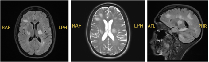
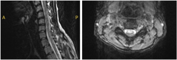
MRI of the brain with and without contrast demonstrated multiple T2/FLAIR hyperintense lesions involving both cerebral hemispheres. The report described associated T1 hypointense enhancing lesions involving the bilateral thalami, left cerebellar peduncle, and left insular/temporal regions, concerning for active inflammatory demyelination. MRI of the cervical spine demonstrated a suspected T2 hyperintense lesion in the ventral cord at C3 without gross enhancement. Thoracic spine MRI demonstrated additional subtle T2 hyperintensities suspicious for demyelination without associated enhancement, including a lesion reported at approximately the T2 level. Taken together, the brain and spinal cord abnormalities were highly concerning for a multifocal demyelinating process and radiographically suggestive of MS; however, imaging alone did not establish a definitive MS diagnosis.
Figure 1. Brain MRI demonstrating multifocal T2/FLAIR hyperintense lesions with enhancing lesions reported in the bilateral thalami, left cerebellar peduncle, and left insular/temporal regions. Findings were concerning for active inflammatory demyelination and were radiographically suggestive of MS, but were not independently diagnostic of MS.
Figure 2. Cervical and thoracic spine MRI demonstrating intramedullary lesions at the C3 and T2 levels without significant associated spinal canal stenosis. These findings supported an inflammatory demyelinating differential.
The radiologic differential included MS, acute disseminated encephalomyelitis (ADEM), other inflammatory demyelinating disorders such as neuromyelitis optica spectrum disorder or MOG antibody-associated disease, central nervous system vasculitis, infectious/inflammatory processes, and less likely neoplastic disease
. MS was favored radiographically because of the multifocal brain and spinal cord distribution and the presence of enhancing and nonenhancing abnormalities, while the absence of encephalopathy or a preceding infectious syndrome made classic ADEM less typical. Nevertheless, the absence of cerebrospinal fluid analysis, disease-specific serologic testing, and longitudinal imaging or clinical follow-up prevented definitive exclusion of these alternatives and precluded confirmation of MS
.
2.4. Hospital Course
Corticosteroid treatment was discussed; however, the patient expressed concern regarding treatment while breastfeeding. The patient left the hospital prior to initiation of therapy and before additional diagnostic studies, including lumbar puncture and cerebrospinal fluid analysis, could be completed. Outpatient neurology follow-up was recommended, but subsequent follow-up information was unavailable. Consequently, the final diagnostic status remained an inflammatory demyelinating process highly suggestive of MS rather than confirmed MS.
2.5. Clinical Timeline
Table 1. Clinical Timeline.
Day | Clinical Event |
Day 0 | Sudden death of patient’s sister-in-law |
Day 7 | Onset of headache, dysarthria, facial droop, and extremity weakness |
Day 8 | Presentation to emergency department; initial CT and vascular imaging unremarkable Psychiatry consultation requested due to concern for Functional Neurologic Disorder |
Day 9 | Brain MRI revealed multifocal enhancing demyelinating lesions Cervical and thoracic spinal MRI demonstrated additional spinal cord lesions Findings considered radiographically consistent with demyelinating disease concerning for multiple sclerosis |
Day 10 | Corticosteroid treatment discussed; patient expressed concern regarding treatment while breastfeeding Patient left hospital prior to treatment initiation |
Post-discharge | Outpatient neurology follow-up recommended; subsequent follow-up information unavailable |
3. Discussion
3.1. Diagnostic Overshadowing in Acute Neurologic Presentations
This case highlights the risk of diagnostic overshadowing, in which psychosocial circumstances may bias clinical judgment and contribute to premature attribution of symptoms to a psychiatric cause. The patient’s recent bereavement influenced the initial diagnostic framing and raised consideration of FND while a structural neurologic process remained in the differential. Diagnostic overshadowing is particularly important in acute care environments, where clinicians must rapidly synthesize emotionally significant contextual information while evaluating potentially serious neurologic pathology
.
The temporal relationship between grief and symptom onset created a compelling psychosocial narrative. However, a recent stressor is neither necessary nor sufficient for the diagnosis of FND, and it should not supersede neurologic examination findings or replace an appropriate medical evaluation
| [3] | Espay AJ, Aybek S, Carson A, et al. Current concepts in diagnosis and treatment of functional neurological disorders. JAMA Neurol. 2018; 75(9): 1132-1141.
https://doi.org/10.1001/jamaneurol.2018.1264 |
| [4] | Finkelstein SA, Popkirov S. Functional Neurological Disorder: Diagnostic Pitfalls and Differential Diagnostic Considerations. Neurol Clin. 2023; 41(4): 665-679.
https://doi.org/10.1016/j.ncl.2023.04.001 |
| [5] | Mavroudis I, Kazis D, Kamal FZ, et al. Understanding Functional Neurological Disorder: Recent Insights and Diagnostic Challenges. Int J Mol Sci. 2024; 25(8): 4470.
https://doi.org/10.3390/ijms25084470 |
[3-5]
.
3.2. Distinguishing Functional Neurologic Disorder from Demyelinating Disease
FND may present with weakness, speech abnormalities, sensory changes, gait disturbances, or other symptoms that resemble structural neurologic disease
| [3] | Espay AJ, Aybek S, Carson A, et al. Current concepts in diagnosis and treatment of functional neurological disorders. JAMA Neurol. 2018; 75(9): 1132-1141.
https://doi.org/10.1001/jamaneurol.2018.1264 |
| [4] | Finkelstein SA, Popkirov S. Functional Neurological Disorder: Diagnostic Pitfalls and Differential Diagnostic Considerations. Neurol Clin. 2023; 41(4): 665-679.
https://doi.org/10.1016/j.ncl.2023.04.001 |
| [5] | Mavroudis I, Kazis D, Kamal FZ, et al. Understanding Functional Neurological Disorder: Recent Insights and Diagnostic Challenges. Int J Mol Sci. 2024; 25(8): 4470.
https://doi.org/10.3390/ijms25084470 |
[3-5]
. Importantly, focal or anatomically localizable symptoms do not by themselves exclude FND. Current diagnostic practice instead relies on positive examination findings characteristic of FND, including specific patterns of variability, distractibility, or functional weakness that can be demonstrated during the neurologic examination
| [3] | Espay AJ, Aybek S, Carson A, et al. Current concepts in diagnosis and treatment of functional neurological disorders. JAMA Neurol. 2018; 75(9): 1132-1141.
https://doi.org/10.1001/jamaneurol.2018.1264 |
| [4] | Finkelstein SA, Popkirov S. Functional Neurological Disorder: Diagnostic Pitfalls and Differential Diagnostic Considerations. Neurol Clin. 2023; 41(4): 665-679.
https://doi.org/10.1016/j.ncl.2023.04.001 |
| [5] | Mavroudis I, Kazis D, Kamal FZ, et al. Understanding Functional Neurological Disorder: Recent Insights and Diagnostic Challenges. Int J Mol Sci. 2024; 25(8): 4470.
https://doi.org/10.3390/ijms25084470 |
[3-5]
.
By contrast, this patient demonstrated focal and anatomically localizable neurologic findings, including unilateral facial droop, dysarthria, and progressive extremity weakness. These findings were less consistent with classic functional presentations and more suggestive of an underlying structural neurologic process. Recent emotional trauma alone should not be considered sufficient evidence that neurologic symptoms are psychogenic in origin.
This distinction is clinically significant because misattribution of structural neurologic disease to psychiatric illness may delay diagnosis, postpone treatment initiation, and contribute to worse patient outcomes. Historical studies have demonstrated that patients initially diagnosed with conversion disorder may later be found to have underlying neurologic disease, highlighting the importance of thorough diagnostic evaluation and ongoing reassessment
.
3.3. Limitations of Initial CT Imaging in Demyelinating Disease
Another teaching point is the limited sensitivity of non-contrast CT for inflammatory demyelinating lesions. Initial CT and vascular imaging were reportedly unremarkable despite abnormalities subsequently identified on MRI. MRI is the principal imaging modality for suspected MS and other central nervous system demyelinating disorders because lesion morphology, distribution, and contrast enhancement provide substantially greater diagnostic information
.
In this patient, multifocal supratentorial and infratentorial brain lesions together with cervical and thoracic cord lesions supported an inflammatory demyelinating process. The reported combination of enhancing and nonenhancing lesions increased concern for lesions of differing activity, but the available information was insufficient to independently verify fulfillment of formal dissemination-in-time criteria. Normal CT imaging therefore should not be used to exclude demyelinating disease when the clinical presentation remains concerning; MRI and appropriate neurologic evaluation are warranted when symptoms remain unexplained
.
3.4. Differential Diagnosis of Multifocal Demyelinating-Appearing Lesions
Because CSF studies and longitudinal follow-up were unavailable, the imaging findings must be interpreted within a broader differential. MS was favored radiographically because of multifocal brain and spinal cord involvement and the reported presence of enhancing and nonenhancing lesions. ADEM was also considered; however, the patient’s adult age, absence of encephalopathy, and lack of a preceding infectious illness made the presentation less typical of classic ADEM. Nevertheless, ADEM cannot be excluded solely on these features
. Other inflammatory demyelinating disorders, including neuromyelitis optica spectrum disorder and MOG antibody-associated disease, should also be considered when supported by the clinical presentation and imaging findings, with appropriate antibody testing used to help distinguish these conditions. Central nervous system vasculitis and infectious or systemic inflammatory disorders may also produce multifocal neurologic symptoms and MRI abnormalities, while neoplastic disease was considered less likely by the interpreting radiologists
.
Definitive differentiation among these entities may require CSF analysis, targeted serologic studies, careful review of lesion morphology and distribution, and longitudinal clinical or MRI assessment
. Because these data were not available, the most supportable conclusion in this report is that the imaging demonstrated a multifocal inflammatory demyelinating process highly suggestive of MS rather than confirmed MS.
3.5. Psychosocial Stress and Neuroimmune Activation
The close temporal relationship between acute bereavement and neurologic symptom onset is clinically notable but observational. A recent systematic review and meta-analysis found a small to moderate association between psychological stress and MS onset, inflammatory activity, and disease progression, although limitations across the included studies may have influenced these findings
| [7] | von Drathen S, Gold SM, Peper J, et al. Stress and Multiple Sclerosis - Systematic review and meta-analysis of the association with disease onset, relapse risk and disability progression. Brain Behav Immun. 2024; 120: 620-629.
https://doi.org/10.1016/j.bbi.2024.06.004 |
[7]
. Earlier literature similarly explored stress-related neuroendocrine and immune pathways
| [12] | Mohr DC, Hart SL, Julian L, Cox D, Pelletier D. Association between stressful life events and exacerbation in multiple sclerosis: A meta-analysis. BMJ. 2004; 328(7442): 731.
https://doi.org/10.1136/bmj.38041.724421.55 |
| [13] | Artemiadis AK, Anagnostouli MC, Alexopoulos EC. Stress as a risk factor for multiple sclerosis onset or relapse: A systematic review. Neuroepidemiology. 2011; 36(2): 109-120.
https://doi.org/10.1159/000323953 |
[12, 13]
.
These associations do not establish that bereavement caused, triggered, or precipitated the patient’s demyelinating disease. At the individual-patient level, the temporal sequence could be coincidental or reflect mechanisms that cannot be determined from a single case. Psychosocial stress and trauma have also been discussed in the FND literature
| [14] | Roelofs K, Spinhoven P. Trauma and medically unexplained symptoms: Towards an integration of cognitive and neurobiological accounts. Clin Psychol Rev. 2007; 27(7): 798-820.
https://doi.org/10.1016/j.cpr.2007.07.004 |
| [15] | Keynejad RC, Frodl T, Kanaan R, et al. Stress and functional neurological disorders: Mechanistic insights. J Neurol Neurosurg Psychiatry. 2019; 90(7): 813-821.
https://doi.org/10.1136/jnnp-2018-318297 |
[14, 15]
, further underscoring that the presence of a stressor is diagnostically nonspecific. The appropriate clinical lesson is therefore not causal, but diagnostic: emotionally salient events should be considered as context without being allowed to substitute for objective neurologic assessment.
3.6. Importance of Interdisciplinary Collaboration
This case further illustrates the value of interdisciplinary collaboration between psychiatry and neurology. Psychiatry consultation did not simply serve to exclude psychiatric disease; rather, continued multidisciplinary reassessment preserved diagnostic openness and supported escalation to advanced neurologic evaluation. Psychiatric and neurologic disorders can coexist, and collaborative assessment may help reduce cognitive bias and premature diagnostic closure in complex neuropsychiatric presentations.
4. Conclusions
This case illustrates how acute psychosocial stressors can shape diagnostic framing in a patient presenting with new-onset neurologic symptoms. Although recent bereavement initially raised consideration of FND, subsequent MRI demonstrated multifocal brain and spinal cord lesions concerning for inflammatory demyelination and highly suggestive of MS. A definitive diagnosis of MS was not established because CSF studies and longitudinal follow-up were unavailable.
Several clinical lessons emerge. FND should be diagnosed through positive functional signs rather than psychosocial stress or exclusion alone, and focal symptoms do not independently rule FND in or out. Normal CT imaging does not exclude demyelinating disease, and persistent or unexplained neurologic abnormalities may warrant MRI and specialist evaluation. Finally, interdisciplinary collaboration between psychiatry and neurology can help preserve diagnostic flexibility and reduce premature closure. The temporal association between bereavement and symptom onset in this case should be interpreted as observational rather than causal.
This report highlights the importance of maintaining diagnostic vigilance while recognizing the complex interplay between psychosocial stress, cognitive bias, and neurologic disease.
Abbreviations
CNS | Central Nervous System |
CSF | Cerebrospinal Fluid |
CT | Computed Tomography |
FND | Functional Neurologic Disorder |
HPA | Hypothalamic-Pituitary-Adrenal |
MOGAD | Myelin Oligodendrocyte Glycoprotein Antibody-Associated Disease |
MRI | Magnetic Resonance Imaging |
MS | Multiple Sclerosis |
NMOSD | Neuromyelitis Optica Spectrum Disorder |
T2/FLAIR | T2-weighted Fluid-Attenuated Inversion Recovery |
Acknowledgments
The authors would like to acknowledge the multidisciplinary psychiatry and neurology teams involved in the patient’s care.
Author Contributions
Jessica Cho: Conceptualization, Investigation, Writing – original draft, Writing – review & editing
Priya Choudhary: Investigation, Writing – review & editing
Andy Ngo: Supervision, Validation, Writing – review & editing
Aneeqa Ahmed: Supervision, Validation, Writing – review & editing
Mahfoud Saddi: Supervision, Validation, Writing – review & editing
Mujeeb Shad: Supervision, Validation, Writing – review & editing
Data Availability Statement
The data supporting the findings of this case report are contained within the manuscript. Additional identifying patient information has been omitted to preserve patient confidentiality.
Conflicts of Interest
The authors declare no conflicts of interest.
References
| [1] |
Thompson AJ, Banwell BL, Barkhof F, et al. Diagnosis of multiple sclerosis: 2017 revisions of the McDonald criteria. Lancet Neurol. 2018; 17(2): 162-173.
https://doi.org/10.1016/S1474-4422(17)30470-2
|
| [2] |
McDonald WI, Compston A, Edan G, et al. Recommended diagnostic criteria for multiple sclerosis: Guidelines from the International Panel on the diagnosis of multiple sclerosis. Ann Neurol. 2001; 50(1): 121-127.
https://doi.org/10.1002/ana.1032
|
| [3] |
Espay AJ, Aybek S, Carson A, et al. Current concepts in diagnosis and treatment of functional neurological disorders. JAMA Neurol. 2018; 75(9): 1132-1141.
https://doi.org/10.1001/jamaneurol.2018.1264
|
| [4] |
Finkelstein SA, Popkirov S. Functional Neurological Disorder: Diagnostic Pitfalls and Differential Diagnostic Considerations. Neurol Clin. 2023; 41(4): 665-679.
https://doi.org/10.1016/j.ncl.2023.04.001
|
| [5] |
Mavroudis I, Kazis D, Kamal FZ, et al. Understanding Functional Neurological Disorder: Recent Insights and Diagnostic Challenges. Int J Mol Sci. 2024; 25(8): 4470.
https://doi.org/10.3390/ijms25084470
|
| [6] |
Croskerry P. The importance of cognitive errors in diagnosis and strategies to minimize them. Acad Med. 2003; 78(8): 775-780.
https://doi.org/10.1097/00001888-200308000-00003
|
| [7] |
von Drathen S, Gold SM, Peper J, et al. Stress and Multiple Sclerosis - Systematic review and meta-analysis of the association with disease onset, relapse risk and disability progression. Brain Behav Immun. 2024; 120: 620-629.
https://doi.org/10.1016/j.bbi.2024.06.004
|
| [8] |
Miller DH, Weinshenker BG, Filippi M, et al. Differential diagnosis of suspected multiple sclerosis: A consensus approach. Lancet Neurol. 2008; 7(9): 841-852.
https://doi.org/10.1016/S1474-4422(08)70173-6
|
| [9] |
Filippi M, Rocca MA. MR imaging of multiple sclerosis. Radiology. 2011; 259(3): 659-681.
https://doi.org/10.1148/radiol.11101362
|
| [10] |
Stone J, Smyth R, Carson A, et al. Systematic review of misdiagnosis of conversion symptoms and “hysteria.” BMJ. 2005; 331(7523): 989.
https://doi.org/10.1136/bmj.38628.466898.55
|
| [11] |
Stone J, Carson A, Duncan R, et al. Who is referred to neurology clinics? - The diagnoses made in 3781 new patients. Clin Neurol Neurosurg. 2010; 112(9): 747-751.
https://doi.org/10.1016/j.clineuro.2010.05.011
|
| [12] |
Mohr DC, Hart SL, Julian L, Cox D, Pelletier D. Association between stressful life events and exacerbation in multiple sclerosis: A meta-analysis. BMJ. 2004; 328(7442): 731.
https://doi.org/10.1136/bmj.38041.724421.55
|
| [13] |
Artemiadis AK, Anagnostouli MC, Alexopoulos EC. Stress as a risk factor for multiple sclerosis onset or relapse: A systematic review. Neuroepidemiology. 2011; 36(2): 109-120.
https://doi.org/10.1159/000323953
|
| [14] |
Roelofs K, Spinhoven P. Trauma and medically unexplained symptoms: Towards an integration of cognitive and neurobiological accounts. Clin Psychol Rev. 2007; 27(7): 798-820.
https://doi.org/10.1016/j.cpr.2007.07.004
|
| [15] |
Keynejad RC, Frodl T, Kanaan R, et al. Stress and functional neurological disorders: Mechanistic insights. J Neurol Neurosurg Psychiatry. 2019; 90(7): 813-821.
https://doi.org/10.1136/jnnp-2018-318297
|
Cite This Article
-
APA Style
Cho, J., Choudhary, P., Ngo, A., Ahmed, A., Saddi, M., et al. (2026). When Grief Mimics Conversion Disorder: New-Onset Focal Neurologic Deficits Revealing Demyelinating Disease. American Journal of Psychiatry and Neuroscience, 14(4), 93-99. https://doi.org/10.11648/j.ajpn.20261404.12
 Copy
|
 Download
ACS Style
Cho, J.; Choudhary, P.; Ngo, A.; Ahmed, A.; Saddi, M., et al. When Grief Mimics Conversion Disorder: New-Onset Focal Neurologic Deficits Revealing Demyelinating Disease. Am. J. Psychiatry Neurosci. 2026, 14(4), 93-99. doi: 10.11648/j.ajpn.20261404.12
Copy
|
Download
AMA Style
Cho J, Choudhary P, Ngo A, Ahmed A, Saddi M, et al. When Grief Mimics Conversion Disorder: New-Onset Focal Neurologic Deficits Revealing Demyelinating Disease. Am J Psychiatry Neurosci. 2026;14(4):93-99. doi: 10.11648/j.ajpn.20261404.12
Copy
|
Download
-
@article{10.11648/j.ajpn.20261404.12,
  author = {Jessica Cho and Priya Choudhary and Andy Ngo and Aneeqa Ahmed and Mahfoud Saddi and Mujeeb Shad},
  title = {When Grief Mimics Conversion Disorder: New-Onset Focal Neurologic Deficits Revealing Demyelinating Disease},
  journal = {American Journal of Psychiatry and Neuroscience},
  volume = {14},
  number = {4},
  pages = {93-99},
  doi = {10.11648/j.ajpn.20261404.12},
  url = {https://doi.org/10.11648/j.ajpn.20261404.12},
  eprint = {https://article.sciencepublishinggroup.com/pdf/10.11648.j.ajpn.20261404.12},
  abstract = {Acute neurologic presentations in the setting of profound emotional trauma can create significant diagnostic complexity, particularly when symptoms overlap with Functional Neurologic Disorder (FND), formerly referred to as Conversion Disorder. In these situations, clinicians may be vulnerable to diagnostic overshadowing, in which emotionally salient psychosocial factors bias interpretation of objective neurologic findings and lead to premature psychiatric attribution. This risk may be further amplified when initial non-contrast neuroimaging is unrevealing. We present the case of a 26-year-old previously healthy female who developed severe headache, dysarthria, right-sided facial droop, and bilateral extremity weakness one week after the sudden death of her sister-in-law. Initial vascular imaging and non-contrast computed tomography (CT) were unremarkable, raising concern for FND in the context of acute bereavement, and psychiatry was consulted to evaluate for a possible functional or psychiatric etiology. Subsequent multidisciplinary reassessment involving psychiatry and neurology prompted advanced neuroimaging. Magnetic resonance imaging (MRI) of the brain demonstrated multifocal T2/FLAIR hyperintense lesions, including enhancing lesions in the bilateral thalami, left cerebellar peduncle, and left insular/temporal regions; cervical and thoracic spinal cord T2 hyperintensities were also identified without reported enhancement. Collectively, these findings were concerning for an active demyelinating process and highly suggestive of multiple sclerosis (MS), but a definitive diagnosis was not established because cerebrospinal fluid studies and subsequent neurologic follow-up were unavailable. This case highlights several important clinical lessons: acute emotional stressors should not substitute for a neurologic evaluation when objective deficits are present; FND is a rule-in diagnosis supported by positive functional signs rather than psychosocial stress alone; normal CT imaging does not exclude demyelinating disease; and interdisciplinary evaluation may help reduce premature diagnostic closure. The temporal association between bereavement and symptom onset in this case is observational and does not establish that stress caused or precipitated demyelinating disease.},
 year = {2026}
}
Copy
|
Download
-
TY  - JOUR
T1  - When Grief Mimics Conversion Disorder: New-Onset Focal Neurologic Deficits Revealing Demyelinating Disease
AU  - Jessica Cho
AU  - Priya Choudhary
AU  - Andy Ngo
AU  - Aneeqa Ahmed
AU  - Mahfoud Saddi
AU  - Mujeeb Shad
Y1  - 2026/10/20
PY  - 2026
N1  - https://doi.org/10.11648/j.ajpn.20261404.12
DO  - 10.11648/j.ajpn.20261404.12
T2  - American Journal of Psychiatry and Neuroscience
JF  - American Journal of Psychiatry and Neuroscience
JO  - American Journal of Psychiatry and Neuroscience
SP  - 93
EP  - 99
PB  - Science Publishing Group
SN  - 2330-426X
UR  - https://doi.org/10.11648/j.ajpn.20261404.12
AB  - Acute neurologic presentations in the setting of profound emotional trauma can create significant diagnostic complexity, particularly when symptoms overlap with Functional Neurologic Disorder (FND), formerly referred to as Conversion Disorder. In these situations, clinicians may be vulnerable to diagnostic overshadowing, in which emotionally salient psychosocial factors bias interpretation of objective neurologic findings and lead to premature psychiatric attribution. This risk may be further amplified when initial non-contrast neuroimaging is unrevealing. We present the case of a 26-year-old previously healthy female who developed severe headache, dysarthria, right-sided facial droop, and bilateral extremity weakness one week after the sudden death of her sister-in-law. Initial vascular imaging and non-contrast computed tomography (CT) were unremarkable, raising concern for FND in the context of acute bereavement, and psychiatry was consulted to evaluate for a possible functional or psychiatric etiology. Subsequent multidisciplinary reassessment involving psychiatry and neurology prompted advanced neuroimaging. Magnetic resonance imaging (MRI) of the brain demonstrated multifocal T2/FLAIR hyperintense lesions, including enhancing lesions in the bilateral thalami, left cerebellar peduncle, and left insular/temporal regions; cervical and thoracic spinal cord T2 hyperintensities were also identified without reported enhancement. Collectively, these findings were concerning for an active demyelinating process and highly suggestive of multiple sclerosis (MS), but a definitive diagnosis was not established because cerebrospinal fluid studies and subsequent neurologic follow-up were unavailable. This case highlights several important clinical lessons: acute emotional stressors should not substitute for a neurologic evaluation when objective deficits are present; FND is a rule-in diagnosis supported by positive functional signs rather than psychosocial stress alone; normal CT imaging does not exclude demyelinating disease; and interdisciplinary evaluation may help reduce premature diagnostic closure. The temporal association between bereavement and symptom onset in this case is observational and does not establish that stress caused or precipitated demyelinating disease.
VL  - 14
IS  - 4
ER  -
Copy
|
Download





 Share This Article
 Twitter
 Linked In
 Facebook
