The development of polymer implant materials for use as drug carriers is an urgent task today. The aim of this work was to study the bioavailability of methyluracil when immobilized on a polyurethane carrier in vitro, as well as to study the biocompatibility of the obtained material when conducting model operations on experimental animals in vivo. As a result of the conducted studies, it was shown that the bioavailability of methyluracil immobilized on a polyurethane carrier is 78.5%, its prolonged release into the model environment is observed for 84 days. At the same time, more than 50% of the injected methyluracil was released by the 14th day of the study, which can significantly contribute to increasing the efficiency of regenerative processes at the implantation site, especially in the initial stages of the postoperative period. The conducted model operations on experimental animals made it possible to establish that the developed composite material with methyluracil is biocompatible and bioactive. Implantation of polymer samples with prolonged release of methyluracil contributed to the reduction of alteration and exudation phenomena in the implant placement area, activation of regeneration processes, and the formation of a mature and thin capsule around the implant already in the early stages of the study.
This is an Open Access article, distributed under the terms of the Creative Commons Attribution 4.0 International License (http://creativecommons.org/licenses/by/4.0/), which permits unrestricted use, distribution and reproduction in any medium or format, provided the original work is properly cited.
The development of polymer implant materials for use as drug carriers requires an interdisciplinary approach, including chemistry, biology, pharmacology and bioengineering
[1]
Stewart SA, Domínguez-Robles J, Donnelly RF, Larrañeta E. Implantable Polymeric Drug Delivery Devices: Classification, Manufacture, Materials, and Clinical Applications. Polymers. 2018; 10(12), 1379.
. It is known that polymer carriers allow to significantly improve the efficiency of drug delivery, ensuring local and controlled drug delivery directly to the desired area of the body, minimizing systemic side effects and toxicity, significantly increasing the bioavailability of the active substance
[2
-4], which opens up new opportunities for personalized medicine and increasing the effectiveness of treatment. A number of requirements are imposed on polymer implant materials, which they must meet. Among the main ones are biocompatibility and safety, the absence of an immune response
[5]
Galatenko N. A., Rozhnova R. A. Biologically active polymeric materials for medicine, Kyiv, Naukova Dumka. 2013, 211 p.
[6]
Gryn S. V., Synyugina A. T. Polymers for medical purposes, Kyiv. 2023, 88 p.
[5, 6]
. In recent years, polyurethanes (PU), due to their chemical stability, high biocompatibility and low cytotoxicity, have been widely used in biomedical purposes
[7]
Wienen D., Gries T., Cooper S. L., Heath D. E. An overview of polyurethane biomaterials and their use in drug delivery. Journal of Controlled Release. 2023, 363, 376-388.
Cui M., Chai Z., Lu Y., Zhu J., Chen J. Developments of polyurethane in biomedical applications: A review, Resources Chemicals and Materials. 2023, 2(4), 262-276.
Sobczak, M.; Kędra, K. Biomedical polyurethanes for anti-cancer drug delivery systems: a brief, comprehensive review. Int. J. Mol. Sci. 2022, 23, 8181.
Drożdż K., Gołda-Cępa M., Brzychczy-Włoch M. Polyurethanes as Biomaterials in Medicine: Advanced Applications, Infection Challenges, and Innovative Surface Modification Methods. Advancements of Microbiology. 2025, 4(63), 223-238.
. PU are a class of polymers that can act as drug carriers, and depending on their composition, they can be given the necessary properties and specified parameters
[11]
Wang W., Wang C. Polyurethane for biomedical applications: A review of recent developments. In the design and manufacture of medical devices. 2012. 115-151.
Bose N., Rajappan K. Summary on polyurethane-based drug delivery system in perspective for future implantable drug system. International Journal of Polymeric Materials and Polymeric Biomaterials. 2024, 73(18), 1629-1648.
One of the drugs that is promising for the purposes of immobilization on a PU carrier, in our opinion, is methyluracil, which belongs to the group of tissue regeneration stimulants. It is widely used in medicine to accelerate wound healing, tissue repair and improve metabolic processes
[13]
Svitlana V. Shishkina, Anna M. Shaposhnik and Victoriya V. Dyakonenko et al. New Polymorphic Modifications of 6-Methyluracil: An Experimental and Quantum Chemical Study. ACS Omega. 2023. Vol. 8(23): 20661-20674.
. Methyluracil has immunomodulatory and anti-inflammatory properties, is often used in medicine to stimulate cellular metabolism and protein synthesis, which contributes to the rapid healing of wounds and damaged tissues. As part of implant materials, methyluracil can be used to improve biocompatibility and accelerate tissue regeneration processes during implant use. Therefore, the current task is the development of polymeric materials with methyluracil as implants for use in various fields of medicine, in particular, in reconstructive surgery.
Considering the above, the purpose of this work was to study the bioavailability of methyluracil when immobilized on a PU carrier in vitro, as well as to study the biocompatibility of the obtained material when performing model operations on experimental animals in vivo.
2. Materials and Methods
2.1. Materials
The starting materials for obtaining composite materials were: polymer base - oligoetheretherethanediisocyanate, polymerization accelerator (PA-606/2) - 2,4,6-tris(dimethylaminomethyl)phenol. Methyluracil - a drug with regenerating, anti-inflammatory and immunostimulating properties - was used as a biologically active substance for immobilization on a polymer carrier. Polymer compositions with methyluracil in an amount of 2 wt. % were obtained, as well as PU samples without methyluracil as a control. Polymer compositions were obtained by sequential mechanical mixing of oligoetheretherethanediisocyanate, methyluracil, PA 606/2 and distilled water at room temperature. The resulting mixture was poured into fluoroplastic molds and dried in a thermostat at a temperature of 70ºС. The cured composite materials had the appearance of fine-pored elastic sponges.
2.2. Drug Release of Methyluracil
It was of great interest to investigate the bioavailability of methyluracil immobilized on a PU carrier, using spectrophotometric determination of the dynamics of methyluracil release into a model environment. For this purpose, the obtained control polymer samples and polymer samples with methyluracil in an amount of 2 wt. % were placed in jars with ground stoppers, 40 ml of distilled water were added and incubated in a thermostat at a temperature of 37±1°C. The obtained extracts were periodically drained and the optical density was recorded on a Specord M40 spectrophotometer in cuvettes with an optical layer thickness of 10 mm. During the study, distilled water was replaced with fresh water. An extract from a polymer sample without methyluracil was used as a control solution. UV absorption spectra of the studied methyluracil solutions before and after incubation of polymer samples under the specified conditions were identical to the spectrum of methyluracil itself.
The absorption spectrum of methyluracil had a maximum at a wavelength of λ=260±1 nm. To verify the implementation of Beer's law and to construct a calibration graph of the dependence of the optical density of solutions on their concentration, a series of aqueous methyluracil solutions with concentrations: 0.00025; 0.0005; 0.00075; 0.001; 0.00125% was prepared. The calibration graph was a straight line in the entire range of studied concentrations.
The amount of methyluracil (MU) released from polymer samples was calculated by the formulas (1) and (2):
(1)
(2)
where:
MU (mg) - the amount of methyluracil released, mg;
С - the concentration of methyluracil found from the calibration graph, %;
V - the volume of the solution in which the leaching took place, ml;
n - the degree of dilution of the solution during the analysis, times.
2.3. Animal Studies
2.3.1. Implantation Test
To study the interaction of the developed polymer materials with the tissues of a living organism, an implantation test was performed and cellular reactions in the tissues surrounding the implants of experimental animals were studied. The experiment was conducted on laboratory rats of the Wistar line weighing 180-210 g. All manipulations with experimental animals were carried out in compliance with the principles set forth in the European Convention for the Protection of Vertebrate Animals used for Experimental and Other Purposes
[14]
European convention for the protection of vertebrate animals used for experimental and other scientific purposes. Council of Europe, Strasbourg. 1986, 53 p.
[14]
and in accordance with the Law of Ukraine “On the Protection of Animals from Cruelty” No. 3447-IV of February 21, 2006. Model operations were performed under aseptic conditions. After processing the surgical field, polymer samples without methyluracil and samples with 2 wt.% methyluracil measuring 5 x 10 mm were placed subcutaneously in the back area of experimental animals without additional fixation, to exclude the influence of the suture material on the wound process. Animals were withdrawn from the experiment by ether overdose for 7, 14, 30 and 90 days.
2.3.2. Histological Evaluation
The experimental material (polymer sample with surrounding connective tissue) was fixed in 10% formalin solution and embedded in paraffin after histological processing according to the standard method
[15]
Bahrii M. M., Dibrova V. A., Popadynets O. H., Hryshchuk M. I. Metodyky morfolohichnykh doslidzhen. Vinnytsia: Nova Knyha. 2016, 328 p.
[15]
. Sections 10-15 μm thick were stained with hematoxylin and eosin. Analysis of cellular reactions and assessment of biocompatibility of polymer materials was carried out by examining histological preparations using light microscopy - microscopes "Mikmed-2", Carl Zeiss Primo Star, microphotography was carried out using a Canon PowerShot A640 camera with a Soligor Adapter Tube for Canon A610/A620 52 mm Tele.
3. Results and Discussion
3.1. Study of Drug Release of Methyluracil from Polyurethane Compositions in Vitro
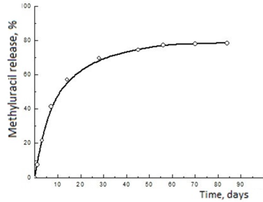
As a result of the conducted studies, it was found that 78.5% of methyluracil immobilized on a PU carrier was released in a sustained manner over 84 days (Figure 1). At the same time, the “shock” dose (more than 50% of the administered methyluracil) was released by the 14th day of the study, which will probably allow to immediately achieve a high therapeutic concentration and lead to a rapid achievement of the therapeutic effect, increasing the efficiency of regenerative processes at the implantation site in the initial stages of the postoperative period.
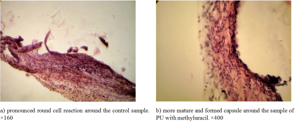
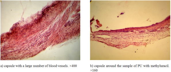
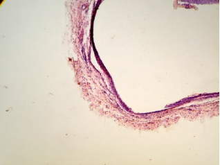
As a result of the work, was established that the bioavailability of methyluracil immobilized on a polyurethane carrier is 78.5%, and its prolonged release into the model environment is observed for 84 days. At the same time, more than 50% of the injected methyluracil was released by the 14th day of the study, which can significantly contribute to increasing the efficiency of regenerative processes at the implantation site, especially in the initial stages of the postoperative period. Model operations on experimental animals made it possible to establish that the developed composite material with methyluracil is biocompatible and bioactive. Implantation of polymer samples with prolonged release of methyluracil contributed to the reduction of alteration and exudation phenomena in the implant placement area, activation of regeneration processes and the formation of a mature and thin capsule around the implant already in the early stages of the study.
Stewart SA, Domínguez-Robles J, Donnelly RF, Larrañeta E. Implantable Polymeric Drug Delivery Devices: Classification, Manufacture, Materials, and Clinical Applications. Polymers. 2018; 10(12), 1379.
Pratap Singh L., Patro L. R., Sayeed M., Rani mandadi S., Pant N. C., Panda C., Aparna T. N., Vodeti R., Das C. Polymeric Drug Delivery Systems: Chemical Design, Functionalization and Biomedical Applications. J. Chem. Rev. 2025, 7(3), 421-451.
Sharma P, Jain V, Tailang M. Selection and Role of Polymers for Designing of a Drug Carrier [Internet]. Drug Carriers. IntechOpen; 2022. Available from:
Galatenko N. A., Rozhnova R. A. Biologically active polymeric materials for medicine, Kyiv, Naukova Dumka. 2013, 211 p.
[6]
Gryn S. V., Synyugina A. T. Polymers for medical purposes, Kyiv. 2023, 88 p.
[7]
Wienen D., Gries T., Cooper S. L., Heath D. E. An overview of polyurethane biomaterials and their use in drug delivery. Journal of Controlled Release. 2023, 363, 376-388.
Cui M., Chai Z., Lu Y., Zhu J., Chen J. Developments of polyurethane in biomedical applications: A review, Resources Chemicals and Materials. 2023, 2(4), 262-276.
Sobczak, M.; Kędra, K. Biomedical polyurethanes for anti-cancer drug delivery systems: a brief, comprehensive review. Int. J. Mol. Sci. 2022, 23, 8181.
Drożdż K., Gołda-Cępa M., Brzychczy-Włoch M. Polyurethanes as Biomaterials in Medicine: Advanced Applications, Infection Challenges, and Innovative Surface Modification Methods. Advancements of Microbiology. 2025, 4(63), 223-238.
Wang W., Wang C. Polyurethane for biomedical applications: A review of recent developments. In the design and manufacture of medical devices. 2012. 115-151.
Bose N., Rajappan K. Summary on polyurethane-based drug delivery system in perspective for future implantable drug system. International Journal of Polymeric Materials and Polymeric Biomaterials. 2024, 73(18), 1629-1648.
Svitlana V. Shishkina, Anna M. Shaposhnik and Victoriya V. Dyakonenko et al. New Polymorphic Modifications of 6-Methyluracil: An Experimental and Quantum Chemical Study. ACS Omega. 2023. Vol. 8(23): 20661-20674.
European convention for the protection of vertebrate animals used for experimental and other scientific purposes. Council of Europe, Strasbourg. 1986, 53 p.
[15]
Bahrii M. M., Dibrova V. A., Popadynets O. H., Hryshchuk M. I. Metodyky morfolohichnykh doslidzhen. Vinnytsia: Nova Knyha. 2016, 328 p.
Kuliesh, D., Nechaeva, L., Hrytsenko, V., Rozhnova, R. (2025). Study of the Bioavailability of Methyluracil in Vitro and Its Biocompatibility in Vivo in the Composition of Polyurethane Implant Material. American Journal of Polymer Science and Technology, 11(1), 1-6. https://doi.org/10.11648/j.ajpst.20251101.11
Kuliesh, D.; Nechaeva, L.; Hrytsenko, V.; Rozhnova, R. Study of the Bioavailability of Methyluracil in Vitro and Its Biocompatibility in Vivo in the Composition of Polyurethane Implant Material. Am. J. Polym. Sci. Technol.2025, 11(1), 1-6. doi: 10.11648/j.ajpst.20251101.11
Kuliesh D, Nechaeva L, Hrytsenko V, Rozhnova R. Study of the Bioavailability of Methyluracil in Vitro and Its Biocompatibility in Vivo in the Composition of Polyurethane Implant Material. Am J Polym Sci Technol. 2025;11(1):1-6. doi: 10.11648/j.ajpst.20251101.11
@article{10.11648/j.ajpst.20251101.11,
  author = {Dmytro Kuliesh and Ludmila Nechaeva and Vira Hrytsenko and Rita Rozhnova},
  title = {Study of the Bioavailability of Methyluracil in Vitro and Its Biocompatibility in Vivo in the Composition of Polyurethane Implant Material
},
  journal = {American Journal of Polymer Science and Technology},
  volume = {11},
  number = {1},
  pages = {1-6},
  doi = {10.11648/j.ajpst.20251101.11},
  url = {https://doi.org/10.11648/j.ajpst.20251101.11},
  eprint = {https://article.sciencepublishinggroup.com/pdf/10.11648.j.ajpst.20251101.11},
  abstract = {The development of polymer implant materials for use as drug carriers is an urgent task today. The aim of this work was to study the bioavailability of methyluracil when immobilized on a polyurethane carrier in vitro, as well as to study the biocompatibility of the obtained material when conducting model operations on experimental animals in vivo. As a result of the conducted studies, it was shown that the bioavailability of methyluracil immobilized on a polyurethane carrier is 78.5%, its prolonged release into the model environment is observed for 84 days. At the same time, more than 50% of the injected methyluracil was released by the 14th day of the study, which can significantly contribute to increasing the efficiency of regenerative processes at the implantation site, especially in the initial stages of the postoperative period. The conducted model operations on experimental animals made it possible to establish that the developed composite material with methyluracil is biocompatible and bioactive. Implantation of polymer samples with prolonged release of methyluracil contributed to the reduction of alteration and exudation phenomena in the implant placement area, activation of regeneration processes, and the formation of a mature and thin capsule around the implant already in the early stages of the study.},
 year = {2025}
}
TY  - JOUR
T1  - Study of the Bioavailability of Methyluracil in Vitro and Its Biocompatibility in Vivo in the Composition of Polyurethane Implant Material

AU  - Dmytro Kuliesh
AU  - Ludmila Nechaeva
AU  - Vira Hrytsenko
AU  - Rita Rozhnova
Y1  - 2025/08/20
PY  - 2025
N1  - https://doi.org/10.11648/j.ajpst.20251101.11
DO  - 10.11648/j.ajpst.20251101.11
T2  - American Journal of Polymer Science and Technology
JF  - American Journal of Polymer Science and Technology
JO  - American Journal of Polymer Science and Technology
SP  - 1
EP  - 6
PB  - Science Publishing Group
SN  - 2575-5986
UR  - https://doi.org/10.11648/j.ajpst.20251101.11
AB  - The development of polymer implant materials for use as drug carriers is an urgent task today. The aim of this work was to study the bioavailability of methyluracil when immobilized on a polyurethane carrier in vitro, as well as to study the biocompatibility of the obtained material when conducting model operations on experimental animals in vivo. As a result of the conducted studies, it was shown that the bioavailability of methyluracil immobilized on a polyurethane carrier is 78.5%, its prolonged release into the model environment is observed for 84 days. At the same time, more than 50% of the injected methyluracil was released by the 14th day of the study, which can significantly contribute to increasing the efficiency of regenerative processes at the implantation site, especially in the initial stages of the postoperative period. The conducted model operations on experimental animals made it possible to establish that the developed composite material with methyluracil is biocompatible and bioactive. Implantation of polymer samples with prolonged release of methyluracil contributed to the reduction of alteration and exudation phenomena in the implant placement area, activation of regeneration processes, and the formation of a mature and thin capsule around the implant already in the early stages of the study.
VL  - 11
IS  - 1
ER  -
 (1)
(1)  (2)
(2)