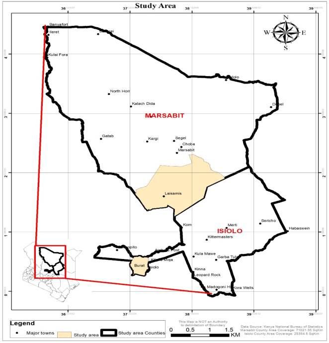
Camel rearing systems in the Arid and Semi Arid lands are undergoing significant changes, particularly around trading centers. More intensive camel production practices are replacing traditional extensive production systems. A cross-sectional study was conducted in Burat Ward, Isiolo County for intensive production systems and Laisamis, Marsabit County (extensive production systems). The aim of the study was to investigate the prevalence of common peri-parturient diseases and assess whether production systems and physiological status influence variations in serum levels of glucose, calcium, and magnesium. Data collection methods included focus group discussions, structured interviews, and blood sample analysis. The data were analyzed using descriptive statistics, mean comparisons, and Analysis of Variance. Results revealed that the prevalence of common diseases were higher in Laisamis (13.32-27%) than in Burat (4.59-12.06%). Likewise, gross mortality was higher in Laisamis (37.39%) than Burat (7.09%). Serum glucose levels were significantly lower in peri-parturient camels (3.91 and 4.45 mmol/L) compared to those in ordinary physiological status (6.09 mmol/L). Calcium levels remained consistent across physiological statuses and production systems (10.62-11.39 mg/dl). Magnesium levels were similar across physiological statuses but varied depending on the production system, they were higher in Burat (2.91-3.08 mg/dl) than Laisamis (2.46-2.71mg/dl). Most of the camels had below, normal and above normal levels of serum glucose, calcium and magnesium respectively. This was an indication that magnesium levels are influenced by dietary availability rather than physiological status. Malnutrition was the leading cause of death around parturition, primarily driven by negative energy balance. Blood glucose levels were found to depend on the physiological status of the camel, while calcium levels are tightly regulated by homeostatic mechanisms. Magnesium levels, however, depend on dietary intake. The study recommends improving camel nutrition during late pregnancy and early lactation to reduce the risk of metabolic and nutritional disorders.
| Published in | Animal and Veterinary Sciences (Volume 13, Issue 1) |
| DOI | 10.11648/j.avs.20251301.14 |
| Page(s) | 22-30 |
| Creative Commons |
This is an Open Access article, distributed under the terms of the Creative Commons Attribution 4.0 International License (http://creativecommons.org/licenses/by/4.0/), which permits unrestricted use, distribution and reproduction in any medium or format, provided the original work is properly cited. |
| Copyright |
Copyright © The Author(s), 2025. Published by Science Publishing Group |
Peri Parturient, Production System, Diseases, Disorders, Glucose, Magnesium, Calcium
Locations | Burat (n=108) | Laisamis (n=110) | Total (n=218) | ||||
|---|---|---|---|---|---|---|---|
Number and Percentage | No. | % | No. | % | No. | % | |
Abortion | Yes | 17 | 15.7 | 83 | 75.5 | 100 | 45.7 |
No | 91 | 84.3 | 27 | 24.5 | 118 | 54.3 | |
Dystocia | Yes | 26 | 24.1 | 48 | 43.6 | 74 | 33.8 |
No | 82 | 75.9 | 62 | 56.4 | 144 | 66.2 | |
Retained placenta | Yes | 23 | 21.3 | 57 | 51.8 | 80 | 36.5 |
No | 85 | 78.7 | 53 | 48.2 | 138 | 63.5 | |
Downer camel | Yes | 28 | 25.9 | 43 | 39.1 | 72 | 32.9 |
No | 80 | 74.1 | 67 | 60.9 | 146 | 67.1 | |
Agalactia | Yes | 29 | 26.9 | 44 | 40 | 74 | 33.8 |
No | 79 | 73.1 | 66 | 60 | 144 | 66.2 | |
Mastitis | Yes | 53 | 49.1 | 62 | 56.4 | 115 | 53 |
No | 55 | 50.9 | 48 | 43.6 | 103 | 47 | |
Burat | Laisamis | p/value | |
|---|---|---|---|
Disease Morbidities | |||
Abortion (n=80) | 4.59±1.29 | 27.00±2.92 | 0.000 |
Dystocia (n=91) | 6.90±1.44 | 13.40±2.29 | 0.020 |
Retained placenta (n=91) | 6.00±1.51 | 16.38±2.46 | 0.000 |
Malnutrition/Downer (n=90) | 6.96±1.24 | 13.32±2.42 | 0.017 |
Agalactia (n=90) | 6.78±1.33 | 14.10±2.47 | 0.012 |
Mastitis (n=87) | 12.06±1.63 | 16.64±2.30 | 0.099 |
Disease Mortalities | |||
Abortion (n=103) | 0.00±0.00 | 12.37±2.13 | 0.000 |
Dystocia (n=102) | 1.80±0.59 | 9.89±1.99 | 0.000 |
Retained placenta (n=102) | 1.18±0.48 | 9.93±1.96 | 0.000 |
Malnutrition/Downer (n=103) | 3.98±0.81 | 10.74±2.05 | 0.003 |
Agalactia (n=84) | 0.55±0.39 | 8.04±2.18 | 0.001 |
Mastitis (n=103) | 0.12±0.12 | 8.76±1.89 | 0.000 |
Percentage of dead to surviving | 7.63% | 59.73 | |
Gross mortality | 7.09% | 37.39% | |
Serum nutritional elements level | Physiological state and the wards | |||||
|---|---|---|---|---|---|---|
Late pregnancy | Early lactation | Ordinary | ||||
Burat | Laisamis | Burat | Laisamis | Burat | Laisamis | |
Glucose | ||||||
Below normal | 97 | 97 | ND | 63.6 | ||
Normal | 3 | 3 | ND | 27.3 | ||
Above normal | ND | 9.1 | ||||
Calcium | ||||||
Below normal | 6.1 | |||||
Normal | 90.9 | 100 | 87.9 | 81.8 | 100 | 66.3 |
Above normal | 9 | 12.1 | 18 | 27.6 | ||
Magnesium | ||||||
Below normal | 0 | 3 | 3 | |||
Normal | 6.1 | 9.1 | 9.1 | 6 | 18.2 | 21.2 |
Above normal | 93.9 | 90.9 | 90.9 | 91 | 81.8 | 75.8 |
Ward | Status | Glucose mmol/L | Calcium mg/dl | Magnesium mg/dl |
|---|---|---|---|---|
Burat | Late pregnancy (n=33) | 3.91±1.44a | 10.76±1.15ab | 2.95±0.53ab |
Early lactation (n=33) | 4.45±1.20a | 10.78±1.32ab | 2.91±0.53ab | |
Ordinary status (n=33) | 6.09±1.79b | 10.62±1.24b | 3.08±0.60ab | |
Laisamis | Late pregnancy (n=33) | ND | 10.75±1.15ab | 2.57±0.36cde |
Early lactation (n=33) | ND | 11.39±1.16a | 2.71±0.43bcd | |
Ordinary status (n=33) | ND | 11.34±2.18a | 2.46±0.27ce |
| [1] | Kenya National Bureau of Statistics (2019). National Population and Housing Census. Nairobi: Government Printers. |
| [2] | Delgado, C., Rosegrant, M., Steinfeld, H., Ehui, S., & Courbois., C. (1999). Livestock to 2020: The next food revolution. Washington D. C.: International Food Policy Research Institute. |
| [3] | Latino, L. R., Pica-Ciamarra, U., & Wisser, D. (2020). Africa: The livestock revolution urbanizes. Global Food Security, 26 (100399), 1-18. |
| [4] | Faye, B. (2014). The Camel today: Assets and potentials. Anthropozoologica, 49(2), 167–176. |
| [5] | Yosef, T., Mengistu, U., Solomon, A., Mohammed, Y., & Kefelegn, K. (2013). Camel and cattle population dynamics and livelihood diversification as a response to climate change in pastoral areas of Ethiopia. Livestock Research for Rural Development, 9(25) 1-7. |
| [6] | Kagunyu, A. W., & Wanjohi, J. (2014). Camel rearing replacing cattle production among the Borana community in Isiolo County of Northern Kenya, as climate variability bites. Pastoralism, 4(1), 1-13. |
| [7] | Hussien, A. A., Ali, S. M., & Tahir, A. (2011). Town Camels: Pastoral innovation in fast changing world. A case study from Gode Town, Somali regional state, Ethiopia. International conference of the future of pastoralism, 21-23rd march. Brighton. |
| [8] | Noor, I. M., Bebe, B. O., & Guliye, A. Y. (2012). Analysis of an emerging peri-urban camel production in Isiolo County, Northern Kenya. Journal of Camelid Science 41-61, 41-61. |
| [9] | Elitok, B., & Cirac, A. C. (2018). Clinical, hematological and blood biochemical features of Camels. Medcrave Online Journal of Immunology, 288-295. |
| [10] | Bzuneh, E., Alemneh, T., & Getabalew, M. (2020). Milk fever (parturient paresis) and its economic impact in dairy cattle production. Journal of Veterinary Medicine and Research 7(3) 1-8. |
| [11] | Blowey, R., Boyd, H., & Eddy, R. (2004). Bovine Medicine-Diseases and Husbandry of Cattle. Oxford: Blackwell Science Ltd. |
| [12] | Kumar, S., Purohit, G. N., & Pushp, M. K. (2016). Retention of placenta in a female Camel: A case report. Theriogenology insight - An International Journal of Reproduction in all animals, 6(1), 53-55. |
| [13] | Muhammad, S., Farooq, A., Akhtar, M., & Hayat, C. S. (2005). Parturient udder oedema in dromedary Camel (Camelus dromedaries). Pakistan Veterinary Journal, 25(2),100. |
| [14] | Magersa, B. (2010). An Epidemiology study of major Camel diseases in the Borana Lowland, Southern Ethiopia. Dryland Co-ordination report. |
| [15] | Osman, A. O., El-Metwaly, H., Wahba, A., & Hefny, S. (2016). Studies on causes of abortion in Maghrabian Camels. Egyptian Journal of Agricultural Research, 94(4)955-967. |
| [16] | Pfeiffer, D. U. (2010). Veterinary epidemiology: An introduction. Oxford, UK: Wiley Blackwell. |
| [17] | Poloju, K. K., Naidu, V. R., Rollakanti, C. R., Kishore, R., & Joe, A. (2021). A new method of data collection using the Kobo toolbox. Journal of Positive School Psychology, 6(4), 1527-1535. |
| [18] | Renjini, A., & Dileep, D. (2017). Spectrophotometry and spectrometry- Concept and applications. International Journal of Advance Research and Innovative Ideas in Education, 2(4), 96-100. |
| [19] | Dingwell, R T, Kelton D. F & Leslie, K. E. (2003). Management of the dry cow in control of peripartum disease and mastitis. Veterinary Clinics of North American Food Animal Practice. 19: 235-65. |
| [20] | Nikolic, J. A., Kulscar M., Nedic O, Janosi S & Huszenica G., (2003). Periparturient endocrine and metabolic changes in healthy cows affected by mastitis. Journal of Veterinary Medicine, Physiology Pathology and Clinical Medicine 50(1): 22-9. |
| [21] | Nipane, S., Kawitkar, S., Dhok, A., Chopde, S., Jawale, M., & Lende, S. (2022). Nutritional effect on immunity in animals. International Journal of Livestock Research, 12(4), 1-7. |
| [22] | Nagy P., Reiczigel, J., Barua, R., Das Gupta, A., & Juhász J. (2023). Pregnancy and parturition in dromedary camels III. Incidence, timing and factors affecting abortions and perinatal mortality under intensive management. Theriogenology. 197(1) 322-333. |
| [23] | Ahad, A. A., Megersa, B. & Edao, B. M. (2024). Brucellosis in camel, small ruminants, and Somali pastoralists in Eastern Ethiopia: A One Health approach. Frontiers in Veterinary Science, 11(1276275)1-11 |
| [24] | Aiello, S. E., Moses, M. A., & Allen, D. G. (Eds.). (2016). The Merck veterinary manual (Eleventh edition). Merck & Co., Inc. |
| [25] | Cheng, W. N., & Han, S. G. (2020). Bovine mastitis: Risk factors, therapeutic strategies, and alternative treatments — A review. Asian-Australasian Journal of Animal Sciences, 33(11), 1699–1713. |
| [26] | Wilson, R., & Dioli, M. (2021). History of Trypanosomosis in the one-humped Camel and development of its treatment and cure, with special Reference to Sudan. Medical Research Archives, 9(7), 1-21. |
| [27] | Bittner, L., Krämer, K., Wöckel, A., Snedec, T., Delling, C., Böttcher, D., Köller, G., Baumgartner, W., Richardt, W., & Starke, A. (2021). Malnutrition as the cause of recumbency in suckler cows associated with Trypanosoma theileria infection. Acta Veterinaria Scandinavica, 63(2), 1-8. |
| [28] | Puerto-Parada M, Bilodeau MÈ, Francoz D, Desrochers A, Nichols S, Babkine M, Arango-Sabogal JC, & Fecteau G. (2021). Survival and prognostic indicators in downer dairy cows presented to a referring hospital: A retrospective study. Journal of Veterinary Internal Medicine. 35(5), 2534-2543. |
| [29] | Ali, A., Derar, D., Tharwat, M., Zeitoun, M. M., & Alsobyil, F. A. (2016) Dystocia in dromedary camels: Prevalence, forms, risks and hematobiochemical changes. Animal Reproduction Science, 170(1)149-156, |
| [30] | Purohit, G. N. (2012). Dystocia in camelids: The causes and approaches of management. Open Journal of Animal Sciences, 2(02), 99–105. |
| [31] | Islam, S., Ferdous, J., Rahman, M. K., Akter, S., Hassan, M. M., & Islam, A. (2019). Reference values for hematological and serum biochemical parameters of dromedary Camel (Camelus dromedarius) in sub-tropical climate of Bangladesh. Advances in Animal and Veterinary Sciences, 7(4). 232-237 |
| [32] | Chand, B., Subedi, D., & Poudel, S. K. (2020). Effect of delayed serum separation on glucose concentration of dogs at room temperature. Advances in Animal and Veterinary Sciences, 8(8) 800-803. |
| [33] | Faye, B., & Bengoumi, M. (2018). Camel clinical biochemistry and hematology. New York: Springer International Publishers. |
| [34] | Badakhshan, Y., & Mirmahmoudi, R. (2016). Blood metabolites of one-humped camel (Camelus dromedarius) versus sheep during summer heat stress. Iranian Journal of Veterinary Medicine 10(1) 65-71. |
| [35] | Qureshi, A. S., Deeba, F., & Asrar, R. (2020). Calcium and phosphorus: Backbone of Camel health—A review. ECronicon Veterinary Science 5(3)1-9. |
| [36] | Temesgen D, Mohammed Y. K. & Beneberu S (2012). Critical macro and micro minerals concentration in the blood serum of camel (Camelus dromedarius) in Jijiga district, Eastern Ethiopia. Livestock Research for Rural Development. 24(60), 1-9. Retrieved from |
| [37] | Shoeib, S., Sayed-Ahmed, M., & El-khodery, S. (2019). Hypomagnesemic tetany in camel calves (camelus dromedarius): Clinical consequences and treatment outcomes. Slovenian Veterinary Research. 56(22).589–594 |
| [38] | Martens, H., Leonhard-Marek, S., Röntgen, M., & Stumpff, F. (2018). Magnesium homeostasis in cattle: Absorption and excretion. Nutrition Research Reviews, 31(1), 114–130. |
| [39] | Musalia L. M, Kirimi, J. G., Changwony, D., Thiakunu, F., Arimi, J, & Huka, G. (2020). Validation of climate smart forage preference by Camel (Camelus dromedarius) In Isiolo and Marsabit County, Kenya. Proceedings of Tharaka University 2nd Research E-Conference, 18-20th November 2020. |
| [40] | Kuria, S. G., Wahome, R. G., Gachuiri, C. K. & Wanyoike, M. M. (2004). Evaluation of forages as mineral sources for Camels in Western Marsabit, Kenya. South African Journal Animal Science, 34(3): 183-184. |
APA Style
Thiakunu, F., Kirimi, J., Arimi, J. (2025). Common Peri Parturient Diseases, Disorders and Levels of Serum Nutritional Elements of One Humped Female Camel (Camelus dromedaries) in Northern Kenya. Animal and Veterinary Sciences, 13(1), 22-30. https://doi.org/10.11648/j.avs.20251301.14
ACS Style
Thiakunu, F.; Kirimi, J.; Arimi, J. Common Peri Parturient Diseases, Disorders and Levels of Serum Nutritional Elements of One Humped Female Camel (Camelus dromedaries) in Northern Kenya. Anim. Vet. Sci. 2025, 13(1), 22-30. doi: 10.11648/j.avs.20251301.14
AMA Style
Thiakunu F, Kirimi J, Arimi J. Common Peri Parturient Diseases, Disorders and Levels of Serum Nutritional Elements of One Humped Female Camel (Camelus dromedaries) in Northern Kenya. Anim Vet Sci. 2025;13(1):22-30. doi: 10.11648/j.avs.20251301.14
@article{10.11648/j.avs.20251301.14,
  author = {Florence Thiakunu and James Kirimi and Joshua Arimi},
  title = {Common Peri Parturient Diseases, Disorders and Levels of Serum Nutritional Elements of One Humped Female Camel (Camelus dromedaries) in Northern Kenya},
  journal = {Animal and Veterinary Sciences},
  volume = {13},
  number = {1},
  pages = {22-30},
  doi = {10.11648/j.avs.20251301.14},
  url = {https://doi.org/10.11648/j.avs.20251301.14},
  eprint = {https://article.sciencepublishinggroup.com/pdf/10.11648.j.avs.20251301.14},
  abstract = {Camel rearing systems in the Arid and Semi Arid lands are undergoing significant changes, particularly around trading centers. More intensive camel production practices are replacing traditional extensive production systems. A cross-sectional study was conducted in Burat Ward, Isiolo County for intensive production systems and Laisamis, Marsabit County (extensive production systems). The aim of the study was to investigate the prevalence of common peri-parturient diseases and assess whether production systems and physiological status influence variations in serum levels of glucose, calcium, and magnesium. Data collection methods included focus group discussions, structured interviews, and blood sample analysis. The data were analyzed using descriptive statistics, mean comparisons, and Analysis of Variance. Results revealed that the prevalence of common diseases were higher in Laisamis (13.32-27%) than in Burat (4.59-12.06%). Likewise, gross mortality was higher in Laisamis (37.39%) than Burat (7.09%). Serum glucose levels were significantly lower in peri-parturient camels (3.91 and 4.45 mmol/L) compared to those in ordinary physiological status (6.09 mmol/L). Calcium levels remained consistent across physiological statuses and production systems (10.62-11.39 mg/dl). Magnesium levels were similar across physiological statuses but varied depending on the production system, they were higher in Burat (2.91-3.08 mg/dl) than Laisamis (2.46-2.71mg/dl). Most of the camels had below, normal and above normal levels of serum glucose, calcium and magnesium respectively. This was an indication that magnesium levels are influenced by dietary availability rather than physiological status. Malnutrition was the leading cause of death around parturition, primarily driven by negative energy balance. Blood glucose levels were found to depend on the physiological status of the camel, while calcium levels are tightly regulated by homeostatic mechanisms. Magnesium levels, however, depend on dietary intake. The study recommends improving camel nutrition during late pregnancy and early lactation to reduce the risk of metabolic and nutritional disorders.},
 year = {2025}
}
TY  - JOUR
T1  - Common Peri Parturient Diseases, Disorders and Levels of Serum Nutritional Elements of One Humped Female Camel (Camelus dromedaries) in Northern Kenya
AU  - Florence Thiakunu
AU  - James Kirimi
AU  - Joshua Arimi
Y1  - 2025/02/11
PY  - 2025
N1  - https://doi.org/10.11648/j.avs.20251301.14
DO  - 10.11648/j.avs.20251301.14
T2  - Animal and Veterinary Sciences
JF  - Animal and Veterinary Sciences
JO  - Animal and Veterinary Sciences
SP  - 22
EP  - 30
PB  - Science Publishing Group
SN  - 2328-5850
UR  - https://doi.org/10.11648/j.avs.20251301.14
AB  - Camel rearing systems in the Arid and Semi Arid lands are undergoing significant changes, particularly around trading centers. More intensive camel production practices are replacing traditional extensive production systems. A cross-sectional study was conducted in Burat Ward, Isiolo County for intensive production systems and Laisamis, Marsabit County (extensive production systems). The aim of the study was to investigate the prevalence of common peri-parturient diseases and assess whether production systems and physiological status influence variations in serum levels of glucose, calcium, and magnesium. Data collection methods included focus group discussions, structured interviews, and blood sample analysis. The data were analyzed using descriptive statistics, mean comparisons, and Analysis of Variance. Results revealed that the prevalence of common diseases were higher in Laisamis (13.32-27%) than in Burat (4.59-12.06%). Likewise, gross mortality was higher in Laisamis (37.39%) than Burat (7.09%). Serum glucose levels were significantly lower in peri-parturient camels (3.91 and 4.45 mmol/L) compared to those in ordinary physiological status (6.09 mmol/L). Calcium levels remained consistent across physiological statuses and production systems (10.62-11.39 mg/dl). Magnesium levels were similar across physiological statuses but varied depending on the production system, they were higher in Burat (2.91-3.08 mg/dl) than Laisamis (2.46-2.71mg/dl). Most of the camels had below, normal and above normal levels of serum glucose, calcium and magnesium respectively. This was an indication that magnesium levels are influenced by dietary availability rather than physiological status. Malnutrition was the leading cause of death around parturition, primarily driven by negative energy balance. Blood glucose levels were found to depend on the physiological status of the camel, while calcium levels are tightly regulated by homeostatic mechanisms. Magnesium levels, however, depend on dietary intake. The study recommends improving camel nutrition during late pregnancy and early lactation to reduce the risk of metabolic and nutritional disorders.
VL  - 13
IS  - 1
ER  -
Department of Animal Science, Meru University of Science and Technology, Meru, Kenya
Department of Livestock and Fisheries Development, Meru County Government, Meru, Kenya
Department of Food Science, Meru University of Science and Technology, Meru, Kenya
Information