Cirrhosis is a serious, progressive disease and constitutes a public health problem. The objective of this study was to examine the role of ultrasound in the diagnosis of cirrhosis at the Hospital of Mali. This was a prospective cross-sectional study conducted from February 2024 to February 2025. The study included all patients admitted to the department for abdominal ultrasound as part of the diagnosis of liver cirrhosis. Data were analyzed using SPSS version 21.0. Patient participation was voluntary. Patient confidentiality and anonymity were guaranteed. We identified 121 cases of cirrhosis diagnosed among 3,142 abdominal ultrasounds performed, representing a prevalence of 3.85%. Male patients accounted for 70% of cases. The mean age was 51.34 ± 13.86 years. The predominant clinical symptom was abdominal pain in 89.3% of cases. Hepatomegaly with a sharp lower border was recorded in 80.88% of cases. On ultrasound, hepatomegaly was present in 55% of patients. The echogenicity of the liver was heterogeneous in 96.7% of cases. The liver margins were irregular in 73% of cases. Hepatic dysmorphism was present in 74% of cases. Nodules were present in 60% of patients, and portal vein dilation in 58.7% of patients. Cirrhosis remains a common and serious condition. Ultrasound is an essential tool for screening and diagnosis.
This is an Open Access article, distributed under the terms of the Creative Commons Attribution 4.0 International License (http://creativecommons.org/licenses/by/4.0/), which permits unrestricted use, distribution and reproduction in any medium or format, provided the original work is properly cited.
Cirrhosis, Ultrasound Diagnosis, Medical Imaging, Mali Hospital
1. Introduction
Cirrhosis is a chronic fibrotic liver disease characterized by the transformation of normal liver architecture through a diffuse process of concentric fibrosis that encases regenerative nodules
[1]
Koama A, Tiemtore–Kambou BMA, Guingane A, Sieba IFN, Ouedraogo NAN, Napon M, et al. Morpho-biometric as-pects of liver cirrhosis and prevalence of portal hypertension in chronic carriers of the hepatitis B virus in Bogodogo (Burkina Faso). J Afr D’Imagerie Médicale. 2022; 14(3): 3.
[1]
. There are many types of cirrhosis, including post-hepatitis, alcoholic, steatohepatitis associated with metabolic dysfunction, and mixed cirrhosis, as well as congestive, biliary, and parasitic cirrhosis. Its etiological factors and morbidity vary by geographic region
[2]
Zhu JA, Hu B. Ultrasonography in predicting and screening liver cirrhosis in children: A preliminary study. World J Gastroenterol. 15 oct 2003; 9(10): 2348-2349.
Sarliève P, Delabrousse E, Saillet N, Rodière E, Michalakis D, Kastler B. DIG36 Prevalence of various signs of hepatic dysmorphia in cirrhosis. J Radiol. 1 sept 2004; 85(9): 1499.
Sarliève P, Delabrousse E, Saillet N, Rodière E, Michalakis D, Kastler B. DIG36 Prevalence of various signs of hepatic dysmorphia in cirrhosis. J Radiol. 1 sept 2004; 85(9): 1499.
. In Africa, the incidence of cirrhosis was 27.63% in Burkina Faso and 32.07% in Niger
[6]
Touré ES. Epidemiological, etiological, clinical, and therapeutic aspects of cirrhosis at the National Hospital of Niamey [thesis]. Université de Bamako; 2008.
[7]
Somé EN, Guingané NA, Lompo TI, Sombié R. Liver Cirrhosis: Epidemiological and Diagnostic Aspects at the Yalgado Ouédraogo University Hospital. Rev Afr Sci Soc Santé Publique. 13 juill 2021; 3(1): 53-64.
[6, 7]
. In Mali, a recent study conducted in 2025 at the referral health center in Commune V reported a prevalence of 4.1% for cirrhosis
[8]
Zikoume S. Cirrhosis at the referral health center in District V: epidemiological and clinical aspects. USTTB. [Master’s thesis], Bamako, 2025; N°193: 93.
[8]
.
Ultrasound is a noninvasive imaging method that uses ultrasound waves. It is the first-line method for screening for liver cirrhosis
[9]
Liu GJ, Lu MD. Diagnosis of liver cirrhosis with contrast-enhanced ultrasound. World J Radiol. 28 janv 2010; 2(1): 32-36.
. It allows for the assessment of morphology (shape and contours), echostructure (homogeneous, heterogeneous, nodular), and signs of portal hypertension (splenomegaly, shunts, flow abnormalities, and signs of thrombi)
[10]
Kanté S. Ultrasound findings of CHC in the Department of Radiology and Nuclear Medicine at Point G University Hos-pital. 2013.
[11]
Lebigot J, Elkhiry M, Boursier J, Bertrais S, Fouchard-Hubert I, Oberti F, et al. Diagnostic de cirrhose : l’echodoppler est toujours un examen performant! J Radiol. 1 oct 2009; 90(10): 1422.
. According to a prospective study conducted among patients suspected of having cirrhosis who underwent a liver biopsy, ultrasound had a sensitivity of 91% and a specificity of 94% for diagnosis
[12]
Simonovský V. The diagnosis of cirrhosis by high resolution ultrasound of the liver surface. Br J Radiol. janv 1999; 72(853): 29-34.
This increase in the incidence of cirrhosis demonstrates that early diagnosis and follow-up of affected patients are essential for improving patient care. Among the available diagnostic tools, abdominal ultrasound plays a key role due to its accessibility, non-invasive nature, and relatively low cost. This study may help optimize the use of ultrasound in the early detection of cirrhosis and its complications. It is in this context that we initiated this work. The objective of this study was to examine the role of ultrasound in the diagnosis of cirrhosis in the Medical Imaging Department of the Hospital of Mali.
2. Patients and Methods
Our study was conducted in the Medical Imaging Department of Mali Hospital and the Hepatogastroenterology Department of Gabriel Touré University Hospital. It was a prospective cross-sectional study conducted from February 2024 to February 2025. The study included all patients admitted to this department for an abdominal ultrasound as part of the diagnosis of liver cirrhosis. Data were entered and analyzed using SPSS version 21.0. Patient participation was voluntary, and consent was obtained from the parents or guardians of patients under the age of 18. Patient confidentiality and anonymity were ensured.
Our study included all patients referred by the Hepatogastroenterology Department of Gabriel Touré University Hospital for whom a diagnosis of cirrhosis had been confirmed. Patients with conditions other than cirrhosis were excluded. Abdominal ultrasound was performed in the Medical Imaging Department.
The examinations were performed using a FUJIFILM ARIETTA 50 ultrasound machine equipped with four probes: a 1–5 MHz convex probe, a 5–13 MHz linear probe, a 2–10 MHz transvaginal probe, and a 1–5 MHz cardiac probe. The ultrasounds were performed by a radiologist a physician specializing in ultrasound.
3. Results
3.1. Frequency
We identified 121 cases of cirrhosis diagnosed among 3,142 abdominal ultrasounds performed, representing an incidence of 3.85%.
3.2. Socio-demographic Data
Table 1. Distribution of patients according to socio-demographic data.
Sociodemographic data
n=121
%
Gender
Male
85
70
Female
36
30
Age group (years)
41-50
29
24,0
51-60
30
24,8
Occupation
Farmer
44
36,4
Houswife
31
25,6
In this study, male patients were in the majority, with a sex ratio of 2.36. The mean age was 51.34 ± 13 years.
The other occupations were primarily: manual laborers (7.4%), drivers (5.8%), and teachers (2.5%).
3.3. Clinical Data
Table 2. Distribution of patients according to clinical data.
Clinical data
n=121
%
Medical history
History of blood transfusions
16
13,2
Hypertension
41
33,9
Diabetes
24
19,8
History of familial liver disease
21
17,3
History of urinary schistosomiasis
19
15,7
Toxic habits
Alcohol
19
15,7
Tobacco
53
43,8
Hepatotoxic medications
76
62,8
Clinical signs
Abdominal pain
108
89,3
Jaundice
103
85,1
Gastrointestinal bleeding
33
27,3
Ascites
98
81
Physical signs
Hepatomegaly
68
56,2
Splenomegaly
39
32,2
Dullness
89
73,5
Collateral venous circulation
18
14,9
Clinical characteristics of hepatomegaly
Painful
45
37,2
Hard
43
35,6
Firm
25
20,7
Sharp lower edge
55
45,6
Nodular
52
42,9
Concept of gastrointestinal bleeding
Hematemesis
18
14,9
Melena
9
7.43
Rectal bleeding
6
4,27
Hepatomegaly with a sharp lower border was observed in 45.6% of cases.
3.4. Ultrasound Data
Table 3. Distribution of patients based on ultrasound-derived liver morphological data.
Données échographiques
n=121
%
Size of liver (cm)
< 15
54
45,0
> 15
67
55,0
Structure du foie
Heterogeneous
117
96,7
Homogeneous
3
2,5
Diffuse steatosis
1
0,8
Liver margins
Irregular
88
73,0
Regular
33
27,0
Dysmorphia
Yes
90
74,0
No
31
26,0
Presence of nodules
Yes
73
60,0
No
48
40,0
Size of nodules (cm)
n=73
< 3
27
37,0
> 3
46
63,0
Number of nodules
Single
5
7,0
Multiple
68
93,0
All patients exhibited at least two of the HTP signs (100% of cases).
The absence of respiratory modulation in 33.9% of cases.
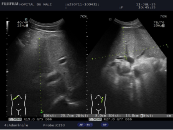
Figure 3. Abdominal ultrasound showing transverse and oblique views of the liver and spleen, revealing dilation of the portal trunk.
4. Discussion
4.1. Prevalence
We identified 121 cases of cirrhosis diagnosed among 3,142 abdominal ultrasounds performed, representing a prevalence of 3.85%. Diarra et al.
[14]
Diarra M, Konaté A, Soukho A, Dicko M, Kallé A, Doumbia K, et al. Progressive aspects of cirrhosis in a hepatogas-troenterology department at Mali. Mali Med. 2010; 25(1): 42-46.
[14]
reported a hospital prevalence of 2.35% in their study on the progression of cirrhosis. Sehounou et al.
[15]
Trop M. Liver Cirrhosis in Cotonou (Republic of Benin): Clinical Features and Factors Associated with Death. Tropical Medicine. 2010; 70(4): 375-378.
[15]
found a prevalence of 22.6% in their study on liver cirrhosis in Benin. Zikoume S.
[8]
Zikoume S. Cirrhosis at the referral health center in District V: epidemiological and clinical aspects. USTTB. [Master’s thesis], Bamako, 2025; N°193: 93.
[8]
reported a prevalence of 4.1% for cirrhosis in his study. In our context, this prevalence of cirrhosis reflects the high prevalence of hepatitis B and C.
4.2. Socio-demographic Data
In this study, males were in the majority, with a sex ratio of 2.33. This result was higher than that reported by Martin et al.
[16]
Martin J, Khatri G, Gopal P, Singal AG. Accuracy of Ultrasound and Noninvasive Markers of Fibrosis to Identify Pa-tients with Cirrhosis. Dig Dis Sci. juin 2015; 60(6): 1841-1847.
, who found a slight male predominance with a sex ratio of 1.15 in their study on the accuracy of ultrasound and non-invasive markers of fibrosis in identifying patients with cirrhosis. Diarra et al.
[14]
Diarra M, Konaté A, Soukho A, Dicko M, Kallé A, Doumbia K, et al. Progressive aspects of cirrhosis in a hepatogas-troenterology department at Mali. Mali Med. 2010; 25(1): 42-46.
[14]
found a sex ratio of 1.47 in favor of men, based on the progression of cirrhotic disease. These results reflect both behavioral and biological risk factors, highlighting the importance of targeting the male population in liver disease prevention and screening programs.
The 51–60 age group was the most common, accounting for 24.8% of cases, with a mean age of 51.34 ± 13.86 years. These results were similar to those reported by Driouiche et al
[18]
Mbendi CN, Nkodila A, Zingondo JCB, Manangama CN, Taty PL, Ngoma JA, et al. Aspects épidémio-clinique et évolutif de la Cirrhose du foie à Kinshasa: Etude Multicentrique: Multicentric study on epidemiological, clinical and progressive aspects of liver cirrhosis in Kinshasa.
[18]
in Morocco, who found a mean age of 54.4 years, as well as to the work of Mohammad et al
[19]
Dupuis M, Spahr L, Giostra E, Elkrief L. Portal vein thrombosis in patients with cirrhosis. Rev Med Suisse. Aug 30, 2017; 572: 1470–3.
[19]
in Nigeria, where the mean age ranged between 45 and 55 years. These results demonstrate the chronic nature of the disease, its gradual and slow onset, as well as its late detection in resource-limited settings such as Mali.
4.3. Clinical Data
In this study, the predominant clinical manifestations were abdominal pain (89.3%), jaundice (85.1%), and ascites. These Figures are higher than those reported by Mbendi et al.
[20]
Han SK, Kim MY, Kang SH, Baik SK. Application of ultrasound for the diagnosis of cirrhosis/portal hypertension. J Med Ultrason. July 1, 2022; 49(3): 321–31.
, where abdominal pain (44.9%), jaundice (42.3%), and ascites (64.7%) were observed in patients. In the study by Diarra et al.
[14]
Diarra M, Konaté A, Soukho A, Dicko M, Kallé A, Doumbia K, et al. Progressive aspects of cirrhosis in a hepatogas-troenterology department at Mali. Mali Med. 2010; 25(1): 42-46.
[14]
, ascites was the reason for consultation in 49.1% of cases. In our context, these proportions indicate a diagnostic delay often linked, on the one hand, to the population’s lack of information regarding cirrhosis and, on the other hand, to the sociocultural and economic conditions of the majority of the population.
Physical signs included dullness (73.5%), hepatomegaly (56.2%), and splenomegaly (32.2%). These results were similar to those observed by Mbendi et al.
[20]
Han SK, Kim MY, Kang SH, Baik SK. Application of ultrasound for the diagnosis of cirrhosis/portal hypertension. J Med Ultrason. July 1, 2022; 49(3): 321–31.
in Kinshasa, who found dullness in 64.7% and hepatomegaly in 54.1% of cases. Diarra et al.
[14]
Diarra M, Konaté A, Soukho A, Dicko M, Kallé A, Doumbia K, et al. Progressive aspects of cirrhosis in a hepatogas-troenterology department at Mali. Mali Med. 2010; 25(1): 42-46.
[14]
reported ascites and jaundice as physical signs found in 70.2% and 54.4% of cases, respectively, during their study. The observation of these signs underscores the importance of a comprehensive clinical evaluation, confirmed by paraclinical tests such as ultrasound, in diagnosing complications of cirrhosis.
4.4. Ultrasound Findings (Morphological and Spectral)
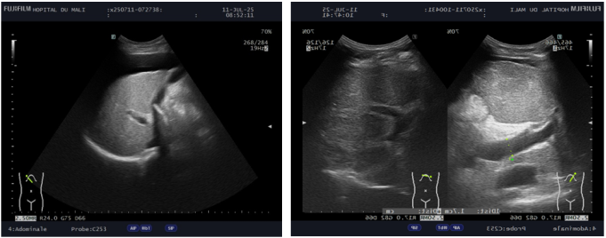
On ultrasound, hepatomegaly was present in 55% of patients. The liver’s echostructure was heterogeneous in 96.7% of cases, and the margins were irregular (73%). Hepatic dysmorphism was present in 74% of patients, and nodules in 60%. Koama et al.
[1]
Koama A, Tiemtore–Kambou BMA, Guingane A, Sieba IFN, Ouedraogo NAN, Napon M, et al. Morpho-biometric as-pects of liver cirrhosis and prevalence of portal hypertension in chronic carriers of the hepatitis B virus in Bogodogo (Burkina Faso). J Afr D’Imagerie Médicale. 2022; 14(3): 3.
[1]
reported that, on ultrasound, the right lobe of the liver was atrophic in 19% of cases. Hepatomegaly was present in 11% of cases. The liver margins were irregular in 55% of cases. The liver echostructure was micronodular and granular in 71% of cases in their study on the morphometric aspects of the cirrhotic liver and the prevalence of portal hypertension among chronic hepatitis B virus carriers in Burkina Faso. In the study by Mohammad et al
[17]
Maàji SM, Yakubu A, Odunko DD. Pattern of abnormal ultrasonographic findings in patients with clinical suspicion of chronic liver disease in Sokoto and its environs. Asian Pac J Trop Dis. 1 avr 2013; 3(3): 202-206.
in Nigeria, hepatic nodules were observed in 15% of patients. These characteristic ultrasound signs of liver cirrhosis confirm the indispensable role of this examination in the screening and diagnosis of this condition.
In our study, the indirect signs of portal hypertension were primarily characterized by dilation of the portal trunk in 19% of cases, followed by the presence of partial or total thrombosis in 33.1% and the presence of retrograde hepatic flow in 32.1% of cases. A prevalence of 28% had been reported by Dupuis et al. in their study
[19]
Dupuis M, Spahr L, Giostra E, Elkrief L. Portal vein thrombosis in patients with cirrhosis. Rev Med Suisse. Aug 30, 2017; 572: 1470–3.
[19]
. All these results were consistent with those in the literature, reflecting disease decompensation
[19]
Dupuis M, Spahr L, Giostra E, Elkrief L. Portal vein thrombosis in patients with cirrhosis. Rev Med Suisse. Aug 30, 2017; 572: 1470–3.
[20]
Han SK, Kim MY, Kang SH, Baik SK. Application of ultrasound for the diagnosis of cirrhosis/portal hypertension. J Med Ultrason. July 1, 2022; 49(3): 321–31.
. These results demonstrate the direct impact of cirrhosis on hepatic vascularization.
4.5. Complications on Ultrasound
Ascites was the most common complication encountered, accounting for 81% of cases. Kader et al. reported an ascites rate of 94.3% in their study. Most patients in Africa were diagnosed at advanced stages, which explains this increase in complications.
5. Conclusion
These results showed that cirrhosis remains a common and serious condition due to its complications, which can be severe. The majority of patients were men, with a mean age of 51.34 years. Ultrasound findings included hepatomegaly, heterogeneous liver texture, irregular margins, nodules, and portal vein dilation. Ultrasound is an essential tool for screening, diagnosing, and monitoring complications associated with this disease.
Yanogue Aldjouma: Formal Analysis, Writing – original draft
Diarra Hawa: Supervision
Sanogo Souleymane: Supervision
Kone Abdoulaye: Supervision
Goita Youssouf: Investigation
Kouma Alassane: Supervision
Toure Boubacar Mama: Supervision
Maiga Oumou: Supervision
Kamia Boureima: Supervision
Yara Mahamadou: Supervision
Coulibaly Salia: Validation
Sidibe Siaka: Validation
Conflicts of Interest
The authors declare no conflicts of interest.
References
[1]
Koama A, Tiemtore–Kambou BMA, Guingane A, Sieba IFN, Ouedraogo NAN, Napon M, et al. Morpho-biometric as-pects of liver cirrhosis and prevalence of portal hypertension in chronic carriers of the hepatitis B virus in Bogodogo (Burkina Faso). J Afr D’Imagerie Médicale. 2022; 14(3): 3.
[2]
Zhu JA, Hu B. Ultrasonography in predicting and screening liver cirrhosis in children: A preliminary study. World J Gastroenterol. 15 oct 2003; 9(10): 2348-2349.
Sarliève P, Delabrousse E, Saillet N, Rodière E, Michalakis D, Kastler B. DIG36 Prevalence of various signs of hepatic dysmorphia in cirrhosis. J Radiol. 1 sept 2004; 85(9): 1499.
Touré ES. Epidemiological, etiological, clinical, and therapeutic aspects of cirrhosis at the National Hospital of Niamey [thesis]. Université de Bamako; 2008.
[7]
Somé EN, Guingané NA, Lompo TI, Sombié R. Liver Cirrhosis: Epidemiological and Diagnostic Aspects at the Yalgado Ouédraogo University Hospital. Rev Afr Sci Soc Santé Publique. 13 juill 2021; 3(1): 53-64.
[8]
Zikoume S. Cirrhosis at the referral health center in District V: epidemiological and clinical aspects. USTTB. [Master’s thesis], Bamako, 2025; N°193: 93.
[9]
Liu GJ, Lu MD. Diagnosis of liver cirrhosis with contrast-enhanced ultrasound. World J Radiol. 28 janv 2010; 2(1): 32-36.
Kanté S. Ultrasound findings of CHC in the Department of Radiology and Nuclear Medicine at Point G University Hos-pital. 2013.
[11]
Lebigot J, Elkhiry M, Boursier J, Bertrais S, Fouchard-Hubert I, Oberti F, et al. Diagnostic de cirrhose : l’echodoppler est toujours un examen performant! J Radiol. 1 oct 2009; 90(10): 1422.
Diarra M, Konaté A, Soukho A, Dicko M, Kallé A, Doumbia K, et al. Progressive aspects of cirrhosis in a hepatogas-troenterology department at Mali. Mali Med. 2010; 25(1): 42-46.
[15]
Trop M. Liver Cirrhosis in Cotonou (Republic of Benin): Clinical Features and Factors Associated with Death. Tropical Medicine. 2010; 70(4): 375-378.
[16]
Martin J, Khatri G, Gopal P, Singal AG. Accuracy of Ultrasound and Noninvasive Markers of Fibrosis to Identify Pa-tients with Cirrhosis. Dig Dis Sci. juin 2015; 60(6): 1841-1847.
Maàji SM, Yakubu A, Odunko DD. Pattern of abnormal ultrasonographic findings in patients with clinical suspicion of chronic liver disease in Sokoto and its environs. Asian Pac J Trop Dis. 1 avr 2013; 3(3): 202-206.
Mbendi CN, Nkodila A, Zingondo JCB, Manangama CN, Taty PL, Ngoma JA, et al. Aspects épidémio-clinique et évolutif de la Cirrhose du foie à Kinshasa: Etude Multicentrique: Multicentric study on epidemiological, clinical and progressive aspects of liver cirrhosis in Kinshasa.
[19]
Dupuis M, Spahr L, Giostra E, Elkrief L. Portal vein thrombosis in patients with cirrhosis. Rev Med Suisse. Aug 30, 2017; 572: 1470–3.
[20]
Han SK, Kim MY, Kang SH, Baik SK. Application of ultrasound for the diagnosis of cirrhosis/portal hypertension. J Med Ultrason. July 1, 2022; 49(3): 321–31.
Abdoulaye, C. M., Ilias, G., Maba, T. M., Aldjouma, Y., Hawa, D., et al. (2026). The Role of Ultrasound in the Diagnosis of Liver Cirrhosis in the Medical Imaging Department at Mali Hospital. Clinical Medicine Research, 15(2), 19-25. https://doi.org/10.11648/j.cmr.20261502.11
Abdoulaye, C. M.; Ilias, G.; Maba, T. M.; Aldjouma, Y.; Hawa, D., et al. The Role of Ultrasound in the Diagnosis of Liver Cirrhosis in the Medical Imaging Department at Mali Hospital. Clin. Med. Res.2026, 15(2), 19-25. doi: 10.11648/j.cmr.20261502.11
Abdoulaye CM, Ilias G, Maba TM, Aldjouma Y, Hawa D, et al. The Role of Ultrasound in the Diagnosis of Liver Cirrhosis in the Medical Imaging Department at Mali Hospital. Clin Med Res. 2026;15(2):19-25. doi: 10.11648/j.cmr.20261502.11
@article{10.11648/j.cmr.20261502.11,
  author = {Camara Mody Abdoulaye and Guindo Ilias and Traore Mohamed Maba and Yanogue Aldjouma and Diarra Hawa and Sanogo Souleymane and Kone Abdoulaye and Goita Youssouf and Kouma Alassane and Toure Boubacar Mama and Maiga Oumou and Kamia Boureima and Yara Mahamadou and Coulibaly Salia and Sidibe Siaka},
  title = {The Role of Ultrasound in the Diagnosis of Liver Cirrhosis in the Medical Imaging Department at Mali Hospital},
  journal = {Clinical Medicine Research},
  volume = {15},
  number = {2},
  pages = {19-25},
  doi = {10.11648/j.cmr.20261502.11},
  url = {https://doi.org/10.11648/j.cmr.20261502.11},
  eprint = {https://article.sciencepublishinggroup.com/pdf/10.11648.j.cmr.20261502.11},
  abstract = {Cirrhosis is a serious, progressive disease and constitutes a public health problem. The objective of this study was to examine the role of ultrasound in the diagnosis of cirrhosis at the Hospital of Mali. This was a prospective cross-sectional study conducted from February 2024 to February 2025. The study included all patients admitted to the department for abdominal ultrasound as part of the diagnosis of liver cirrhosis. Data were analyzed using SPSS version 21.0. Patient participation was voluntary. Patient confidentiality and anonymity were guaranteed. We identified 121 cases of cirrhosis diagnosed among 3,142 abdominal ultrasounds performed, representing a prevalence of 3.85%. Male patients accounted for 70% of cases. The mean age was 51.34 ± 13.86 years. The predominant clinical symptom was abdominal pain in 89.3% of cases. Hepatomegaly with a sharp lower border was recorded in 80.88% of cases. On ultrasound, hepatomegaly was present in 55% of patients. The echogenicity of the liver was heterogeneous in 96.7% of cases. The liver margins were irregular in 73% of cases. Hepatic dysmorphism was present in 74% of cases. Nodules were present in 60% of patients, and portal vein dilation in 58.7% of patients. Cirrhosis remains a common and serious condition. Ultrasound is an essential tool for screening and diagnosis.},
 year = {2026}
}
TY  - JOUR
T1  - The Role of Ultrasound in the Diagnosis of Liver Cirrhosis in the Medical Imaging Department at Mali Hospital
AU  - Camara Mody Abdoulaye
AU  - Guindo Ilias
AU  - Traore Mohamed Maba
AU  - Yanogue Aldjouma
AU  - Diarra Hawa
AU  - Sanogo Souleymane
AU  - Kone Abdoulaye
AU  - Goita Youssouf
AU  - Kouma Alassane
AU  - Toure Boubacar Mama
AU  - Maiga Oumou
AU  - Kamia Boureima
AU  - Yara Mahamadou
AU  - Coulibaly Salia
AU  - Sidibe Siaka
Y1  - 2026/04/28
PY  - 2026
N1  - https://doi.org/10.11648/j.cmr.20261502.11
DO  - 10.11648/j.cmr.20261502.11
T2  - Clinical Medicine Research
JF  - Clinical Medicine Research
JO  - Clinical Medicine Research
SP  - 19
EP  - 25
PB  - Science Publishing Group
SN  - 2326-9057
UR  - https://doi.org/10.11648/j.cmr.20261502.11
AB  - Cirrhosis is a serious, progressive disease and constitutes a public health problem. The objective of this study was to examine the role of ultrasound in the diagnosis of cirrhosis at the Hospital of Mali. This was a prospective cross-sectional study conducted from February 2024 to February 2025. The study included all patients admitted to the department for abdominal ultrasound as part of the diagnosis of liver cirrhosis. Data were analyzed using SPSS version 21.0. Patient participation was voluntary. Patient confidentiality and anonymity were guaranteed. We identified 121 cases of cirrhosis diagnosed among 3,142 abdominal ultrasounds performed, representing a prevalence of 3.85%. Male patients accounted for 70% of cases. The mean age was 51.34 ± 13.86 years. The predominant clinical symptom was abdominal pain in 89.3% of cases. Hepatomegaly with a sharp lower border was recorded in 80.88% of cases. On ultrasound, hepatomegaly was present in 55% of patients. The echogenicity of the liver was heterogeneous in 96.7% of cases. The liver margins were irregular in 73% of cases. Hepatic dysmorphism was present in 74% of cases. Nodules were present in 60% of patients, and portal vein dilation in 58.7% of patients. Cirrhosis remains a common and serious condition. Ultrasound is an essential tool for screening and diagnosis.
VL  - 15
IS  - 2
ER  -
Department of Medical Imaging, Kati University Hospital, Kati, Mali; Faculty of Medicine and Odontostomatology, Bamako University of Science and Technology, Bamako, Mali
Traore Mohamed Maba
Department of Medical Imaging, Mali Hospital, Bamako, Mali
Yanogue Aldjouma
Department of Medical Imaging, Kati University Hospital, Kati, Mali
Department of Medical Imaging, Mali Hospital, Bamako, Mali; Department of Medical Imaging, Kati University Hospital, Kati, Mali; Department of Neurosurgery, Kati University Hospital, Kati, Mali; Department of Medical Imaging, Mother and Child Luxembourg University Hospital, Bamako, Mali; Faculty of Medicine and Odontostomatology, Bamako University of Science and Technology, Bamako, Mali
Sanogo Souleymane
Department of Medical Imaging, Mother and Child Luxembourg University Hospital, Bamako, Mali; Faculty of Medicine and Odontostomatology, Bamako University of Science and Technology, Bamako, Mali
Kone Abdoulaye
Faculty of Medicine and Odontostomatology, Bamako University of Science and Technology, Bamako, Mali
Goita Youssouf
Department of Medical Imaging, Kati University Hospital, Kati, Mali
Kouma Alassane
Department of Medical Imaging, Mother and Child Luxembourg University Hospital, Bamako, Mali
Toure Boubacar Mama
Department of Medical Imaging, Mali Hospital, Bamako, Mali
Maiga Oumou
Department of Medical Imaging, Mali Hospital, Bamako, Mali
Kamia Boureima
Department of Medical Imaging, Mali Hospital, Bamako, Mali
Yara Mahamadou
Department of Medical Imaging, Mali Hospital, Bamako, Mali
Coulibaly Salia
Department of Medical Imaging, Kati University Hospital, Kati, Mali
Sidibe Siaka
Faculty of Medicine and Odontostomatology, Bamako University of Science and Technology, Bamako, Mali
Abdoulaye, C. M., Ilias, G., Maba, T. M., Aldjouma, Y., Hawa, D., et al. (2026). The Role of Ultrasound in the Diagnosis of Liver Cirrhosis in the Medical Imaging Department at Mali Hospital. Clinical Medicine Research, 15(2), 19-25. https://doi.org/10.11648/j.cmr.20261502.11
Abdoulaye, C. M.; Ilias, G.; Maba, T. M.; Aldjouma, Y.; Hawa, D., et al. The Role of Ultrasound in the Diagnosis of Liver Cirrhosis in the Medical Imaging Department at Mali Hospital. Clin. Med. Res.2026, 15(2), 19-25. doi: 10.11648/j.cmr.20261502.11
Abdoulaye CM, Ilias G, Maba TM, Aldjouma Y, Hawa D, et al. The Role of Ultrasound in the Diagnosis of Liver Cirrhosis in the Medical Imaging Department at Mali Hospital. Clin Med Res. 2026;15(2):19-25. doi: 10.11648/j.cmr.20261502.11
@article{10.11648/j.cmr.20261502.11,
  author = {Camara Mody Abdoulaye and Guindo Ilias and Traore Mohamed Maba and Yanogue Aldjouma and Diarra Hawa and Sanogo Souleymane and Kone Abdoulaye and Goita Youssouf and Kouma Alassane and Toure Boubacar Mama and Maiga Oumou and Kamia Boureima and Yara Mahamadou and Coulibaly Salia and Sidibe Siaka},
  title = {The Role of Ultrasound in the Diagnosis of Liver Cirrhosis in the Medical Imaging Department at Mali Hospital},
  journal = {Clinical Medicine Research},
  volume = {15},
  number = {2},
  pages = {19-25},
  doi = {10.11648/j.cmr.20261502.11},
  url = {https://doi.org/10.11648/j.cmr.20261502.11},
  eprint = {https://article.sciencepublishinggroup.com/pdf/10.11648.j.cmr.20261502.11},
  abstract = {Cirrhosis is a serious, progressive disease and constitutes a public health problem. The objective of this study was to examine the role of ultrasound in the diagnosis of cirrhosis at the Hospital of Mali. This was a prospective cross-sectional study conducted from February 2024 to February 2025. The study included all patients admitted to the department for abdominal ultrasound as part of the diagnosis of liver cirrhosis. Data were analyzed using SPSS version 21.0. Patient participation was voluntary. Patient confidentiality and anonymity were guaranteed. We identified 121 cases of cirrhosis diagnosed among 3,142 abdominal ultrasounds performed, representing a prevalence of 3.85%. Male patients accounted for 70% of cases. The mean age was 51.34 ± 13.86 years. The predominant clinical symptom was abdominal pain in 89.3% of cases. Hepatomegaly with a sharp lower border was recorded in 80.88% of cases. On ultrasound, hepatomegaly was present in 55% of patients. The echogenicity of the liver was heterogeneous in 96.7% of cases. The liver margins were irregular in 73% of cases. Hepatic dysmorphism was present in 74% of cases. Nodules were present in 60% of patients, and portal vein dilation in 58.7% of patients. Cirrhosis remains a common and serious condition. Ultrasound is an essential tool for screening and diagnosis.},
 year = {2026}
}
TY  - JOUR
T1  - The Role of Ultrasound in the Diagnosis of Liver Cirrhosis in the Medical Imaging Department at Mali Hospital
AU  - Camara Mody Abdoulaye
AU  - Guindo Ilias
AU  - Traore Mohamed Maba
AU  - Yanogue Aldjouma
AU  - Diarra Hawa
AU  - Sanogo Souleymane
AU  - Kone Abdoulaye
AU  - Goita Youssouf
AU  - Kouma Alassane
AU  - Toure Boubacar Mama
AU  - Maiga Oumou
AU  - Kamia Boureima
AU  - Yara Mahamadou
AU  - Coulibaly Salia
AU  - Sidibe Siaka
Y1  - 2026/04/28
PY  - 2026
N1  - https://doi.org/10.11648/j.cmr.20261502.11
DO  - 10.11648/j.cmr.20261502.11
T2  - Clinical Medicine Research
JF  - Clinical Medicine Research
JO  - Clinical Medicine Research
SP  - 19
EP  - 25
PB  - Science Publishing Group
SN  - 2326-9057
UR  - https://doi.org/10.11648/j.cmr.20261502.11
AB  - Cirrhosis is a serious, progressive disease and constitutes a public health problem. The objective of this study was to examine the role of ultrasound in the diagnosis of cirrhosis at the Hospital of Mali. This was a prospective cross-sectional study conducted from February 2024 to February 2025. The study included all patients admitted to the department for abdominal ultrasound as part of the diagnosis of liver cirrhosis. Data were analyzed using SPSS version 21.0. Patient participation was voluntary. Patient confidentiality and anonymity were guaranteed. We identified 121 cases of cirrhosis diagnosed among 3,142 abdominal ultrasounds performed, representing a prevalence of 3.85%. Male patients accounted for 70% of cases. The mean age was 51.34 ± 13.86 years. The predominant clinical symptom was abdominal pain in 89.3% of cases. Hepatomegaly with a sharp lower border was recorded in 80.88% of cases. On ultrasound, hepatomegaly was present in 55% of patients. The echogenicity of the liver was heterogeneous in 96.7% of cases. The liver margins were irregular in 73% of cases. Hepatic dysmorphism was present in 74% of cases. Nodules were present in 60% of patients, and portal vein dilation in 58.7% of patients. Cirrhosis remains a common and serious condition. Ultrasound is an essential tool for screening and diagnosis.
VL  - 15
IS  - 2
ER  -