Case Report | | Peer-Reviewed

Operative Technique and Short-term Outcome of Re-do Failed Hypospadias Repair: A Case Report

Received: 3 December 2024     Accepted: 31 December 2024     Published: 21 January 2025
Views:       Downloads:
Abstract

Introduction: A failed hypopadias repair is a difficult case to treat considering the poor quality of surrounding tissues arising from disruption of normal vasculature associated with the underlying disorder as well as scaring from previous repairs. In this case report we describe our experience with the management of late presentation of failed hypospadias repair in a 12 year old boy. Case report: Our patient is a twelve year old ‘small for age’ who presented to our center with failed mid-penile hypospadias repair. He first presented at the age of eight to another hospital where he was evaluated and a diagnosis of mid-penile hypospadias with significant chordee was made. He had the first repair at the age of eight but the repair failed two weeks after surgery. Two years later he had a second repair in same facility which also failed after approximately two weeks. They consequently presented to our center two years later at the age of twelve. Following appropriate evaluation and counseling, he had the 3rd repair during which we ensured a preoperative sterile urine culture, created a urinary diversion via suprapubic cystostomy, the use of a monofilament (monocryl) suture, complete release of chordee after excision of scar tissues, a second intervening layer over the repair, the use of an improvised stent instead of a urethral catheter and appropriate anchorage of the stent to the suprapubic skin using a nylon suture. Patient did well post-operatively and was discharged home 14 days after the repair. Subsequent outpatient visits were uneventful and he was discharged after three months to see us only when necessary. Conclusion: This case report suggests recurrent ventral chordee and wound dehiscence as the possible risk factors for re-operation, while detailing complete release of chordee and appropriate tubularization urethroplasty technique as tips for success.

Published in International Journal of Clinical Urology (Volume 9, Issue 1)
DOI 10.11648/j.ijcu.20250901.12
Page(s) 7-12
Creative Commons

This is an Open Access article, distributed under the terms of the Creative Commons Attribution 4.0 International License (http://creativecommons.org/licenses/by/4.0/), which permits unrestricted use, distribution and reproduction in any medium or format, provided the original work is properly cited.

Copyright

Copyright © The Author(s), 2025. Published by Science Publishing Group

Keywords

Hypospadias, Repair, Recurrent, Late Presentation, Urethroplasty

1. Introduction
Hypospadias is seen in about one in 250-300 live births. It is the most common congenital penile abnormality. The recommended age for hypospadias surgery is between 6 and 12 months, though the general consensus is to ensure complete repair before the age of psychological awareness. Healing at this age is said to occur more promptly with minimal scarring.
Increasing age does not indicate a greater risk for urethroplasty or complications after hypospadias surgery. The multiplicity of repairs predisposes the patient to more failed repairs because the tissues are not healthy coupled with distorted blood supply. Furthermore, it is reported that urethroplasty complications doubled in those undergoing a secondary hypospadias urethroplasty compared with those undergoing a primary repair with the risk increasing to 40% with three or more re-operations.
Hypospadias complications in older children and adults are a challenging reconstructive problem. The outcomes are not assured and patients often require multiple surgeries to achieve a satisfactory outcome. There are a varied reconstructive options for management of the various complications that patients have after failed hypospadias repair.
Most studies on complications after hypospadias surgery have described only the short term complications restricted to the childhood period. Approximately 30% of patients who undergo an initially successful repair in childhood later develop complications in adulthood, suggesting that the results of repair may not be as durable as estimated by studies with short-term follow-up. Some complications that may require repair include meatal stenosis, urethrocutaneous fistulae, urethral stricture, persistent hypospadias, diverticula, and chordee.
Generally for any surgery some complications are inevitable. Certain factors that increase the chance of developing complications includes poor surgical technique, postoperative infection, wound dehiscence, urine extravasation, hematoma, ischemia, and necrosis of the flap or graft used, leading to poor healing of the reconstructed tissue.
2. Case Report
He is a twelve year old ‘small for age’ boy who presented to our center with failed mid-penile hypospadias repair. He was born with abnormal opening of the urethra at the mid-part of ventral surface of the penis with significant chordee. There is no other obvious congenital anomaly. He first presented at the age of eight to another hospital where he was evaluated and a diagnosis of mid-penile hypospadias with significant chordee was made. Both testes were intra-scrotal with well-formed scrotal sac.
He had the first repair at the age of eight but the repair failed two weeks after surgery. Two years later he had a second repair in same facility which also failed after approximately two weeks.
Patient’s caregivers became worried because the boy is now psychologically aware and bothered because of the abnormality. They consequently presented to our center two years later at the age of twelve for counseling on the way forward. We evaluated the patient and counseled both the patient and caregiver on the prognosis of subsequent repair as the ‘first repair’ is always the best chance to get it right. We arrived at an informed decision to proceed with the third repair and observe precautionary measures including:
1. The placement of diversion supra-pubic cystostomy by seldinger technique before repair of the hypospadias.
2. Appropriate suturing technique with an intervening layer.
3. Use of monocryl 4/0 sutures.
4. Use of stent rather than urethral catheter.
5. Anchoring/transfixing of stent to the glandular stay suture.
6. Improvised tegaderm dressing.
7. Prophylactic intravenous meropenem immediately after surgery.
The patient was worked up for surgery. Serum electrolyte, urea and creatinine (EUCr), complete blood count (CBC), urinalysis, urine MCS, clotting profile, viral screening, urologic scan, 2D echo were all within normal limit. The pre-operative picture of the failed hypospadias repair is as shown in Figures 1 and 2.
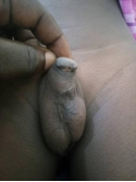
Figure 1. Showing pre-operative picture before the third repair.
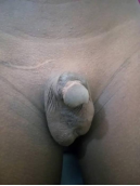
Figure 2. Showing pre-operative picture of the failed hypospadias repair.
Our intra-operative protocol was strictly observed including:
1. Patient had general anesthesia with cuff endotracheal tube intubation (CETT).
2. He was prepped with povidone iodine thrice and draped to expose the surgical field.
3. Diversion safety suprapubic cystostomy via seldinger technique was inserted and connected to a urine bag on continuous drainage. We ensured that the urine was sterile via microscopy culture and sensitivity before surgery.
4. A stay suture was placed on the dorsum of glans penis to aid retraction.
5. The tissue planes was elevated with adrenaline diluted to 1:100000 using spinal needle.
6. The penis was degloved though with difficulty due to scar from previous repair. All scar tissues were completely excised.
7. Following degloving of the penis, we noted a significant 40 degree chordee and performed Nesbit plication at three point on the dorsal summit of the chordee. The first plication was placed at the 12 O’clock position while the other two were placed at the 11 O’clock and 1 O’clock position respectively.
8. We confirmed the orthoplasty with an artificial erection test using normal saline.
9. The urethral plate was adequate so we proceeded with tubulization urethroplasty described by Thiersh Duplay, with good suturing technique using monocryl 4/0 sutures over a urethral stent.
10. With much difficulty though, we were able to raise a second layer over the first layer before proceeding to glanuloplasty and approximating the penis skin to the anatomic position.
11. We anchored the stent using the glanular stay suture by passing the suture through the stent and tying same to prevent mobility of the stent which may distort the fresh repair. We also anchored same stay suture to the supra-pubic region thus protecting the fresh repair from direct pressure contact/impact with the stent. We cleaned the wound before applying the improvised tegaderm dressing over a sufra-tulle gauze.
Post-operatively, we commenced intravenous meropenem 500mg 8hourly for 5 days because we are mindful to avoid any form of sepsis or wound infection, intravenous metronidazole and intravenous fluid for 48 hours before commencing graded oral fluids. Twenty four hours later we allowed the patient take only minimal liquid/semi solid diet like pap, fruit juice, tea and bread for another 72 hours before progression to normal diet. We ensured above graded diet to minimize bowel movement which may predispose the wound to infection and subsequent breakdown. Patient was also placed on syrup oxybutinin to prevent bladder spasm which may encourage peri-catheter/stent urine leak predisposing the wound to infection, adequate analgesia, zinc, vitamin C and supplements.
Pre-operative pack cell volume was 30 and post-operatively we transfused one unit of whole blood slowly over 4-6 hours to encourage wound healing.
We inspected the wound on post-operative day 4 and noted significant progress with no sign of imminent wound breakdown or discharge as shown in Figure 3 below.
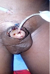
Figure 3. Also showing the diversion suprapubic cystostomy.
We dressed the wound again with improvised tegaderm after cleaning with normal saline and applying sufra-tulle. We then commenced the patient on semi-solid/liquid diet and extended the intravenous antibiotics by 48 hours when we intend to inspect the wound again. Post-operative outcome of the repair were satisfactory with good progress as shown in Figures 4, 5, 6, 7 and 8. We commenced open dressing on post-operative day 7 using povidone iodine ointment thrice daily and after each bowel movement. And he was discharged on post-operative day 14 on oral medications to continue follow up at our outpatient department.
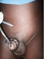
Figure 4. Post-operative day 7. We also commenced open dressing with povidone iodine onitment.
Figure 5. Post-operative day 9 with diversion seldinger SPC still in situ.
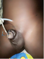
Figure 6. Post-operative day 12. The urethral stent still transfixed with the glandular stay suture.
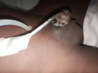
Figure 7. Post-operative day 14. He was discharged today to continue open dressing at home.
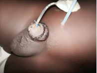
Figure 8. Post-operative day 14. Lateral view still showing the diversion SPC.
Our decision to keep the patient for 14 days was necessary to avoid retrogression of the progress achieved by enforcing bed rest. We were mindful that he is likely to engage in activity with his younger siblings or friends that may lead to inadvertent contact with the wound hence endangering the wound to trauma, infection and subsequent breakdown.
His outpatient visit was uneventful. We removed the urethral stent after 4 weeks and spigoted the suprapubic cystostomy to observe the urine flow for another 7 days. See Figure 9 below. Subsequent visit was uneventful and he was discharged after three months to see us only when necessary.
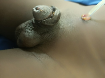
Figure 9. Stent remove with good outcome.
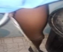
Figure 10. Good urine stream achieved following removal of urethral stent.
3. Discussion
Contemporary hypospadias surgery involves achievement of normal anatomy and aesthetically satisfactory penile appearance as well as functional results. Several techniques have been described for primary hypospadias repair and the treatment of those with previously failed or unsatisfactory hypospadias repair. However, studies looking at the long-term effects of hypospadias repair in childhood using modern techniques are scarce.
In this case report, our patient was 12 years. He was almost at puberty and apparently already experienced some psychological side effects of the previous failed repairs. Some studies have shown that the advanced age may lead to psychological problems in addition to those of erection and the risk of infection, especially in patients older than 15 years of age.
There are also long-term issues with micturition, ejaculation, and erectile function that have been published. However in terms of re-operations, one study showed that age was not associated with numerous re-operations. Thus, there was no extra disadvantage in delayed recurrent ventral chordee correction after puberty.
Initial meatal position has been traditionally factored when dealing with complications. However, contrary to prior belief, the position was not significantly associated with numerous re-operations for complications in our data.
Our index case, had a mid-penile hypospadias with adequate urethral plate width and we repaired it using the urethral plate tubularization technique described by Thiersch-Duplay. This suggest that when dealing with complications, greater attention should be paid to the primary surgical techniques and not necessarily the initial meatal position.
In our patient, he had a lot of scar tissues over the penis due to the previous surgeries. We had to carefully and painstakingly excise all the scar tissues. As in most reported cases, a patient with failed hypospadias repair presenting in adulthood will have a more densely scarred penis, with less vascular and less pliable tissues to work with. Therefore, repeated attempts at surgical repair in those complicated cases are less likely to succeed. This is evident in a study by Hensle et al. where in an adult group of patients who underwent hypospadias repair, complications were noted in 37.5% of patients who had no previous hypospadias surgery, 41.67% of patients who had undergone one or more procedures in childhood but in whom local tissue was relatively intact and 63.6% of patients who underwent multiple unsuccessful hypospadias repairs with various degrees of penile deformity and loss of local tissue. Our patient had extensive scarring and we were mindful of poor healing with greater risk of infection hence we used monocryl sutures instead of vicryl and commence intravenous meropenem post operatively. Studies concluded that although the number of surgeries for the correction of primary hypospadias may represent a risk factor for surgical failure, it was stricture length, not the number of previous operations needed for primary hypospadias repair that was significantly associated with a higher risk of failure . Our patient had adequate urethral plate and that made it easy for our team to achieve good tubularization with good technique.
Severe recurrent ventral curvature conferred the highest risk of numerous re-operations followed by urethral stricture, then wound dehiscence . In our index case, patient had recurrent ventral chordee, and wound dehiscence as the possibly identified risk factors for failure. There was no urethral stricture identified.
In most studies recurrent ventral chordee was under reported and often recurred after puberty. One of the study showed that severe recurrent ventral chordee usually occurs in older children (median age of 9.1 years vs overall median age of 2.2 years) . However our patient was 12 years old, which is still at his early pubertal stage. Thus long-term follow-up will be essential to identify the possibility of progressive recurrent ventral chordee and we hope he will not be lost to follow up.
Talking about the optimal algorithm for re-operations, there is still no consensus. Our patient had release of ventral chordee after complete excision of the scar tissues, tubularization urethroplasty, glanuloplasty, meatoplasty and skin cover. Nozohoor proposed an algorithm for failed hypospadias in adults that detailed the treatment for urethral stricture but did not include urethral tissue transection for severe recurrent ventral chordee correction. We did not transect the urethral plate but rather performed a Nesbit plication at three points in the summit. Another author detailed recurrent ventral chordee correction as priority and the subsequent technique for urethroplasty depended on the health of urethral plate and local skin tissue . We totally agree because it is functionally and aesthetically arguable in favor of the author.
4. Conclusion
We have presented the operative technique of a short-term outcome for a re-do failed hypospadias repair. This case report suggested recurrent ventral chordee and wound dehiscence as the possible risk factors for re-operation, while detailing complete release of chordee and appropriate tubularization urethroplasty technique as tips for success.
Abbreviations

EUCr

Electrolyte, Urea and Creatinine

CBC

Complete Blood Count

MCS

Microscopy, Culture and Sensitivity

CETT

Cuffed Endotracheal Tube Intubation

Author Contributions
Obiatuegwu Kenenna: Conceptualization, Funding acquisition, Project administration, Writing – original draft, Writing – review & editing
Magnus Felix E: Formal Analysis, Writing – original draft
Atim Terkaa: Validation, Writing – review & editing
Otabor Christopher: Project administration, Resources, Software
Conflicts of Interest
The authors declare no conflicts of interest.
References
[1] Aldamanhori R, Chapple CR. Management of the patient with failed hypospadias surgery presenting in adulthood [version 1; peer review: 3 approved]. F1000 Research 2017, 6 (F1000 Faculty Rev):
[2] Timing of elective surgery on the genitalia of male children with particular reference to the risks, benefits, and psychological effects of surgery and anesthesia. American Academy of Pediatrics. Pediatrics. 1996; 97(4): 590–594.
[3] Snodgrass W, Villanueva C, Bush N: Primary and reoperative hypospadias repair in adults--are results different than in children? J Urol. 2014; 192(6): 1730–1733.
[4] Craig JR, Wallis C, Brant WO, Hotaling JM, Myers JB. Management of adults with prior failed hypospadias surgery. Transl Androl Urol 2014; 3(2): 196-204.
[5] Hoy NY, Rourke KF: Better Defining the Spectrum of Adult Hypospadias: Examining the Effect of Childhood Surgery on Adult Presentation. Urology. 2017; 99: 281–286.
[6] Barbagli G, De Angelis M, Palminteri E, et al.: Failed hypospadias repair presenting in adults. Eur Urol. 2006; 49(5): 887–894; discussion 895.
[7] Springer A. Assessment of outcome in hypospadias surgery – a review. Front. Pediatr. 2014; 2: 2.
[8] Halaseh SA, Halaseh S, Ashour M. Hypospadias: A Comprehensive Review Including Its Embryology, Etiology and Surgical Techniques. Cureus. 2022; 14(7): e27544.
[9] Mundy, A. R. Failed hypospadias repair presenting in adults. Eur Urol. 2006; 49: 774-776.
[10] Hensle, T. W. Tennenbaum, S. Y. Reiley, E. A. Hypospadias repair in adults: adventures and misadventures. J Urol. 2001; 165: 77-79.
[11] Ru, W. Shen, J. Tang, D. Width proportion of the urethral plate to the glans can serve as an appraisal index of the urethral plate in hypospadias repair Int J Urol. 2018; 25: 649-653.
[12] Stecker JF Jr, Horton CE, Devine CJ Jr, et al.: Hypospadias cripples. Urol Clin North Am. 1981; 8(3): 539–544. PMID: 7034357.
[13] Hensle TW, Tennenbaum SY, Reiley EA, et al.: Hypospadias repair in adults: adventures and misadventures. J Urol. 2001; 165(1): 77–9.
[14] Barbagli G, Fossati N, Larcher A, et al.: Pd60-04 Predictive Factors Of Success In Adult Patients Treated For Urethral Stricture After Primary Hypospadias Repair Failure: A Multivariable Analysis Of A Single-Surgeon Series. J Urol. 2017; 197(4): e1184.
[15] Wei R, Daxing T, Dehua w, Jia W, hongjuan T, Qiang S. Identification of risk factors associated with numerous reoperations following primary hypospadias repair. JPUrol, 2021; 17(1p61. E1-61. E5).
[16] Nozohoor Ekmark, A. Svensson, H. Arnbjornsson, E. Failed hypospadias repair: an algorithm for secondary reconstruction using remaining local tissue. J Plast Reconstr Aesthet Surg. 2015; 68: 1600-1609.
Cite This Article
  • APA Style

    Kenenna, O., E, M. F., Terkaa, A., Christopher, O. (2025). Operative Technique and Short-term Outcome of Re-do Failed Hypospadias Repair: A Case Report. International Journal of Clinical Urology, 9(1), 7-12. https://doi.org/10.11648/j.ijcu.20250901.12

    Copy | Download

    ACS Style

    Kenenna, O.; E, M. F.; Terkaa, A.; Christopher, O. Operative Technique and Short-term Outcome of Re-do Failed Hypospadias Repair: A Case Report. Int. J. Clin. Urol. 2025, 9(1), 7-12. doi: 10.11648/j.ijcu.20250901.12

    Copy | Download

    AMA Style

    Kenenna O, E MF, Terkaa A, Christopher O. Operative Technique and Short-term Outcome of Re-do Failed Hypospadias Repair: A Case Report. Int J Clin Urol. 2025;9(1):7-12. doi: 10.11648/j.ijcu.20250901.12

    Copy | Download

  • @article{10.11648/j.ijcu.20250901.12,
      author = {Obiatuegwu Kenenna and Magnus Felix E and Atim Terkaa and Otabor Christopher},
      title = {Operative Technique and Short-term Outcome of Re-do Failed Hypospadias Repair: A Case Report
    },
      journal = {International Journal of Clinical Urology},
      volume = {9},
      number = {1},
      pages = {7-12},
      doi = {10.11648/j.ijcu.20250901.12},
      url = {https://doi.org/10.11648/j.ijcu.20250901.12},
      eprint = {https://article.sciencepublishinggroup.com/pdf/10.11648.j.ijcu.20250901.12},
      abstract = {Introduction: A failed hypopadias repair is a difficult case to treat considering the poor quality of surrounding tissues arising from disruption of normal vasculature associated with the underlying disorder as well as scaring from previous repairs. In this case report we describe our experience with the management of late presentation of failed hypospadias repair in a 12 year old boy. Case report: Our patient is a twelve year old ‘small for age’ who presented to our center with failed mid-penile hypospadias repair. He first presented at the age of eight to another hospital where he was evaluated and a diagnosis of mid-penile hypospadias with significant chordee was made. He had the first repair at the age of eight but the repair failed two weeks after surgery. Two years later he had a second repair in same facility which also failed after approximately two weeks. They consequently presented to our center two years later at the age of twelve. Following appropriate evaluation and counseling, he had the 3rd repair during which we ensured a preoperative sterile urine culture, created a urinary diversion via suprapubic cystostomy, the use of a monofilament (monocryl) suture, complete release of chordee after excision of scar tissues, a second intervening layer over the repair, the use of an improvised stent instead of a urethral catheter and appropriate anchorage of the stent to the suprapubic skin using a nylon suture. Patient did well post-operatively and was discharged home 14 days after the repair. Subsequent outpatient visits were uneventful and he was discharged after three months to see us only when necessary. Conclusion: This case report suggests recurrent ventral chordee and wound dehiscence as the possible risk factors for re-operation, while detailing complete release of chordee and appropriate tubularization urethroplasty technique as tips for success.
    },
     year = {2025}
    }
    

    Copy | Download

  • TY  - JOUR
    T1  - Operative Technique and Short-term Outcome of Re-do Failed Hypospadias Repair: A Case Report
    
    AU  - Obiatuegwu Kenenna
    AU  - Magnus Felix E
    AU  - Atim Terkaa
    AU  - Otabor Christopher
    Y1  - 2025/01/21
    PY  - 2025
    N1  - https://doi.org/10.11648/j.ijcu.20250901.12
    DO  - 10.11648/j.ijcu.20250901.12
    T2  - International Journal of Clinical Urology
    JF  - International Journal of Clinical Urology
    JO  - International Journal of Clinical Urology
    SP  - 7
    EP  - 12
    PB  - Science Publishing Group
    SN  - 2640-1355
    UR  - https://doi.org/10.11648/j.ijcu.20250901.12
    AB  - Introduction: A failed hypopadias repair is a difficult case to treat considering the poor quality of surrounding tissues arising from disruption of normal vasculature associated with the underlying disorder as well as scaring from previous repairs. In this case report we describe our experience with the management of late presentation of failed hypospadias repair in a 12 year old boy. Case report: Our patient is a twelve year old ‘small for age’ who presented to our center with failed mid-penile hypospadias repair. He first presented at the age of eight to another hospital where he was evaluated and a diagnosis of mid-penile hypospadias with significant chordee was made. He had the first repair at the age of eight but the repair failed two weeks after surgery. Two years later he had a second repair in same facility which also failed after approximately two weeks. They consequently presented to our center two years later at the age of twelve. Following appropriate evaluation and counseling, he had the 3rd repair during which we ensured a preoperative sterile urine culture, created a urinary diversion via suprapubic cystostomy, the use of a monofilament (monocryl) suture, complete release of chordee after excision of scar tissues, a second intervening layer over the repair, the use of an improvised stent instead of a urethral catheter and appropriate anchorage of the stent to the suprapubic skin using a nylon suture. Patient did well post-operatively and was discharged home 14 days after the repair. Subsequent outpatient visits were uneventful and he was discharged after three months to see us only when necessary. Conclusion: This case report suggests recurrent ventral chordee and wound dehiscence as the possible risk factors for re-operation, while detailing complete release of chordee and appropriate tubularization urethroplasty technique as tips for success.
    
    VL  - 9
    IS  - 1
    ER  - 

    Copy | Download

Author Information
  • Division of Urology, Department of Surgery, Federal Medical Centre, Abuja, Nigeria; Department of Surgery, Alliance Hospital and Services Limited, Abuja, Nigeria; Department of Surgery, Baze University, Abuja, Nigeria

    Research Fields: Andrology, Renal transplantation, Endourology, Reconstructive urology, Uro-oncology

  • Division of Urology, Department of Surgery, Chivar Specialist Hospital and Urology Centre Limited, Abuja, Nigeria

    Research Fields: Endourology, Laser surgeries, Reconstructive urology, Uro-oncolgy, renal stone

  • Division of Urology, Department of Surgery, University of Abuja Teaching Hospital, Gwagwalada, Nigeria

    Research Fields: Endourology, Reconstructive urology, Prostate cancer, Laser surgery, General urology.

  • Department of Surgery, Alliance Hospital and Services Limited, Abuja, Nigeria

    Research Fields: General surgery, Oethopaedics, Spine surgery