Urethral polyps represent a rare cause of lower urinary tract obstruction, documented primarily in pediatrics and more specifically in boys during the first decade of life. We report the case of a 21-year-old patient with no significant past medical history, presenting with a vulvar mass showing progressive enlargement associated with dysuria and mechanical pain, evolving since the age of 10. Clinical examination revealed a 1.5 cm pedunculated, erythematous, and tender mass protruding from the urethral meatus. Cystoscopy confirmed a bleeding-upon-contact polyp implanted on the posterior wall of the meatus, adjacent to healthy bladder mucosa. Complete surgical excision with a surgical bade no 11 was performed under spinal anesthesia, followed by cauterization and urethral catheterization for 48 hours. Histopathological analysis concluded a benign fibro-epithelial polyp without atypia or signs of malignancy. The immediate and six-month postoperative outcomes were excellent, with complete resolution of symptoms. This case, exceptional due to the patient's age and long clinical history, contrasts with available large pediatric series and the few reported female pediatric cases. It underscores the necessity of considering this diagnosis even in young adult women presenting with any urethral mass and confirms the curative effectiveness of simple and complete surgical excision for this benign but potentially debilitating pathology.
This is an Open Access article, distributed under the terms of the Creative Commons Attribution 4.0 International License (http://creativecommons.org/licenses/by/4.0/), which permits unrestricted use, distribution and reproduction in any medium or format, provided the original work is properly cited.
Urethral Polyp, Young Woman, Fibro-Epithelial, Urethral Prolapse, Surgical Excision
1. Introduction
Urethral polyps represent a rare cause of lower urinary tract obstruction, documented primarily in pediatrics and more specifically in boys during the first decade of life
[1]
Beluffi G, Berton F, Gola G, et al. Urethral polyp in a 1-month-old child. Pediatr Radiol 2005; 35: 691-693.
[3]
De Castro R, Campobasso P, Belloli G et al. Solitary polyp of posterior urethra in children. Eur J Pediatr Surg. 1993; 3: 92-96.
[8]
Jain P, Shah H, Parelkar SV, et al. Posterior urethral polyps and review of literature. Indian Journal of Urology 2007; 23: 206.
[1, 3, 8]
. Their discovery in young women is exceptional, with literature reporting only very rare prepubertal cases
[4]
Ben-meir D, Yin M, Chow C. W, et al. Urethral polyps in prepubertal girls. The Journal of Urology. Epub ahead of print October 2005.
De Castro R, Campobasso P, Belloli G et al. Solitary polyp of posterior urethra in children. Eur J Pediatr Surg. 1993; 3: 92-96.
[3]
. This diagnostic rarity, confronted with the commonality of symptoms such as dysuria or a perineal mass, can lead to delayed management. We report the singular case of a 21-year-old patient with a fibro-epithelial urethral polyp evolving since childhood, whose late presentation raises questions about the natural history of these lesions, often considered congenital
[1]
Beluffi G, Berton F, Gola G, et al. Urethral polyp in a 1-month-old child. Pediatr Radiol 2005; 35: 691-693.
[3]
De Castro R, Campobasso P, Belloli G et al. Solitary polyp of posterior urethra in children. Eur J Pediatr Surg. 1993; 3: 92-96.
[1, 3]
. Through this clinical case and a targeted literature review, we illustrate the epidemiological, diagnostic, and therapeutic particularities of this rare entity.
2. Case Presentation
A 21-year-old patient, with no known significant past medical history, was seen in our clinic for a vulvar mass that had been progressively increasing in size since the age of 10, associated with dysuria and vulvar pain exacerbated by physical exertion. No hematuria, recurrent urinary tract infection, or other associated symptomatology was reported.
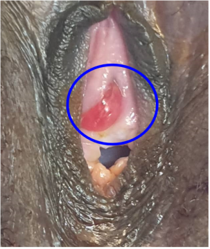
Clinical examination revealed a pedunculated, erythematous mass, measuring 1.5 cm, protruding at the level of the urethral meatus and tender to palpation (Figure 1).
Figure 1. Pedunculated mass (circled in blue), erythematous, protruding at the urethral meatus.
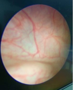
Urethro-cystoscopy showed normal bladder mucosa (Figure 2) and a pedunculated mass on the posterior wall of the urethral meatus, bleeding upon contact and suggestive of a urethral polyp.
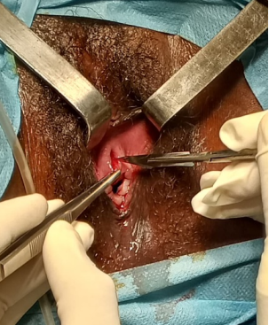
Surgical excision was performed under spinal anesthesia using a No. 11 Surgical Blade (Figure 3), followed by cauterization and urethral catheterization for 48 hours. Histopathological analysis confirmed a benign fibro-epithelial polyp, without atypia or signs of malignancy.
Figure 3. Surgical excision using a No. 11 Surgical Blade.
The outcome was favorable with complete disappearance of symptoms. At six months, the patient was asymptomatic and without recurrence.
3. Discussion
Our observation presents several particularities: the young age of the patient (21 years), the long evolution since childhood, and the symptomatology dominated by pain and dysuria rather than severe obstructive symptoms or recurrent urinary tract infections. This presentation allows for an in-depth discussion by comparing our data with the available literature.
3.1. Epidemiology and Age-Related Particularities
Urethral polyps are a rare pathology, predominantly described in pediatrics. The vast majority of published large series concern male children, with a peak frequency between 5 and 10 years
[1]
Beluffi G, Berton F, Gola G, et al. Urethral polyp in a 1-month-old child. Pediatr Radiol 2005; 35: 691-693.
[2]
Kimche D, Lask D. Congenital Polyp of the Prostatic Urethra. Journal of Urology 1982; 127: 134-134.
[3]
De Castro R, Campobasso P, Belloli G et al. Solitary polyp of posterior urethra in children. Eur J Pediatr Surg. 1993; 3: 92-96.
[8]
Jain P, Shah H, Parelkar SV, et al. Posterior urethral polyps and review of literature. Indian Journal of Urology 2007; 23: 206.
[1, 2, 3, 8]
. The series by De Castro et al. involving 17 cases included only boys
[3]
De Castro R, Campobasso P, Belloli G et al. Solitary polyp of posterior urethra in children. Eur J Pediatr Surg. 1993; 3: 92-96.
[3]
. The rarity of this entity in females, and even more so in young women, is extreme. The literature review conducted by Ben-Meir et al. in 2005 had identified only 11 published cases of urethral polyps in prepubertal girls, 5 of which came from their own institution
[4]
Ben-meir D, Yin M, Chow C. W, et al. Urethral polyps in prepubertal girls. The Journal of Urology. Epub ahead of print October 2005.
. The case we report in a 21-year-old patient is therefore quite exceptional and suggests that minimal congenital lesions, potentially present since childhood, can remain paucisymptomatic and only become apparent later due to local irritative or traumatic factors.
3.2. Clinical Presentation and Differential Diagnosis
The clinical picture of our patient, associating a visible mass, dysuria, and pain triggered by exertion, is consistent with descriptions in the literature, although pediatric series more frequently report genital bleeding (as in 11/12 cases in the series by Ndour et al.
[5]
Ndour O, Malle K, Faye Fall A. L, Ndoye N. A, Nibagora, J, G Ngom, Ndoye M. Urethral mucosal prolapse in girls. Afr J Urol. (2017) 23, 359-363
Beluffi G, Berton F, Gola G, et al. Urethral polyp in a 1-month-old child. Pediatr Radiol 2005; 35: 691-693.
[2]
Kimche D, Lask D. Congenital Polyp of the Prostatic Urethra. Journal of Urology 1982; 127: 134-134.
[8]
Jain P, Shah H, Parelkar SV, et al. Posterior urethral polyps and review of literature. Indian Journal of Urology 2007; 23: 206.
[1, 2, 8]
.
In a young woman, the differential diagnosis for a bleeding and painful urethral mass must be carefully established:
1) Urethral prolapse: Much more common in prepubertal girls, it presents as a circular swelling, centered by the urethral orifice, often ulcerated and bleeding on contact
[5]
Ndour O, Malle K, Faye Fall A. L, Ndoye N. A, Nibagora, J, G Ngom, Ndoye M. Urethral mucosal prolapse in girls. Afr J Urol. (2017) 23, 359-363
4) Malignant tumors: Such as botryoid rhabdomyosarcoma, although rare, must be excluded, especially in the case of a rapidly growing lesion.
3.3. Diagnostic Methods
In our case, the diagnosis was made by clinical examination and cystoscopy. As shown by various series, ultrasound and voiding cystourethrography (VCUG) are key examinations for positive diagnosis. VCUG can reveal a mobile filling defect in the posterior urethra, often associated with a distended bladder and signs of chronic obstruction (trabeculated bladder, diverticula)
[1]
Beluffi G, Berton F, Gola G, et al. Urethral polyp in a 1-month-old child. Pediatr Radiol 2005; 35: 691-693.
[6]
Du L, Zhu Y, Li Y, Sun Y. Ultrasonic manifestation of urethral polyp in a girl. Transl Pediatr 2020; 9(3): 262-265 |
. Finally, cystoscopy remains the gold standard examination, allowing visualization of the lesion, its point of implantation (usually at the verumontanum in males), and for obtaining biopsies.
3.4. Therapeutic Management and Follow-up
The standard treatment is complete excision of the lesion. Several techniques are described, the choice of which depends on the size of the lesion, the patient's age, and the operator's experience:
1) Transurethral endoscopic resection: This is the most commonly reported method, often with fulguration of the base, and it yields excellent results without recurrence in the vast majority of cases
[3]
De Castro R, Campobasso P, Belloli G et al. Solitary polyp of posterior urethra in children. Eur J Pediatr Surg. 1993; 3: 92-96.
[8]
Jain P, Shah H, Parelkar SV, et al. Posterior urethral polyps and review of literature. Indian Journal of Urology 2007; 23: 206.
[3, 8]
.
2) Fulguration or laser vaporization (Nd: YAG): An effective alternative for vascularized lesions
[1]
Beluffi G, Berton F, Gola G, et al. Urethral polyp in a 1-month-old child. Pediatr Radiol 2005; 35: 691-693.
[1]
.
3) Open surgical excision (via cystotomy): Reserved for very large lesions or when endoscopic access is difficult, as was necessary in the 1-month-old infant reported by Beluffi et al.
[1]
Beluffi G, Berton F, Gola G, et al. Urethral polyp in a 1-month-old child. Pediatr Radiol 2005; 35: 691-693.
[1]
.
4) Surgical Blade excision: As in our observation, this simple technique is perfectly suited for accessible distal and pedunculated lesions.
The short duration of postoperative urethral catheterization (48h) that we adopted is similar to protocols of other teams and was sufficient to prevent urinary retention or stenosis
[5]
Ndour O, Malle K, Faye Fall A. L, Ndoye N. A, Nibagora, J, G Ngom, Ndoye M. Urethral mucosal prolapse in girls. Afr J Urol. (2017) 23, 359-363
. The prognosis is excellent, with complete resolution of symptoms and no tendency for recurrence after complete excision, confirming the benign nature of these lesions
[1]
Beluffi G, Berton F, Gola G, et al. Urethral polyp in a 1-month-old child. Pediatr Radiol 2005; 35: 691-693.
[3]
De Castro R, Campobasso P, Belloli G et al. Solitary polyp of posterior urethra in children. Eur J Pediatr Surg. 1993; 3: 92-96.
[4]
Ben-meir D, Yin M, Chow C. W, et al. Urethral polyps in prepubertal girls. The Journal of Urology. Epub ahead of print October 2005.
3.5. Histopathological Aspects and Etiopathogenic Hypotheses
Histological analysis of our specimen concluded a benign fibro-epithelial polyp. This description is unanimously reported in the literature: it is a lesion composed of a central core of more or less edematous and inflammatory fibrovascular tissue, covered by a transitional or squamous epithelium
[1]
Beluffi G, Berton F, Gola G, et al. Urethral polyp in a 1-month-old child. Pediatr Radiol 2005; 35: 691-693.
[3]
De Castro R, Campobasso P, Belloli G et al. Solitary polyp of posterior urethra in children. Eur J Pediatr Surg. 1993; 3: 92-96.
[4]
Ben-meir D, Yin M, Chow C. W, et al. Urethral polyps in prepubertal girls. The Journal of Urology. Epub ahead of print October 2005.
The etiology remains debated. The most widely accepted theory is a congenital, probably hamartomatous origin, related to the persistence of embryonic tissue (such as the Müllerian tubercle) or a defect in the fusion of urogenital structures
[3]
De Castro R, Campobasso P, Belloli G et al. Solitary polyp of posterior urethra in children. Eur J Pediatr Surg. 1993; 3: 92-96.
[8]
Jain P, Shah H, Parelkar SV, et al. Posterior urethral polyps and review of literature. Indian Journal of Urology 2007; 23: 206.
[3, 8]
. This hypothesis is supported by the young age of most patients and the frequent association with other congenital anomalies of the urinary tract (vesicoureteral reflux, posterior urethral valves, pelvic kidney) found in nearly 50% of cases in the series by De Castro et al.
[3]
De Castro R, Campobasso P, Belloli G et al. Solitary polyp of posterior urethra in children. Eur J Pediatr Surg. 1993; 3: 92-96.
[3]
. Association with Beckwith-Wiedemann syndrome has also been described
[1]
Beluffi G, Berton F, Gola G, et al. Urethral polyp in a 1-month-old child. Pediatr Radiol 2005; 35: 691-693.
[1]
.
4. Conclusion
The fibro-epithelial urethral polyp in young women is an exceptional entity, whose rarity contrasts with its relative frequency in male pediatrics. Our observation, with symptom onset in childhood and late revelation in adulthood, underscores the potentially congenital nature of this lesion. The diagnosis, suspected clinically upon finding a pedunculated and bleeding urethral mass, is confirmed by endoscopy and histology. The differential diagnosis must primarily rule out urethral prolapse in younger girls, and leiomyoma or malignant pathology in older women.
Complete surgical excision, achievable by various techniques (endoscopic, laser, or surgical blade), ensures definitive and curative treatment, without a tendency for recurrence, as evidenced by the six-month follow-up of our patient.
Sawadogo, H., Yameogo, C. A. M. K. D., Pare, A., Ouedraogo, F., Millogo, J. D. L. C., et al. (2025). Urethral Polyp in a 21-Year-Old Young Woman: A Case Report and Review of the Literature. International Journal of Clinical Urology, 9(2), 154-157. https://doi.org/10.11648/j.ijcu.20250902.18
Sawadogo, H.; Yameogo, C. A. M. K. D.; Pare, A.; Ouedraogo, F.; Millogo, J. D. L. C., et al. Urethral Polyp in a 21-Year-Old Young Woman: A Case Report and Review of the Literature. Int. J. Clin. Urol.2025, 9(2), 154-157. doi: 10.11648/j.ijcu.20250902.18
Sawadogo H, Yameogo CAMKD, Pare A, Ouedraogo F, Millogo JDLC, et al. Urethral Polyp in a 21-Year-Old Young Woman: A Case Report and Review of the Literature. Int J Clin Urol. 2025;9(2):154-157. doi: 10.11648/j.ijcu.20250902.18
@article{10.11648/j.ijcu.20250902.18,
  author = {Hassami Sawadogo and Clotaire Alexis Marie Kiemdiba Donega Yameogo and Abdoul-Karim Pare and Fatao Ouedraogo and Jean de la Croix Millogo and Brahima Kirakoya and Adama Ouattara and Fasnewinde Aristide Kabore},
  title = {Urethral Polyp in a 21-Year-Old Young Woman: A Case Report and Review of the Literature
},
  journal = {International Journal of Clinical Urology},
  volume = {9},
  number = {2},
  pages = {154-157},
  doi = {10.11648/j.ijcu.20250902.18},
  url = {https://doi.org/10.11648/j.ijcu.20250902.18},
  eprint = {https://article.sciencepublishinggroup.com/pdf/10.11648.j.ijcu.20250902.18},
  abstract = {Urethral polyps represent a rare cause of lower urinary tract obstruction, documented primarily in pediatrics and more specifically in boys during the first decade of life. We report the case of a 21-year-old patient with no significant past medical history, presenting with a vulvar mass showing progressive enlargement associated with dysuria and mechanical pain, evolving since the age of 10. Clinical examination revealed a 1.5 cm pedunculated, erythematous, and tender mass protruding from the urethral meatus. Cystoscopy confirmed a bleeding-upon-contact polyp implanted on the posterior wall of the meatus, adjacent to healthy bladder mucosa. Complete surgical excision with a surgical bade no 11 was performed under spinal anesthesia, followed by cauterization and urethral catheterization for 48 hours. Histopathological analysis concluded a benign fibro-epithelial polyp without atypia or signs of malignancy. The immediate and six-month postoperative outcomes were excellent, with complete resolution of symptoms. This case, exceptional due to the patient's age and long clinical history, contrasts with available large pediatric series and the few reported female pediatric cases. It underscores the necessity of considering this diagnosis even in young adult women presenting with any urethral mass and confirms the curative effectiveness of simple and complete surgical excision for this benign but potentially debilitating pathology.
},
 year = {2025}
}
TY  - JOUR
T1  - Urethral Polyp in a 21-Year-Old Young Woman: A Case Report and Review of the Literature

AU  - Hassami Sawadogo
AU  - Clotaire Alexis Marie Kiemdiba Donega Yameogo
AU  - Abdoul-Karim Pare
AU  - Fatao Ouedraogo
AU  - Jean de la Croix Millogo
AU  - Brahima Kirakoya
AU  - Adama Ouattara
AU  - Fasnewinde Aristide Kabore
Y1  - 2025/10/27
PY  - 2025
N1  - https://doi.org/10.11648/j.ijcu.20250902.18
DO  - 10.11648/j.ijcu.20250902.18
T2  - International Journal of Clinical Urology
JF  - International Journal of Clinical Urology
JO  - International Journal of Clinical Urology
SP  - 154
EP  - 157
PB  - Science Publishing Group
SN  - 2640-1355
UR  - https://doi.org/10.11648/j.ijcu.20250902.18
AB  - Urethral polyps represent a rare cause of lower urinary tract obstruction, documented primarily in pediatrics and more specifically in boys during the first decade of life. We report the case of a 21-year-old patient with no significant past medical history, presenting with a vulvar mass showing progressive enlargement associated with dysuria and mechanical pain, evolving since the age of 10. Clinical examination revealed a 1.5 cm pedunculated, erythematous, and tender mass protruding from the urethral meatus. Cystoscopy confirmed a bleeding-upon-contact polyp implanted on the posterior wall of the meatus, adjacent to healthy bladder mucosa. Complete surgical excision with a surgical bade no 11 was performed under spinal anesthesia, followed by cauterization and urethral catheterization for 48 hours. Histopathological analysis concluded a benign fibro-epithelial polyp without atypia or signs of malignancy. The immediate and six-month postoperative outcomes were excellent, with complete resolution of symptoms. This case, exceptional due to the patient's age and long clinical history, contrasts with available large pediatric series and the few reported female pediatric cases. It underscores the necessity of considering this diagnosis even in young adult women presenting with any urethral mass and confirms the curative effectiveness of simple and complete surgical excision for this benign but potentially debilitating pathology.

VL  - 9
IS  - 2
ER  -