Background: Extracutaneous or deep-seated mycosis of the breast tissue is rather rare, however, it has been described in some parts of the world. Available evidence suggested that a decline in reporting might imply neglect or a low index of suspicion. The aim of this study was to report the features of unusual chronic granulomatous fungal disease that presented to our health facility with features of breast cancer in the first half of year 2025. Case Presentation: A 37 year-old breast-feeding mother who presented to General Surgery Outpatient Clinic on referral from a health center with a history of left breast lump of 5 months duration. She had fever at the onset of the illness, associated with breast pain. The swelling had progressed to form multiple “boil” which ruptured and discharged some relatively clear fluid with associated changes in the skin and thickening of the left breast. There were no other remarkable symptoms. She had had antibiotic treatment and a biopsy taken before referral. Significant findings on physical examination included: gross breast asymmetry with the left breast being bigger, showed hyperemic areas, serous discharge, peau de orange with irregularly shaped scar (previous biopsy scar). There was a mass located in the outer upper and inner upper quadrant, measuring 6cm by 8cm with surrounding discharging sinuses. The breast ultrasound scan revealed left-sided complex heterogenous solid mass 5.4cm x 3.5cm with irregular outline; the left axillary lymph node was enlarged and measured 3.2cm x 1.8cm. The tissue histopathology report of Tru Cut biopsy revealed Chronic non-specific/mastitis idiopathic granulomatous mastitis. Conclusion: The symptoms and signs of breast cancer and breast mycosis share some similarities. Breast fungal disease is rare but does occur, and increased awareness is expected for early diagnosis.
| Published in | Journal of Surgery (Volume 13, Issue 5) |
| DOI | 10.11648/j.js.20251305.15 |
| Page(s) | 146-151 |
| Creative Commons |
This is an Open Access article, distributed under the terms of the Creative Commons Attribution 4.0 International License (http://creativecommons.org/licenses/by/4.0/), which permits unrestricted use, distribution and reproduction in any medium or format, provided the original work is properly cited. |
| Copyright |
Copyright © The Author(s), 2025. Published by Science Publishing Group |
Breast Cancer, Case Report, Chronic Granulomatous Fungal Disease, Masquerading, Port Harcourt, Nigeria
| [1] | Espinel-Ingroff A. History of medical mycology in the United States. Clinical microbiology reviews. 1996; 9(2): 235-72. |
| [2] | Ainsworth GC, Ainsworth GC. Introduction to the history of medical and veterinary mycology: Cambridge University Press; 2002. |
| [3] | Borgers M, Degreef H, Cauwenbergh G. Fungal infections of the skin: infection process and antimycotic therapy. Current drug targets. 2005; 6(8): 849-62. |
| [4] | Crawford F, Hollis S. Topical treatments for fungal infections of the skin and nails of the foot. Cochrane Database of Systematic Reviews. 2007(3): 1-154. |
| [5] | Berger AP, Ford BA, Brown-Joel Z, Shields BE, Rosenbach M, Wanat KA. Angioinvasive fungal infections impacting the skin: Diagnosis, management, and complications. Journal of the American Academy of Dermatology. 2019; 80(4): 883-98. e2. |
| [6] | Rezabek GH, Friedman AD. Superficial fungal infections of the skin: Diagnosis and current treatment recommendations. Drugs. 1992; 43(5): 674-82. |
| [7] | Anaissie E. Opportunistic mycoses in the immunocompromised host: experience at a cancer center and review. Clinical Infectious Diseases. 1992; 14(Supplement_1): S43-S53. |
| [8] | Basiri JS, Khaksar AA. Deep-seated fungal infections in immunocompromised patients in Iran. Iranian journal of allergy, asthma and immunology. 2005; 4(1): 27-32. |
| [9] | Walsh TJ. Management of immunocompromised patients with evidence of an invasive mycosis. Hematology/oncology clinics of North America. 1993; 7(5): 1003-26. |
| [10] | Kershenovich R, Sherman S, Reiter O, Huss SR, Didkovsky E, Mimouni D, et al. A unique clinicopathological manifestation of fungal infection: a case series of deep dermatophytosis in immunosuppressed patients. American Journal of Clinical Dermatology. 2017; 18: 697-704. |
| [11] | Salfelder K, Schwarz J. Mycotic Pseudotumors of the Breast: Report of Four Cases. Archives of Surgery. 1975; 110(6): 751-4. |
| [12] | Rappaport H, Thomas LB. Mycosis fungoides: the pathology of extracutaneous involvement. Cancer. 1974; 34(4): 1198-229. |
| [13] | MacFadden DR, Gold WL, Richardson SE, Bunce PE. A breast mass in a 56-year-old man. CMAJ. 2011; 183(16): 1875-8. |
| [14] | Jain BK, Sehgal VN, Jagdish S, Ratnakar C, Smile SR. Primary actinomycosis of the breast: a clinical review and a case report. The Journal of dermatology. 1994; 21(7): 497-500. |
| [15] | Anandi V, Vishwanathan P, Sasikala S, Rangarajan M, Subramaniyan C, Chidambaram N. Fusarium solani breast abscess. Indian Journal of Medical Microbiology. 2005; 23(3): 198-199. |
| [16] | Sachin MRCNM, Singla KG, Prakash AGSRH. Disseminated Emergomyces pasteurianus Infection in India: A Case Report and a Review. Mycopathologia. 2020; 185, 193–200. |
| [17] | Durry M, Loho LL. Mycetoma of the Breast: A Case Report. Medical Scope Journal. 2023; 5(2): 266-9. |
| [18] | Opri F. Mammary mycoses. Chemotherapy. 1982; 28(Suppl 1): 61-5. |
| [19] | Klimko NN, Khostelidi SN, Volkova AG, Popova MO, Bogomolova TS, Zuborovskaya LS, et al. Mucormycosis in haematological patients: case report and results of prospective study in Saint Petersburg, Russia. Mycoses. 2014; 57: 91-6. |
| [20] | Błażewicz I, Olszewska B, Stawczyk-Macieja M, Jaœkiewicz M, Nowicki R, Sokołowska-Wojdyło M. The incidences of other primary cancers in patients with mycosis fungoides and Sézary syndrome. Advances in Dermatology and Allergology/Postępy Dermatologii i Alergologii. 2021; 38(2): 289-94. |
| [21] | Guglietta S, Li X, Saxena D. Role of Fungi in Tumorigenesis: Promises and Challenges. Annual Review of Pathology: Mechanisms of Disease. 2025; 20(1): 459-82. |
| [22] | Somashekhar S, Jaiswal R, Kumar R, Ashok B, Rakshit S, Rauthan A, et al. Mucormycosis of the Breast in a Patient With Breast Carcinoma After COVID-19 Pneumonia. European Journal of Breast Health. 2022; 18(3): 289-291. |
| [23] | Velez-Haro JM, Pérez-Rodríguez F, Velázquez-Márquez S, Ramírez Medina H, Velázquez-Márquez N. Mycology in Oncology: Exploring the Role of the Mycobiome in Human Cancer, Etiology, Progression, Epidemiology, Mycoses, and Mycotoxins. Pathogens Associated with the Development of Cancer in Humans: OMICs, Immunological, and Pathophysiological Studies. 2024: 303-48. |
| [24] | Makhubela VG, Khaba MC. Mycotic pseudotumor of the breast secondary to cryptococcal infection: report of three rare cases and literature review. International Journal of Surgical Pathology. 2022; 30(4): 432-6. |
| [25] | Liao L. Inequality in breast cancer: Global statistics from 2022 to 2050. The Breast. 2025; 79: 103851. |
| [26] | Wilkinson L, Gathani T. Understanding breast cancer as a global health concern. The British journal of radiology. 2022; 95(1130): 20211033. |
| [27] | Kadam SS, Kadam T. Case Report: Rare Presentation of Aspergillosis of Breast Masquerading as Breast Carcinoma. Carcinogenesis and Chemotherapy. 2023; 2(2). |
| [28] | Garcia-Rodiguez JA, Pattullo A. Idiopathic granulomatous mastitis: a mimicking disease in a pregnant woman: a case report. BMC Research Notes. 2013; 6: 1-4. |
| [29] | Tuli R, O'Hara BJ, Hines J, Rosenberg AL, editors. Idiopathic granulomatous mastitis masquerading as carcinoma of the breast: a case report and review of the literature. International Seminars in Surgical Oncology; 2007: Springer. |
| [30] | Illman JE, Terra SB, Clapp AJ, Hunt KN, Fazzio RT, Shah SS, et al. Granulomatous diseases of the breast and axilla: radiological findings with pathological correlation. Insights into imaging. 2018; 9: 59-71. |
| [31] | Bassey TE, Okekemba I, Egbara WO, Owai P, Okafor LE, Elem DE, et al. Cutaneous manifestations of deep mycoses in Nigeria: a systematic review. Therapeutic Advances in Infectious Disease. 2024; 11: 20499361241286973. |
| [32] | Olaofe OO, Soremekun AI, Olopade BO, Oladele JO. Fungal and Parasitic Diseases Seen in Tissue Biopsies at a Tertiary Health Center in Southwest Nigeria in a Five-Year Period. International Journal of TROPICAL DISEASE & Health. 2023; 44(11): 17-26. |
| [33] | Jiménez-García N, Fernández Sánchez F, Guillén Rodríguez CM, Castilla Castellano MdM, Del Arco Jiménez A. Breast abscess due to Nannizziopsis obscura in an immunocompromised renal transplant patient after travel to Nigeria: case report and review. BMC Infectious Diseases. 2023; 23(1): 50. |
| [34] | Belmokhtar Z, Djaroud S, Matmour D, Merad Y. Atypical and Unpredictable Superficial Mycosis Presentations: A Narrative Review. Journal of Fungi. 2024; 10(4): 295. |
| [35] | Dogra S, Narang T. Emerging atypical and unusual presentations of dermatophytosis in India. Clinical Dermatology Review. 2017; 1(Suppl 1): S12-S8. |
| [36] | Gnat S, Łagowski D, Nowakiewicz A. Major challenges and perspectives in the diagnostics and treatment of dermatophyte infections. Journal of applied microbiology. 2020; 129(2): 212-32. |
| [37] | Douglas P. Overdiagnosis and overtreatment of nipple and breast candidiasis: A review of the relationship between diagnoses of mammary candidiasis and Candida albicans in breastfeeding women. Women's Health. 2021; 17: 17455065211031480. |
| [38] | Hanna L, Cruz SA. Candida mastitis: a case report. The Permanente Journal. 2011; 15(1): 62-64. |
| [39] | Merad Y, Derrar H, Belkacemi M, Drici A, Belmokhtar Z, Drici Sr AE-M. Candida albicans mastitis in breastfeeding woman: an under recognized diagnosis. Cureus. 2020; 12(12). |
| [40] | Saini R, Saini S, Sharma S. Oral sex, oral health and orogenital infections. Journal of global infectious diseases. 2010; 2(1): 57-62. |
| [41] | Murphy MJ. Getting It On: Sexual Behavior and Sexual Health. Living Out Loud: Routledge; 2018. p. 95-142. |
| [42] | Kontoyiannis PD, Koons GL, Hicklen RS, Mikos AG, Kontoyiannis DP, editors. Insect Bite–Associated Invasive Fungal Infections. Open Forum Infectious Diseases; 2019: Oxford University Press US. |
APA Style
Ijah, R. F. O. A., Wagozie, V., Lawson, S., Nwineh, A. (2025). Chronic Granulomatous Fungal Disease Masquerading as Breast Cancer in Port Harcourt, Nigeria: A Case Report and Literature Review. Journal of Surgery, 13(5), 146-151. https://doi.org/10.11648/j.js.20251305.15
ACS Style
Ijah, R. F. O. A.; Wagozie, V.; Lawson, S.; Nwineh, A. Chronic Granulomatous Fungal Disease Masquerading as Breast Cancer in Port Harcourt, Nigeria: A Case Report and Literature Review. J. Surg. 2025, 13(5), 146-151. doi: 10.11648/j.js.20251305.15
@article{10.11648/j.js.20251305.15,
  author = {Rex Friday Ogoronte Alderton Ijah and Victor Wagozie and Stephenson Lawson and Ambassador Nwineh},
  title = {Chronic Granulomatous Fungal Disease Masquerading as Breast Cancer in Port Harcourt, Nigeria: A Case Report and Literature Review
},
  journal = {Journal of Surgery},
  volume = {13},
  number = {5},
  pages = {146-151},
  doi = {10.11648/j.js.20251305.15},
  url = {https://doi.org/10.11648/j.js.20251305.15},
  eprint = {https://article.sciencepublishinggroup.com/pdf/10.11648.j.js.20251305.15},
  abstract = {Background: Extracutaneous or deep-seated mycosis of the breast tissue is rather rare, however, it has been described in some parts of the world. Available evidence suggested that a decline in reporting might imply neglect or a low index of suspicion. The aim of this study was to report the features of unusual chronic granulomatous fungal disease that presented to our health facility with features of breast cancer in the first half of year 2025. Case Presentation: A 37 year-old breast-feeding mother who presented to General Surgery Outpatient Clinic on referral from a health center with a history of left breast lump of 5 months duration. She had fever at the onset of the illness, associated with breast pain. The swelling had progressed to form multiple “boil” which ruptured and discharged some relatively clear fluid with associated changes in the skin and thickening of the left breast. There were no other remarkable symptoms. She had had antibiotic treatment and a biopsy taken before referral. Significant findings on physical examination included: gross breast asymmetry with the left breast being bigger, showed hyperemic areas, serous discharge, peau de orange with irregularly shaped scar (previous biopsy scar). There was a mass located in the outer upper and inner upper quadrant, measuring 6cm by 8cm with surrounding discharging sinuses. The breast ultrasound scan revealed left-sided complex heterogenous solid mass 5.4cm x 3.5cm with irregular outline; the left axillary lymph node was enlarged and measured 3.2cm x 1.8cm. The tissue histopathology report of Tru Cut biopsy revealed Chronic non-specific/mastitis idiopathic granulomatous mastitis. Conclusion: The symptoms and signs of breast cancer and breast mycosis share some similarities. Breast fungal disease is rare but does occur, and increased awareness is expected for early diagnosis.
},
 year = {2025}
}
TY  - JOUR
T1  - Chronic Granulomatous Fungal Disease Masquerading as Breast Cancer in Port Harcourt, Nigeria: A Case Report and Literature Review

AU  - Rex Friday Ogoronte Alderton Ijah
AU  - Victor Wagozie
AU  - Stephenson Lawson
AU  - Ambassador Nwineh
Y1  - 2025/10/14
PY  - 2025
N1  - https://doi.org/10.11648/j.js.20251305.15
DO  - 10.11648/j.js.20251305.15
T2  - Journal of Surgery
JF  - Journal of Surgery
JO  - Journal of Surgery
SP  - 146
EP  - 151
PB  - Science Publishing Group
SN  - 2330-0930
UR  - https://doi.org/10.11648/j.js.20251305.15
AB  - Background: Extracutaneous or deep-seated mycosis of the breast tissue is rather rare, however, it has been described in some parts of the world. Available evidence suggested that a decline in reporting might imply neglect or a low index of suspicion. The aim of this study was to report the features of unusual chronic granulomatous fungal disease that presented to our health facility with features of breast cancer in the first half of year 2025. Case Presentation: A 37 year-old breast-feeding mother who presented to General Surgery Outpatient Clinic on referral from a health center with a history of left breast lump of 5 months duration. She had fever at the onset of the illness, associated with breast pain. The swelling had progressed to form multiple “boil” which ruptured and discharged some relatively clear fluid with associated changes in the skin and thickening of the left breast. There were no other remarkable symptoms. She had had antibiotic treatment and a biopsy taken before referral. Significant findings on physical examination included: gross breast asymmetry with the left breast being bigger, showed hyperemic areas, serous discharge, peau de orange with irregularly shaped scar (previous biopsy scar). There was a mass located in the outer upper and inner upper quadrant, measuring 6cm by 8cm with surrounding discharging sinuses. The breast ultrasound scan revealed left-sided complex heterogenous solid mass 5.4cm x 3.5cm with irregular outline; the left axillary lymph node was enlarged and measured 3.2cm x 1.8cm. The tissue histopathology report of Tru Cut biopsy revealed Chronic non-specific/mastitis idiopathic granulomatous mastitis. Conclusion: The symptoms and signs of breast cancer and breast mycosis share some similarities. Breast fungal disease is rare but does occur, and increased awareness is expected for early diagnosis.

VL  - 13
IS  - 5
ER  -
Department of Surgery, Rivers State University Teaching Hospital, Port Harcourt, Nigeria
Department of Surgery, Rivers State University Teaching Hospital, Port Harcourt, Nigeria
Department of Medical Microbiology, Rivers State University Teaching Hospital, Port Harcourt, Nigeria
Department of Medical Microbiology, Rivers State University Teaching Hospital, Port Harcourt, Nigeria
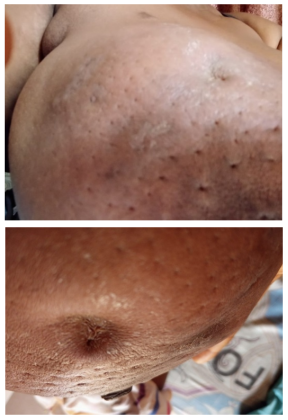
Figure 1. Left Breast showing Peau de orang and some punctate indurations on the skin of the breast.
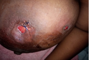
Figure 2. Left Breast showing some discharging ulcers and suture at biopsy site.

Figure 3. Left Breast showing some discharging ulcers and suture at biopsy site.

Figure 4. The left breast during follow-up showing resolution of clinical features.
Information

