Research Article | | Peer-Reviewed

Characterization and Antimicrobial Activity of Silver Nanoparticles Biosynthesised from Some Medicinal Plant Extracts of Benin

Received: 16 December 2024     Accepted: 3 January 2025     Published: 10 February 2025
Views:       Downloads:
Abstract

The use of plant extract as a bio reductant for the synthesis of silver nanoparticles has attracted the attention of several researchers due to its rapid, non-pathogenic and economical protocol. This innovative approach in Benin offers an alternative in medical therapy face of antimicrobial resistance, which is a real public health problem. This study aims to characterize biosynthesized silver nanoparticles (AgNPs) and evaluate the antibacterial activity of synthesized silver nanoparticle from the aqueous extracts of the leaves of Caesalpinia bonduc, Dialium guineense, Momordica charantia, Moringa oleifera, Pavetta corymbosa, Psidium guajava, derived from the flora of Benin. The leaves of plants was collected, authenticated and extracted by water. The synthesized AgNPs by the aqueous extracts were characterized using UV-Vis spectroscopy, X-ray diffraction (XRD), scanning electron microscopy (SEM), and Fourier transform infrared (FTIR) analysis. These characterization techniques allowed to determine the size, shape, crystalline nature, morphology, and the functional groups responsible for the reduction and stabilization of the nanoparticles. The antibacterial activity of AgNPs was determined against six different nosocomial bacteria by the standard disk diffusion method. The results confirmed the successful biosynthesis of AgNPs from the leaves of the six plants as indicated by a colour change from light yellow to brown and grey black. The UV-Vis spectroscopic analysis presented a surface plasmon resonance spectrum with absorption maxima ranging from 340 to 500 nm. XRD analysis demonstrated that the synthesized AgNPs possess a crystalline structure from 1 to 2 μm. In addition, the antimicrobial activities of AgNPs synthesized as reducing agents and stabilizers were investigated against nosocomial bacteria, which are nosocomial infectious agent. Collectively, the findings from this study clearly indicate that the aqueous extracts of the six plants have significant potential for the biosynthesis of silver nanoparticles. The bioactive compounds in the plant extracts were effective in synthesizing AgNPs, and this biological efficiency suggests the potential for incorporating these biosynthesized silver nanoparticles into food and pharmaceutical products.

Published in American Journal of Nano Research and Applications (Volume 13, Issue 1)
DOI 10.11648/j.nano.20251301.11
Page(s) 1-15
Creative Commons

This is an Open Access article, distributed under the terms of the Creative Commons Attribution 4.0 International License (http://creativecommons.org/licenses/by/4.0/), which permits unrestricted use, distribution and reproduction in any medium or format, provided the original work is properly cited.

Copyright

Copyright © The Author(s), 2025. Published by Science Publishing Group

Keywords

Silver Nanoparticles, Benin Medicinal Plant, Aqueous Extract, Antimicrobial Activity

1. Introduction
Nowadays, nanotechnologies intersect with various scientific disciplines including electronics, mechanics, chemistry, optics, biology. Broadly defined, nanotechnology encompasses the science and technology involved in manipulating and manufacturing of materials and artefacts at the nanoscale. At this scale, matter exhibits unique and advantageous properties of atoms or simple molecules .
Noble precious metals, such as silver (Ag), gold (Au) and copper (Cu) are rare metallic chemical elements known for their high resistance to corrosion and oxidation. Among these, silver nanoparticles are particularly prominent in the field of nanomaterials due to their unique properties, including their size, shape, and surface characteristics. These properties make them versatile for a wide range of applications, from biology and medicine to catalysis . Silver nanoparticles can be synthesized through various physicochemical processes such as vapor deposition and chemical reduction. However, some of these process especially photochemical methods, tend to be toxic, costly, and not environmentally sustainable.
In recent years, the scientific community has explored alternative methods for synthesizing environmentally friendly nanoparticles that do not involve toxic substances. One such approach is green synthesis, which utilizes living organisms (such as bacteria, fungi, yeast, and plants) as bioreactors for nanoparticle production . This method has gained significant attention due to its ease of implementation, the accessibility of raw materials, and, notably, its biological activity. Currently, there is a growing focus on using medicinal plants as biological reducers for nanoparticle synthesis. Medicinal plants have always been used to improve human health and/or for a partial or total cure of an illness. In recent decades, scientific research has only confirmed the validity of the therapeutic virtues of most medicinal plants used empirically for thousands of years . Despite the large number of new pharmaceutical drugs available on the market, medicinal plants remain widely used in both developed and underdeveloped countries. These plants have long been valuable sources of biologically active substances. Hence, many drugs currently in clinical use are either derived from natural products or synthetic analogues of these substances.
Benin has remarkable ethnopharmacological potential, which deserves to be explored further in the scientific literature. In 1989, a mission by Adjanohoun et al. listed almost 501 plant species used in traditional medicine, a significant number of which are dedicated to the treatment of infectious diseases, particularly diarrheal diseases. The country's rich plant biodiversity is highlighted by numerous ethnopharmacological surveys , which focus on medicinal plants used to treat various diseases, such as Dialium guineense , Moringa oleifera , Psidium guajava , Momordica charantia , Caesalpinia bonduc and Pavetta corymbose . Recent data have shown the efficacy of extracts from these plants against infective diseases in human health, with inhibitory power ranging from 99.96% to 100%. However, it is interesting to note that, as to the knowledge, no study has yet explored the use of these plants for the biosynthesis of nanoparticles. This choice of research could not only enrich the understanding of the medicinal properties of these species, but also open new perspectives in the field of nanotechnology and modern medicine. This study aims to characterize silver nanoparticles biosynthesized from the aqueous extracts of the leaves of Caesalpinia bonduc, Dialium guineense, Momordica charantia, Moringa oleifera, Pavetta corymbosa, Psidium guajava, derived from the flora of Benin.
2. Methodology
2.1. Vegetal Material
The plants used in this study were collected all from the wild and local markets in southern Benin. Their identification was carried out at the University of Abomey-Calavi (Benin). The dried samples were then brought y in the microbiology laboratory at Anadolu University in Turkey, where they were cut into small pieces and crushed in a plant grinder. Following this, the aqueous extract of each leaf was prepared.
After collecting the plant leaves, they were authenticated by botanists from the National Herbarium of Benin at the University of Abomey-Calavi. Upon arrival at the laboratory, the leaves were quickly washed with tap water and then thoroughly rinsed with distilled water. The plant material was air-dried on the bench and stored away from light and dust to preserve its quality until use. The dried plant parts were then ground into fine powders using a laboratory blender and stored in sealed glass jars at a temperature of 30°C until they were needed for further analysis.
2.2. Preparation and Extraction of Vegetal Material
The aqueous extraction was performed according to the method described by kumar with a slight modification . Fifty grams of each powder were dissolved in 1000 ml of distilled water in a covered glass flask. The flask was boiled for 20 minutes, then allowed to cool to room temperature for 20-30 minutes. The mixture was filtered three times using Whatman paper (No.: 1). The solvent (water) was then evaporated using an evaporator until it decreased by ten times in volume and was maintained at +4°C .
2.3. Synthesis of Silver Nanoparticles
Silver nanoparticles were synthesized by adding silver nitrate (AgNO3) to different aqueous extracts of the six sampled leaves (M. oleifera, C. bonduc, M. charantia, P. corymbosa, P. guajava and D. Guineense). For the synthesis process, a silver nitrate solution was prepared at a concentration of 1 mM and kept in the dark during use. The extract and silver nitrate solution were mixed in 1:9 ratio in a test tube and vortexed for 2 minutes. The tubes were then heated at 80°C for 5–10 minutes with constant stirring at 120 rpm using a rotary shaker. Upon the development n of a yellow-brown color indicating the formation of silver nanoparticles (AgNPs), the resulting was stored at -4°C away from light .
2.4. Characterization of Silver Nanoparticles
2.4.1. UV/VIS Spectral Analysis
The reduction of pure Ag+ ions was assessed by using UV-Vis spectrum analysis (Biotek/Epoch2) Spectrophotometer: For this analysis 2–3 mL nanoparticle of each AgNP solution was placed in a quartz cuvette with a path length of 1 cm. The absorbance was measured over a wavelength range of 300–700 nm .
2.4.2. Scanning Electron Microscope Analysis (SEM)
The AgNPs solutions suspected of containing silver nanoparticles were allowed to dry at room temperature. The dried samples were then subjected to gold plating by placing the powdered samples onto carbon strips. These samples were subsequently t examined and photographed using scanning electron microscopy (SEM) (FESEM, ZEISS Ultra plus) .
2.4.3. X-ray Diffraction (XRD) Analysis
Phase identification and crystal structure determination of the NPs were performed using X-ray diffraction (XRD). With the help of XRD, the presence of AgNPs in the synthesis product can be confirmed. Confirmation is done by identifying peaks in the XRD spectrum characteristic of the face-centred and structure of metallic silver . For X-ray diffraction measurements, the AgNP solutions were dried in an oven at 60°C for two nights The XRD profiles of the resulting powder samples were then obtained using an X-ray diffractometer (XRD, D8 Advance, Bruker Instrument Co., Ltd., Germany) with copper radiation (Cu-Kα = 1.5406 Å) at a scanning speed of 0.5° per minute.
2.4.4. Fourier Transform Infrared Spectroscopy (FT-IR) Analysis
Fourier Transform Infrared Spectroscopy (FTIR) analyses were performed using a PerkinElmer Fourier Transform IR spectrometer in attenuated total reflection (ATR) mode covering a spectral range of 400 - 4000 cm-1 . This technique was used to identify the functional groups present in a sample as well as the bonds developed following the formation of silver nanoparticles. For the FTIR analysis, disks were prepared by drying the samples as described earlier and then mixing them with potassium bromide (KBr). The FTIR spectra were recorded in transmission mode (PerkinElmer, Spectrum 400) over the range of 400–4000 cm-1.
2.5. Antibacterial Activity
The antibacterial activity of AgNPs was determined against six different nosocomial bacteria (Enterococcus faecium, Staphylococcus aureus, Klebsiella pneumonia, Acinobacter baumannii, Pseudomonas aeruginosa, Enterobacter spp) by the standard disk diffusion method . The bacterial pathogens were obtained from the medicine faculty of Osmangazi university in Turkey and maintained on nutrient agar media. Prior to use, the vitality of the bacteria has been tested. After 20 to 24 hours of incubation at 37°C, cultures that successfully formed a single colony and passed purity control were stored at +4 °C for use in subsequent steps. The antimicrobial activity of plant extracts and silver nanoparticle mixtures was determined by well diffusion method . The overnight grown cultures of tested bacteria were diluted into Mueller Hinton Broth (MBH) to 1 × 10⁻⁷ colony forming unit were used for the assay (corresponding to a McFarland turbidity tube of 0.5). After that, 100 µL of bacterial dilution was pipetted onto the surface of petri dishes containing 20 ml of Müller Hinton agar using a sterile pipette and spread evenly over the medium with a sterile Drigalski spatula. Wells of 6 mm diameter were opened on petri dishes, which were kept in a sterile cabinet with their lids half open for 30 minutes for drying, and they were composed of aqueous extracts of the plant whose antimicrobial activity was to be evaluated, and also synthetised nanoparticle of silver nanoparticles. As positive control, antibiotic solution (chloramphenicol [1 mg/ml] as were added and as a negative control, 50 μl of silver nitrate solution were also added. After overnight incubation at 37°C, the inhibition zones formed around the discs were recorded by measurement using a ruler graduated in mm. All experiments were carried out in 2 parallels and the results were recorded as the mean value.
The minimum inhibitory concentration (MIC) of the AgNPs were determined by the two-fold serial dilution method . Different concentrations of AgNPs (100–3.12 mg/mL) were used for MIC test. Prior to test, initially 200 mg of the AgNPs was added to initial tube containing, 2 mL of Nutrient Broth (NB) media, then 1 mL from it was transferred to next tube which contains 1 mL of only NB media and mixed properly, then the dilution was made till the concentration of the last tube was 3.12 mg/mL. The control tube contains only 1 mL of NB media. Then 10 mL of the tested pathogen was added to each tube. This procedure was repeated for all the tested pathogens. Then all the tubes were mixed properly and were incubated at 37°C overnight in a shaker incubator. The lowest concentration of AgNPs that did not show any visible growth of test organisms was determined as the MIC. Further, the MIC concentration and the next higher concentration were spread on Nutrient Agar (NA) plates and incubated for another 24 h at 37°C. The concentration that did not show any growth of a single bacterial colony on the NA plates was defined as the Minimum Bactericidal Concentration (MBC) value. Both MIC and MBC values were expressed as mg/mL.
3. Results and Discussion
3.1. Biosynthesis and Characterization of Silver Nanoparticles
3.1.1. Colour Change
Silver nanoparticles can be synthesized from various parts of plants, such as leaf extracts , fruit extracts , and seed extracts . In this study, silver nanoparticles were synthesized by adding silver nitrate (AgNO3) to different aqueous extracts of the six sampled leaves (M. oleifera, C. bonduc, M. charantia, P. corymbosa, P. guajava and D. Guineense). The aqueous extraction of Moringa oleifera and Dialium guineense leaves, when combined with silver nitrate, exhibited a yellow coloration on one hand and grey-black coloration on the other hand (Figure 1a and 1f). This aligns with findings from a study by Al-kalifazi (2016), where the addition of AgNO₃ to Moringa oleifera leaf extracts and subsequent heating at 60°C for 1 hour resulted in the formation of silver nanoparticles, indicated by a red-brown colour. Furthermore, aqueous extracts from the leaves of Caesalpinia bonduc, Momordica charantia, Pavetta corymbosa, Psidium guajava, showed a brown coloration upon interaction with silver nitrate (Figure 1b, 1c, 1d, and 1e). Previous research has reported comparable results. For instance, Moteira et al. (2014) observed a colour change when the aqueous extract of Psidium guajava leaves, was added to a 1 mM silver nitrate solution, indicating the formation of silver nanoparticles in another involving Momordica charantia plants. The addition of different volumes of aqueous extract to 10 ml of 1 mM AgNO₃ solution, followed by heating at 60°C, resulted in a colour change, confirming the synthesis of silver nanoparticles . Additionally, Nanoparticles were also successfully synthesized using Caesalpinia gilliesii plant extract .
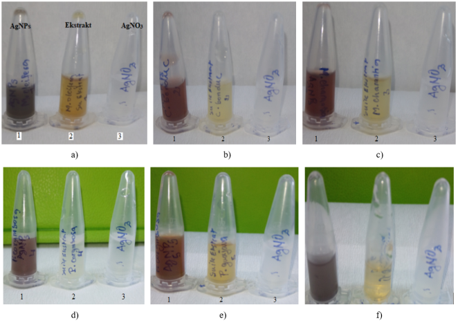
Figure 1. Silver nanoparticles formed from aqueous extract of the leaves of a) Moringa oleifera, b) Caesalpinia bonduc, c) Momordica charantia, d) Pavetta corymbosa, e) Psidium guajava, f) Dialium guineense (1: silver nanoparticle, 2: aqueous extract, 3: silver nitrate).
In this study, the addition of plant extracts to silver nitrate resulted in the reduction of silver ions (Ag+) to silver nanoparticles (Ag-NPs), which was indicated by a colour change. While the negative control containing only the plant extract remained yellow, the dark brown or grey-black coloration observed after adding silver nitrate confirmed the formation of nanoparticles (Figure 1). These colour changes in the solutions are attributed to the phenomenon of surface plasmon resonance (SPR) .
The findings of the study provide a basis for determining suitable plants from Beninese flora for the synthesis of Ag-NPs for 1 day, but no colour.
After adding AgNO₃ to the extracts of different leaves, the mixtures were left in a dark environment for 24 hours, but no colour change was observed, indicating that no reaction had occurred under these conditions. Consequently, the reaction was induced by heating the mixtures at 80°C for 5 minutes, which resulted in a colour change, confirming the formation of Ag-NPs. All subsequent experiments were conducted using this protocol. Similar observations have been reported by other researchers, although with longer heating durations of up to 90 minutes at a lower temperature of 60°C .
3.1.2. UV/VIS Spectral Analysis
The UV-vis spectra of silver nanoparticles (water-NPs) synthesized from M. oleifera, C. bonduc, M. charantia, P. corymbosa, P. guajava and D. guineense showed distinct maximum absorption peaks: 500 nm for M. oleifera, 410 nm for C. bonduc, 500 nm for M. charantia, 340 nm for P. corymbosa, 450 nm for P. guajava and 400 nm for D. guineense, respectively (Table 1; Figure 2). It is important to note that UV-Vis spectrophotometric analysis is generally used to monitor and confirm the formation of AgNPs. From this characterization, it can be deduced that silver nanoparticles synthesized from different plants exhibited maximum absorbance ranging from 394 nm to 500 nm. This difference in wavelengths may be attributed to the phytochemical compounds present in each plant, which differ from one species to another. Smaller AgNPs, typically between 10-50 nm in size, exhibit an absorbance peak near 400 nm, while larger particles, between 100-200 nm, show a broader peak with a maximum that shifts towards longer wavelengths, around 500 nm. This observation supports the formation of silver nanoparticles in the study. In 2015, Sadeghi et al. reported an average nanoparticle size of 25-30 nm . Consequently, our study also reveals that the AgNPs solutions derived from D. guineense and P. corymbosa plants showed a significant absorbance in the UV-visible spectrum, indicating that these solutions gave smaller nanoparticles size than those from other plants. This finding aligns with the earlier observation of a rapid colour change in these solutions compared to the others, indicating a more robust formation of silver nanoparticle.
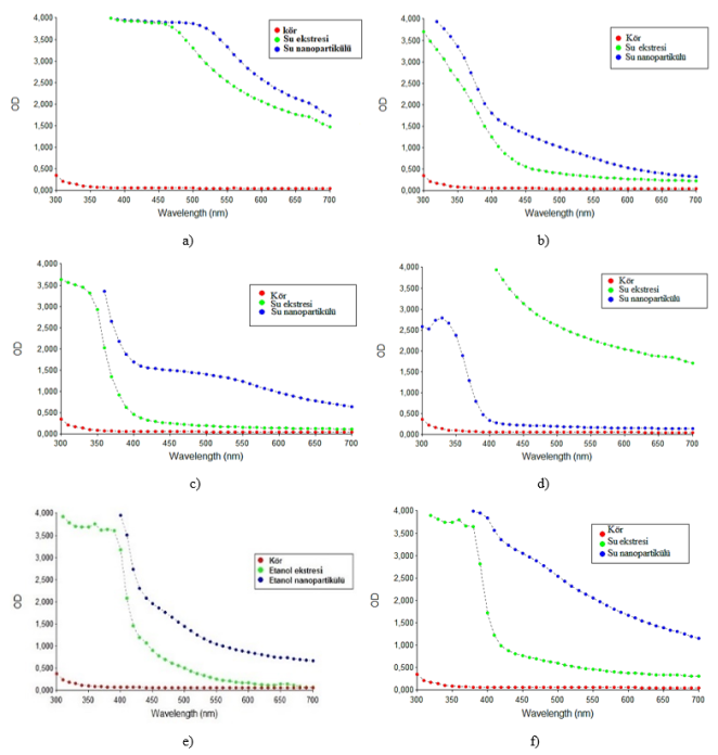
Figure 2. Absorption zone of silver nanoparticles formed from aqueous extract of the leaves of a) Moringa oleifera, b) Caesalpinia bonduc, c) Momordica charantia, d) Pavetta corymbosa, e) Psidium guajava, f) Dialium guineense.
Table 1. Absorption area of silver nanoparticles from different synthesized leaves.

Aqueous extract

UV-Visible absorption zone (nm)

Moringa oleifera

500

Caesalpinia bonduc

410

Momordica chanratia

500

Payetta corymbosa

340

Psidium guajava

450

Dialium guineense

400

When the maximum absorbance occurs between 400 and 450 nm, the nanoparticles have a spherical or near-spherical geometry . Based on Mie theory, small spherical isotopic Ag nanoparticles give a single symmetric surface plasmon resonance (SPR) absorption band within wavelength range of 350 to 500 nm with a peak centred around 410 nm . It is well known that the optical absorption spectra of metal nanoparticles are controlled by (SPRs) which shift to longer wavelengths as the particle size increases due to the effect of the capping material .
3.1.3. Scanning Electron Microscope Analysis (SEM)
In this study, Scanning Electron Microscopy (SEM) analysis revealed aggregates (clusters) and spherical structure of all nanoparticles obtained from the AgNPs extract-solution mixtures (figure 3), confirming the formation of silver nanoparticles (AgNPs). These findings are consistent with reports from several other studies, where AgNPs synthesized from plant extracts, and evaluated by SEM, also exhibited a spherical in shape . The observed aggregation could be attributed to the presence of secondary metabolites in the leaf extracts . Similar results have been reported in the literature on M. oleifera , M. charantia and P. guajava .
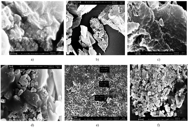
Figure 3. SEM image of silver nanoparticles formed from aqueous extract of leaves of a) Moringa oleifera, b) Caesalpinia bonduc, c) Momordica charantia, d) Pavetta corymbosa, e) Psidium guajava, f) Dialium guineense.
3.1.4. X-ray Diffraction (XRD) Analysis
The crystal structures of the samples were analysed using X-ray diffraction (XRD) (Figure 3). In the spectra, ta sharp peak observed at about 38° corresponds to the characteristic crystal structure of silver nanoparticles.
The XRD analysis of nanoparticles synthesized from plant extracts (Figure 4) shown that the AgNPs obtained through the of silver salt are crystalline in nature. Generally, three characteristic peaks for silver ions were observed within the range of 30° to 60°. The presence of these peaks in the XRD spectra confirms that the synthesized nanoparticles are indeed silver nanoparticles. Specifically, the XRD analysis of nanoparticles obtained from the aqueous extracts of Moringa oleifera showed prominent peaks at approximately 34°, 47°, and 67° (Figure 4). The XRD spectrum of Ag-NPs synthesized from aqueous extracts of Caesalpinia bonduc plants demonstrated that all reflections correspond to pure silver with face-centred cubic (FCC) symmetry. For the AgNPs derived from the aqueous extracts - the intensity of the peaks at 33°, 47°, 68° and 74ᵒ, reflects the high crystallinity of the silver nanoparticles. In contrast, the one synthesized from Momordica charantia extracts plants, showed only two prominent peaks at two points at (32° to 40°). This indicating fewer crystal-line facets Four diffraction peaks at (111), (200), (220) and (311), were observed, which are characteristic of the reflections and the cc FCC structure of metallic silver. The aqueous extracts from Pavetta corymbosa and Psidium guajava each exhibited two diffraction peaks, corresponding to (111), and (200), the body-centred cubic (BCC) structure of metallic silver (Figure 4). For the nanoparticles synthesized from the aqueous extract of Dıalıum guineense leaves, three diffraction peaks were observed at 33°, 47° and 58°. These peaks correspond to the reflections at (111), (200), and (220), respectively, indicating a face-centred cubic structure of metallic silver.
The shape and size of silver nanoparticles produced by leaf extracts in this study were predominantly spherical, in shape, similar to the silver nanoparticles synthesized using other plant sources . The characteristic peaks were observed between 32° and 34° (Figure 4), suggesting that the absence of these peaks in some samples might be due to the crystals being too small to detect (Figure 4). However, analytical methods confirmed that even in samples where these characteristic XRD peaks were not observed a crystalline structure was present .
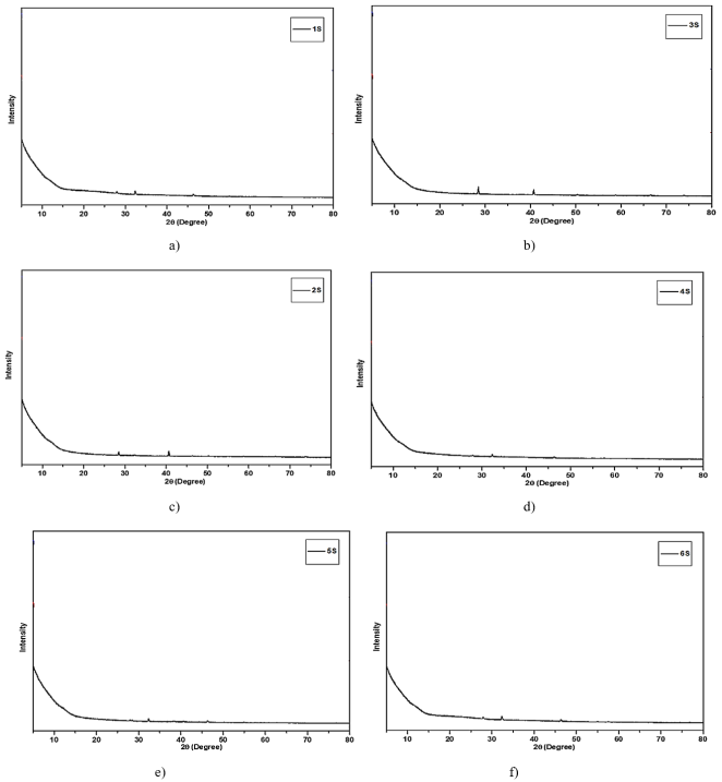
Figure 4. XRD image of silver nanoparticles formed from aqueous extract of leaves of a) Moringa oleifera, b) Caesalpinia bonduc, c) Momordica charantia, d) Pavetta corymbosa, e) Psidium guajava, f) Dialium guineense.
3.1.5. Fourier Transform Infrared Spectroscopy (FT-IR) Analysis
FT-IR has become an important tool for the detection of functional groups between metal particles and biomolecules. In this study, FT-IR measurements were used to identify potential biomolecules involved in the stabilization of the synthesized silver nanoparticles (Figure 5).
In this study, the FT-IR spectra of the samples displayed a peak around 3500 cm-1 corresponding to the O-H stretching vibration. According to Moteriya, silver nanoparticles synthesized using P. guajava exhibited strong peaks at 3722.74, 3599.29, 3315.74, 3009.05, 2305.01, 1844.01, 1491.02, 1311.64, 1039.67 and 661.61 cm-1 in the FT-IR spectrum . The intense bands at 3722.74, 3599.29 cm-1 are characteristic of the primary O-H stretching vibration of free alcohols . Additionally, the peak observed around 2900 cm-1 corresponds to the C-H stretching vibration of fatty acids . The peaks observed in the range of 1645-1750 cm-1 are attributed to the C=O stretching vibrations of proteins. The peak observed around 1450 cm-1 is associated with the C-N stretching vibration of amines, while the peaks at 1844.01 is characteristic of aromatic amines. The peak at 1491.02 is attributed to the C-C (in-ring) stretching of aromatics compounds. The peak at 1311.64 cm-1 corresponds to the symmetric stretching of the N-O bond in N-O compound. Lastly, the peak at 1039.67 is indicative of the CN stretching vibrations of aliphatic amines .
Furthermore, the peak observed at 1417 cm-1 corresponds to the C-O stretching vibration, while the peak at 1384 cm-1 is associated with the C-N stretching vibration of aromatic amine. The peak around 1070 cm-1 indicates the C-OH stretching vibration characteristic of secondary alcohols . Peaks observed below 1000 cm-1 are attributed to the out-of-plane C-H bending vibrations characteristic of aromatic phenols . These results suggest that compounds such as phenolic acids can adsorb onto the surface of AgNPs helping to retain, these substances on the surface and providing stabilization for the nanoparticles . In this study, FTIR measurements were performed to identify possible biomolecules responsible for stabilizing the synthesized silver nanoparticles.
It is well established that proteins can bind to silver nanoparticles through free amine groups or cysteine residues within the proteins and surface-bound proteins help stabilize silver nanoparticles during synthesis.
Additionally, nicotinamide adenine dinucleotide hydrate (NADH)-dependent reductases have been identified as contributors to the biosynthesis of nanoparticles, while polysaccharides play a role in their stabilization . Polyphenols, such as tannic acids, are plant-derived compounds that act as effective reducing agents in the synthesis of silver nanoparticles .
Figure 5. FT-IR image of silver nanoparticles formed from aqueous extract of leaves of a) Moringa oleifera, b) Caesalpinia bonduc, c) Momordica charantia, d) Pavetta corymbosa, e) Psidium guajava, f) Dialium guineense.
3.2. Antibacterial Activity
Many studies have been conducted on the antimicrobial activities of extracts obtained from plant species belonging to numerous families against various pathogens . The present study investigated the antimicrobial activities, through the formation of silver nanoparticles, of some plants such as Moringa oleifera, Caesalpinia bonduc, Momordica charantia, Pavetta corymbosa, Psidium guajava and Dialium guineensen, which are traditionally used against infections, especially in Benin and West Africa. According to this study, silver nanoparticles synthesised from the M. oleifera extracts did not showed any levels of antimicrobial effect on the bacteria tested. Al-Kalifawi and al. reported in 2016, that aqueous extract-AgNPs from the same plant formed an inhibition zone with a diameter of 18 mm on S. aureus . In the same study, it was observed that the aqueous extract did not form a zone of inhibition on the bacteria tested and that other AgNPs obtained using these extracts showed the most effect on the Gram-negative bacterium K. pneumoniae. However, this result was not consistent with our study because K. pneumoniae was not inhibited by the aqueous extract-AgNP.
Table 2. Antimicrobial activity of the M. oleifera extract showing their zones of inhibition.

Inhibitory zone (mm)

Material (50 µl)

E. coli

S. aureus

K. pneumoniae

A. baumani _

P. aeroginosa

E. aerogenes

Leaf extract

-

15

-

15

-

15

AgNP-Leaf

-

-

-

-

-

-

AgNO 3

13

13

15

14

15

-

chloramphenicol

27

29

28

15

15

22

(-): no zone of 'inhibition
Enam and al., showed that the hydroalcoholic extract obtained from Caesalpinia gilliesii formed inhibition zones at different rates, but it was determined that the AgNPs synthesized from these extracts were more effective against all the bacteria tested and formed the most inhibition zones against S. aureus . In our study, the water extract created a significant zone diameter of inhibitory against the S. aureus strain in the antimicrobial activity tests carried out. All inhibitory zones are measured in mm (Table 3).
Table 3. Antimicrobial activity of the C. bonduc extract showing their zones of inhibition.

Inhibitory zones (mm)

Materials (50 µl)

E. coli

S. aureus

K. pneumoniae

baumani

P. aeroginosa

E. aérogènes

Aqueous extract

-

10

-

-

-

-

AgNPs of water

14

14

11

11

11

-

AgNO 3

13

13

15

14

15

-

chloramphenicol

27

29

28

15

15

22

(-): no zone of 'inhibition
The components of the AgNP extract showed superior antimicrobial effect than the extract alone, except for E. coli, and K. pneumoniae that was inhibited by aqueous AgNP extract. No study on Caesalpinia bonduc was found in the literature, our study is the first study conducted with this plant which is one of the important discovery in this Study. According to Krithiga and al. in 2015, the aqueous extract obtained from the plant M. charantia and the synthesised silver nanoparticles have antimicrobial effects on Bacillus subtillis and E. coli, and larger zone diameters were observed in Staphylococcus aureus and Bacillus subtilis compared to E. coli . In this study, the aqueous extract formed a zone of inhibition in all bacteria tested and showed the highest antimicrobial activity against the S. aureus strain with a zone diameter of 19 mm (Table 4).
Table 4. Antimicrobial activity of the M. charantia extract showing their zones of inhibition.

Inhibitory zones (mm)

Matériel d'essai (50 µl)

E. coli

S. aureus

K. pneumoniae

A. baumani

P. aeroginosa

E. aérogènes

Aqueous Extract

14

19

14

16

13

17

ethanol

-

16

-

-

-

16

Méthanol

-

18

-

-

-

17

AgNPs-Aqueous

-

-

11

09

13

09

chloramphenicol

27

29

28

15

15

22

(-): no zone of 'inhibition
However, aqueous extracts-AgNPs synthesised by silver nitrate treatment of extracts obtained from M. charantia leaves with water did not form zones of inhibition on E. coli and S. aureus, whereas they formed smaller zones of inhibition on other bacteria. In another study, Ajitha and al., extracted AgNPs from M. charantia water were generally found to have a larger inhibition zone diameter (7 mm) against Gram-negative bacteria (P. aeroginosa) than Gram-positive bacteria were reported . In our study, the water extract-AgNPs formed a 13 mm diameter zone of inhibition against the P. aeroginosa strain.
David and al., reported that the same aqueous plant extract formed a 10 mm diameter zone of inhibition on K. pneumoniae . Pavetta corymbosa showed no antimicrobial effect on any of the bacteria tested, while the aqueous extract-AgNPs synthesised from it had variable effects on bacteria other than E. coli and E. aerogenes. The diameter of the zone of inhibition formed particularly on P. aeroginosa was found to be equivalent to the standard antibiotic chloramphenicol (15 mm) (Table 5).
Table 5. Antimicrobial activity of the P. corymbosa extract showing their zones of inhibition.

Inhibitory zones (mm)

Material (50 µl)

E.coli

S. aureus

K. pneumoniae

A. baumanii

P. aeroginosa

E.aérogènes

Aqueous Extract

-

-

-

-

-

-

AgNPs-Aqueous

-

11

11

09

15

-

AgNO 3

14

13.5

14

13

15

-

chloramphenicol

27

29

28

15

15

22

(-): no zone of 'inhibition
An important conclusion from this study is that chloramphenicol, the reference antibiotic, and the P. aeroginosa strain are equally effective. There are many studies on the production of silver nanoparticles using the Psidium guajava plant. Moteriya and al. synthesised the water extract-AgNP using aqueous extract of Psidium guajava and investigated its antimicrobial activities on bacteria . In our study, this extract was not effective against E. coli, K. pneumoniae and P. aeruginosa.
Table 6. Antimicrobial activity of the D. guineense extract showing their zones of inhibition.

Inhibitory zones (mm)

Material (50 µl)

E.coli

S. aureus

K. pneumoniae

A. baumanii

P. aeroginosa

E. aérogènes

Aqueous

14

-

-

14

11

11

AgNPs-Aqueous

13

13

12

08

12

-

AgNO 3

14

13

13

13

14

-

chloramphenicol

27

29

28

15

15

22

(-): no zone of 'inhibition
D. guineense extracts prepared from the plant and silver nanoparticles synthesised from them showed variable levels of antimicrobial activity on all the bacteria tested. The only bacterium sensitive to all the extracts and their silver nanoparticles was A. baumanii. It was observed that the plant extracts of P. corymbosa and D. guineense and the nanoparticles synthesised from them used in this study formed a good zone of inhibition. Numerous studies have been carried out on silver nanoparticles in the literature, but these studies have mainly been based on the antimicrobial activities and properties of the nanoparticles. There is very little data on the determination of MIC values. Therefore, in our study, MIC values of plant extracts and silver nanoparticles synthesised from them on microorganisms were also determined. It was determined that some of the silver nanoparticles synthesised had efficacy values close to or even higher than the reference antibiotics. The variation between our results and those in the literature would be linked to complete standardisation, which has not been defined in the study of antimicrobial activity, and the handling techniques may vary from one researcher to another.
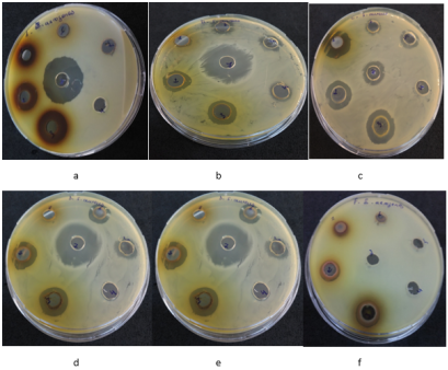
Figure 6. Antimicrobial potentials of different plant extract (a- M. oleifera, b- C. bonduc, c- M. charantia, d- P. corymbose, e- P. guajava, f- D. guineense) showing their zones of inhibition.
4. Conclusion
Nanoparticle biosynthesis methods offer a novel approach to easily synthesize NPs using natural reducing and stabilizing agents, such as the aqueous extracts of plant leaves in our study. This method eliminates the need for chemical reagents making the process environmentally friendly and suitable for pharmaceutical and food applications. For the biosynthesis of silver nanoparticles, our results demonstrated a visible colour change in the mixture confirming the formation of silver nanoparticles. Notably, the nanoparticles with the lowest absorption wavelengths were derived from the leaf extracts of Pavetta corymbosa (340 nm) and Dialium guineense (400 nm) indicating that this plant could serve as valuable resources for future research. In perspective, several aspects of this research warrant further exploration. It would be beneficial to complement this study with the following:
1) An investigation into the yield of dry crude extract for each plant, to better understand the efficiency of the extraction process. An extended antimicrobial study that includes a broader range of pathogenic microorganisms, particularly those associated with foodborne illnesses.
2) An assessment on the AgNPs of the anti-inflammatory and antioxidant activities of the biosynthesized silver nanoparticles need to be carefully study.
3) The development of practical applications on both medical and industrial scales, to harness the full range of beneficial properties of silver nanoparticles for enhancing human well-being and leveraging their advantages across various fields.
Abbreviations

AgNO3

Silver Nitrate

Ag

Silver

Ag+

Silver Ions

AgNPs

Silver Nanoparticles

Au

Gold

Cu

Copper

MBC

Minimum Bactericidal Concentration

MIC

Minimum Inhibitory Concentration

MBH

Mueller Hinton Broth

NA

Nutrient Agar

NB

Nutrient Broth

UV-Vis

Ultra-Violet Visible Spectroscopy

XRD

X-ray Diffraction

SPR

Surface Plasmon Resonance

SEM

Scanning Electron Microscopy

Conflicts of Interest
The authors declare that they do not have any conflicts of interest.
References
[1] Talarska, P., Boruczkowski, M., and Żurawski, J., “Current Knowledge of Silver and Gold Nanoparticles in Laboratory Research-Application, Toxicity, Cellular Uptake,” Nanomaterials (Basel, Switzerland), Vol. 11, No. 9, 2021, p. 2454.
[2] Ramesh, S., and Narayanan, V., “Wet Chemical Synthesis of Cadmium Sulphide Nanoparticles and Its Characterization,” Chemical Science Transactions, 2013. Retrieved 16 November 2024.
[3] Kumar, V., and Pansari, A., “Competitive Advantage through Engagement,” Journal of Marketing Research, Vol. 53, No. 4, 2016, pp. 497–514.
[4] Ibrahim, H. M. M., “Green Synthesis and Characterization of Silver Nanoparticles Using Banana Peel Extract and Their Antimicrobial Activity against Representative Microorganisms,” Journal of Radiation Research and Applied Sciences, Vol. 8, No. 3, 2015, pp. 265–275.
[5] Krishnaraj, C., Jagan, E. G., Rajasekar, S., Selvakumar, P., Kalaichelvan, P. T., and Mohan, N., “Synthesis of Silver Nanoparticles Using Acalypha Indica Leaf Extracts and Its Antibacterial Activity against Water Borne Pathogens,” Colloids and Surfaces B: Biointerfaces, Vol. 76, No. 1, 2010, pp. 50–56.
[6] Lazli, A., Beldi, M., Ghouri, L., and Nouri, N. E. H., “Étude Ethnobotanique et Inventaire Des Plantes Médicinales Dans La Région de Bougous,” Bulletin de la Société royale des sciences de Liège, 2019. Retrieved 16 November 2024.
[7] Adomou, C. A., Dassou, H. G., Houenon, G. H. A., Alladayè, A., and Yedomonhan, H., “Comprendre Les Besoins En Ressources Végétales Des Populations Riveraines Pour Une Gestion Durable de La Forêt Bahazoun Au Sud-Bénin (Afrique de l’Ouest),” International Journal of Biological and Chemical Sciences, Vol. 11, No. 5, 2017, pp. 2040–2057. Retrieved 16 November 2024.
[8] Agbankpé, A. J., Dougnon, T. V., Bankolé, H. S., Yèhouénou, B., Yédomonhan, H., Lègonou, M., and Dougnon, T. J., “Etude Ethnobotanique Des Légumes Feuilles Thérapeutiques Utilisés Dans Le Traitement Des Diarrhées Au Sud-Bénin (Afrique de l’Ouest),” International Journal of Biological and Chemical Sciences, Vol. 8, No. 4, 2014, pp. 1784–1795. Retrieved 16 November 2024.
[9] Belhaddad, A., and Khelifi, F. E. Z., “Etude des mécanismes de résistance aux antibiotiques chez des souches de bactéries isolées au niveau de l‘hôpital de Mohamed Boudiaf de Ouargla.,” Thesis. UNIVERSITE KASDI MERBAH OUARGLA, 2019.
[10] Ali, M., Yahaya, A., Zage, A., and Yusuf, Z., “In-Vitro Antibacterial Activity and Phytochemical Screening of Psidium Guajava on Some Enteric Bacterial Isolates of Public Health Importance,” Journal of Advances in Medical and Pharmaceutical Sciences, Vol. 12, No. 3, 2017, pp. 1–7.
[11] Chokki, M., Zongo, C., Dah-Nouvlessounon, D., Cudălbeanu, M., Noumavo, P., Ghinea, I. O., Furdui, B., Savadogo, A., Dinica, R. M., and Baba-Moussa, L., “Phytochemical Screening and Antimicrobial Activity of Momordica Charantia L. and Morinda Lucida Benth Extracts from Benin,” African Journal of Microbiology Research, Vol. 14, No. 8, 2020, pp. 426–435.
[12] Sasidharan, S., “Caesalpinia Bonduc: A Ubiquitous yet Remarkable Tropical Plant Owing Various Promising Pharmacological and Medicinal Properties with Special References to the Seed,” Los Angeles, Vol. 10, No. 394.
[13] Anyanwu, M., and Okoye, R., “Antimicrobial Activity of Nigerian Medicinal Plants,” J Intercult Ethnopharmacol., 2017. Retrieved 16 November 2024.
[14] Patra, J. K., and Baek, K.-H., “Antibacterial Activity and Synergistic Antibacterial Potential of Biosynthesized Silver Nanoparticles against Foodborne Pathogenic Bacteria along with Its Anticandidal and Antioxidant Effects,” Frontiers in Microbiology, Vol. 8, 2017.
[15] Soshnikova, V., Kim, Y. J., Singh, P., Huo, Y., Markus, J., Ahn, S., Castro-Aceituno, V., Kang, J., Chokkalingam, M., Mathiyalagan, R., and Yang, D. C., “Cardamom Fruits as a Green Resource for Facile Synthesis of Gold and Silver Nanoparticles and Their Biological Applications,” Artificial Cells, Nanomedicine, and Biotechnology, Vol. 46, No. 1, 2018, pp. 108–117.
[16] Krithiga, J., and Briget, M. M., “Synthesis of AgNPs of Momordica Charantia Leaf Extract, Characterization and Antimicrobial Activity,” Pharm. Anal. Acta, Vol. 6, No. 10, 2015, pp. 1–7.
[17] Ghaffari-Moghaddam, M., and Hadi-Dabanlou, R., “Plant Mediated Green Synthesis and Antibacterial Activity of Silver Nanoparticles Using Crataegus Douglasii Fruit Extract,” Journal of Industrial and Engineering Chemistry, Vol. 20, No. 2, 2014, pp. 739–744.
[18] Sadeghi, B., Rostami, A., and Momeni, S. S., “Facile Green Synthesis of Silver Nanoparticles Using Seed Aqueous Extract of Pistacia Atlantica and Its Antibacterial Activity,” Spectrochimica Acta Part A: Molecular and Biomolecular Spectroscopy, Vol. 134, 2015, pp. 326–332.
[19] Emam, M., El Raey, M. A., Eisa, W. H., El-Haddad, A. E., Osman, S. M., El-Ansari, M. A., and Rabie, A.-G. M., “Green Synthesis of Silver Nanoparticles from Caesalpinia Gilliesii (Hook) Leaves: Antimicrobial Activity and in Vitro Cytotoxic Effect against BJ-1 and MCF-7 Cells,” Journal of Applied Pharmaceutical Science, Vol. 7, No. 8, 2017, pp. 226–233.
[20] Lanone, S., and Boczkowski, J., “Les nanomatériaux sont-ils dangereux pour notre santé ?,” Questions de santé publique, No. 10, 2010, pp. 1–4.
[21] Ider, M., “Elaboration et Caractérisation Des Nanomatériaux à Base de Métaux Nobles,” PhD Thesis. Université du Maine; Université Hassan II (Casablanca, Maroc), 2017.
[22] Brause, R., Möltgen, H., and Kleinermanns, K., “Characterization of Laser-Ablated and Chemically Reduced Silver Colloids in Aqueous Solution by UV/VIS Spectroscopy and STM/SEM Microscopy,” Applied Physics B: Lasers and Optics, Vol. 75, Nos. 6–7, 2002, pp. 711–716.
[23] Rao, C. R., and Trivedi, D. C., “Biphasic Synthesis of Fatty Acids Stabilized Silver Nanoparticles: Role of Experimental Conditions on Particle Size,” Materials Chemistry and Physics, Vol. 99, Nos. 2–3, 2006, pp. 354–360.
[24] Ajitha, B., Reddy, Y. A. K., Rajesh, K. M., and Reddy, P. S., “Sesbania Grandiflora Leaf Extract Assisted Green Synthesis of Silver Nanoparticles: Antimicrobial Activity,” Materials Today: Proceedings, Vol. 3, No. 6, 2016, pp. 1977–1984.
[25] Al-Kalifawi, E. J., “Green Synthesis of Silver Nanoparticles Using Leaf Extract of Al-Rawag Tree (Moringa Oleifera Lamarck) Cultivated in Iraq and Efficacy the Antimicrobial Activity,” Mesop Environ J Spicial Issue A, 2016, pp. 39–48. Retrieved 17 November 2024.
[26] David, S. A., Ponvel, K. M., Fathima, M. A., Anita, S., Ashli, J., and Athilakshmi, A., “Biosynthesis of Silver Nanoparticles by Momordica Charantia Leaf Extract: Characterization and Their Antimicrobial Activities,” J. Nat. Prod. Plant Resour, Vol. 4, No. 6, 2014, pp. 1–8. Retrieved 17 November 2024.
[27] Supraja, N., Avinash, B., and Prasad, T., “Green Synthesis and Characterization of Silver Nanoparticles from Momordica Charantia Fruit Extract: Study of Antimicrobial Activities,” Int. J. Pure App. Biosci., Vol. 5, No. 1, 2017, pp. 107–117.
[28] Geetha, V., “Green Synthesis of Silver Nanoparticles from Psidium Guajava Leaves and Its Antibacterial Activity,” International Journal of Bioassays, Vol. 6, 2017, pp. 5441–5443.
[29] Pooja Moteriya, P. M., Hemali Padalia, H. P., and Sumitra Chanda, S. C., “Green Biosynthesis of Silver Nanoparticles Using Psidium Guajava L. Leaf Extract and Antibacterial Activity against Some Pathogenic Microorganisms.,” 2014. Retrieved 17 November 2024.
[30] Ahmed, S., Ahmad, M., Swami, B. L., and Ikram, S., “A Review on Plants Extract Mediated Synthesis of Silver Nanoparticles for Antimicrobial Applications: A Green Expertise,” Journal of advanced research, Vol. 7, No. 1, 2016, pp. 17–28.
[31] Aliyu, A. B., Ibrahim, M. A., Ibrahim, H., Dambatta, M. B., and Oyewale, A. O., “Gc-Ms Analysis of Pavetta Corymbosa Lipophilic Extract and Its Antimicrobial Activity,” Ife Journal of Science, Vol. 19, No. 2, 2017, pp. 363–368.
[32] Zhao, S.-Y., Chen, S.-H., Li, D.-G., Yang, X.-G., and Ma, H.-Y., “A Convenient Phase Transfer Route for Ag Nanoparticles,” Physica E: Low-Dimensional Systems and Nanostructures, Vol. 23, Nos. 1–2, 2004, pp. 92–96.
[33] Akintelu, S. A., Bo, Y., and Folorunso, A. S., “A Review on Synthesis, Optimization, Mechanism, Characterization, and Antibacterial Application of Silver Nanoparticles Synthesized from Plants,” Journal of Chemistry, Vol. 2020, No. 1, 2020, p. 3189043.
[34] Singhal, G., Bhavesh, R., Kasariya, K., Sharma, A. R., and Singh, R. P., “Biosynthesis of Silver Nanoparticles Using Ocimum Sanctum (Tulsi) Leaf Extract and Screening Its Antimicrobial Activity,” Journal of Nanoparticle Research, Vol. 13, No. 7, 2011, pp. 2981–2988.
[35] Anil Kumar, S., Abyaneh, M. K., Gosavi, S. W., Kulkarni, S. K., Pasricha, R., Ahmad, A., and Khan, M. I., “Nitrate Reductase-Mediated Synthesis of Silver Nanoparticles from AgNO3,” Biotechnology Letters, Vol. 29, No. 3, 2007, pp. 439–445.
Cite This Article
  • APA Style

    Yacoubou, A. F., Aydın, M. T. A., Güven, K., Salifou, C. F. A. (2025). Characterization and Antimicrobial Activity of Silver Nanoparticles Biosynthesised from Some Medicinal Plant Extracts of Benin. American Journal of Nano Research and Applications, 13(1), 1-15. https://doi.org/10.11648/j.nano.20251301.11

    Copy | Download

    ACS Style

    Yacoubou, A. F.; Aydın, M. T. A.; Güven, K.; Salifou, C. F. A. Characterization and Antimicrobial Activity of Silver Nanoparticles Biosynthesised from Some Medicinal Plant Extracts of Benin. Am. J. Nano Res. Appl. 2025, 13(1), 1-15. doi: 10.11648/j.nano.20251301.11

    Copy | Download

    AMA Style

    Yacoubou AF, Aydın MTA, Güven K, Salifou CFA. Characterization and Antimicrobial Activity of Silver Nanoparticles Biosynthesised from Some Medicinal Plant Extracts of Benin. Am J Nano Res Appl. 2025;13(1):1-15. doi: 10.11648/j.nano.20251301.11

    Copy | Download

  • @article{10.11648/j.nano.20251301.11,
      author = {Aminath Fidele Yacoubou and Meryem Türkay Aytekin Aydın and Kiymet Güven and Chakirath Folakè Arikè Salifou},
      title = {Characterization and Antimicrobial Activity of Silver Nanoparticles Biosynthesised from Some Medicinal Plant Extracts of Benin
    },
      journal = {American Journal of Nano Research and Applications},
      volume = {13},
      number = {1},
      pages = {1-15},
      doi = {10.11648/j.nano.20251301.11},
      url = {https://doi.org/10.11648/j.nano.20251301.11},
      eprint = {https://article.sciencepublishinggroup.com/pdf/10.11648.j.nano.20251301.11},
      abstract = {The use of plant extract as a bio reductant for the synthesis of silver nanoparticles has attracted the attention of several researchers due to its rapid, non-pathogenic and economical protocol. This innovative approach in Benin offers an alternative in medical therapy face of antimicrobial resistance, which is a real public health problem. This study aims to characterize biosynthesized silver nanoparticles (AgNPs) and evaluate the antibacterial activity of synthesized silver nanoparticle from the aqueous extracts of the leaves of Caesalpinia bonduc, Dialium guineense, Momordica charantia, Moringa oleifera, Pavetta corymbosa, Psidium guajava, derived from the flora of Benin. The leaves of plants was collected, authenticated and extracted by water. The synthesized AgNPs by the aqueous extracts were characterized using UV-Vis spectroscopy, X-ray diffraction (XRD), scanning electron microscopy (SEM), and Fourier transform infrared (FTIR) analysis. These characterization techniques allowed to determine the size, shape, crystalline nature, morphology, and the functional groups responsible for the reduction and stabilization of the nanoparticles. The antibacterial activity of AgNPs was determined against six different nosocomial bacteria by the standard disk diffusion method. The results confirmed the successful biosynthesis of AgNPs from the leaves of the six plants as indicated by a colour change from light yellow to brown and grey black. The UV-Vis spectroscopic analysis presented a surface plasmon resonance spectrum with absorption maxima ranging from 340 to 500 nm. XRD analysis demonstrated that the synthesized AgNPs possess a crystalline structure from 1 to 2 μm. In addition, the antimicrobial activities of AgNPs synthesized as reducing agents and stabilizers were investigated against nosocomial bacteria, which are nosocomial infectious agent. Collectively, the findings from this study clearly indicate that the aqueous extracts of the six plants have significant potential for the biosynthesis of silver nanoparticles. The bioactive compounds in the plant extracts were effective in synthesizing AgNPs, and this biological efficiency suggests the potential for incorporating these biosynthesized silver nanoparticles into food and pharmaceutical products.
    },
     year = {2025}
    }
    

    Copy | Download

  • TY  - JOUR
    T1  - Characterization and Antimicrobial Activity of Silver Nanoparticles Biosynthesised from Some Medicinal Plant Extracts of Benin
    
    AU  - Aminath Fidele Yacoubou
    AU  - Meryem Türkay Aytekin Aydın
    AU  - Kiymet Güven
    AU  - Chakirath Folakè Arikè Salifou
    Y1  - 2025/02/10
    PY  - 2025
    N1  - https://doi.org/10.11648/j.nano.20251301.11
    DO  - 10.11648/j.nano.20251301.11
    T2  - American Journal of Nano Research and Applications
    JF  - American Journal of Nano Research and Applications
    JO  - American Journal of Nano Research and Applications
    SP  - 1
    EP  - 15
    PB  - Science Publishing Group
    SN  - 2575-3738
    UR  - https://doi.org/10.11648/j.nano.20251301.11
    AB  - The use of plant extract as a bio reductant for the synthesis of silver nanoparticles has attracted the attention of several researchers due to its rapid, non-pathogenic and economical protocol. This innovative approach in Benin offers an alternative in medical therapy face of antimicrobial resistance, which is a real public health problem. This study aims to characterize biosynthesized silver nanoparticles (AgNPs) and evaluate the antibacterial activity of synthesized silver nanoparticle from the aqueous extracts of the leaves of Caesalpinia bonduc, Dialium guineense, Momordica charantia, Moringa oleifera, Pavetta corymbosa, Psidium guajava, derived from the flora of Benin. The leaves of plants was collected, authenticated and extracted by water. The synthesized AgNPs by the aqueous extracts were characterized using UV-Vis spectroscopy, X-ray diffraction (XRD), scanning electron microscopy (SEM), and Fourier transform infrared (FTIR) analysis. These characterization techniques allowed to determine the size, shape, crystalline nature, morphology, and the functional groups responsible for the reduction and stabilization of the nanoparticles. The antibacterial activity of AgNPs was determined against six different nosocomial bacteria by the standard disk diffusion method. The results confirmed the successful biosynthesis of AgNPs from the leaves of the six plants as indicated by a colour change from light yellow to brown and grey black. The UV-Vis spectroscopic analysis presented a surface plasmon resonance spectrum with absorption maxima ranging from 340 to 500 nm. XRD analysis demonstrated that the synthesized AgNPs possess a crystalline structure from 1 to 2 μm. In addition, the antimicrobial activities of AgNPs synthesized as reducing agents and stabilizers were investigated against nosocomial bacteria, which are nosocomial infectious agent. Collectively, the findings from this study clearly indicate that the aqueous extracts of the six plants have significant potential for the biosynthesis of silver nanoparticles. The bioactive compounds in the plant extracts were effective in synthesizing AgNPs, and this biological efficiency suggests the potential for incorporating these biosynthesized silver nanoparticles into food and pharmaceutical products.
    
    VL  - 13
    IS  - 1
    ER  - 

    Copy | Download

Author Information
  • Department of Biology, Science Faculty, Eskisehir Technical University, Eskisehir, Turkey; Department of Animal Production and Health of the Polytechnic, School of Abomey-Calavi, Doctoral School of Life and Earth Sciences, University of Abomey-Calavi, Abomey-Calavi, Benin

  • Department of Biology, Science Faculty, Eskisehir Technical University, Eskisehir, Turkey

  • Department of Biology, Science Faculty, Eskisehir Technical University, Eskisehir, Turkey

  • Department of Animal Production and Health of the Polytechnic, School of Abomey-Calavi, Doctoral School of Life and Earth Sciences, University of Abomey-Calavi, Abomey-Calavi, Benin

  • Abstract
  • Keywords
  • Document Sections

    1. 1. Introduction
    2. 2. Methodology
    3. 3. Results and Discussion
    4. 4. Conclusion
    Show Full Outline
  • Abbreviations
  • Conflicts of Interest
  • References
  • Cite This Article
  • Author Information