Absorption, emission, and time-resolved fluorescence maxima of 2,5-dimethylphenol (25DMP) were examined in various solvents, as well as in α-CD and β-CD solutions at pH ~2, pH ~7, and pH ~11. The corresponding nanomaterials were synthesized and characterized using SEM, DSC, FTIR, XRD, and ¹H NMR analyses. At pH ~1 and pH ~7, the absorption/emission maxima and overall spectral profiles of 25DMP in α-CD and β-CD solutions were similar, but differed markedly at pH ~11, suggesting the presence of at least two distinct types of inclusion complexes. PM3 calculations indicate that 25DMP is more deeply embedded within the non-polar region of the β-CD cavity than in α-CD. Solvatochromic studies further show that the absorption and emission maxima of 25DMP display negligible shifts from cyclohexane to water. The fluorescence lifetimes of the 25DMP: CD complexes were greater than those of free 25DMP. The calculated HOMO–LUMO energy gap, total energy, free energy, enthalpy, entropy, dipole moment, and zero-point vibrational energy of the CD: 25DMPcomplex differed significantly from those of the isolated 25DMP, α-CD and β-CD molecules, and both the vertical and horizontal bond lengths between the methyl and hydroxy groups are smaller than the β-CD cavity size confirming the formation of an inclusion complex. SEM images along with DSC, FTIR, XRD, and ¹H NMR data reveal clear differences between Cu nanoparticles, free 25DMP, and the Cu: 25DMP: α-CD and Cu: 25DMP: β-CD nanomaterials. SEM-EDX analysis confirms the presence of 49.95% carbon, 44.03% oxygen, and 3.98% nano-Cu in the prepared nanomaterials.
| Published in | Science Discovery Chemistry (Volume 1, Issue 1) |
| DOI | 10.11648/j.sdc.20260101.15 |
| Page(s) | 41-51 |
| Creative Commons |
This is an Open Access article, distributed under the terms of the Creative Commons Attribution 4.0 International License (http://creativecommons.org/licenses/by/4.0/), which permits unrestricted use, distribution and reproduction in any medium or format, provided the original work is properly cited. |
| Copyright |
Copyright © The Author(s), 2026. Published by Science Publishing Group |
2,5-dimethylphenol, Cyclodextrin, Copper Nano, pH Effects
Concentration of CD x10-3 M | pH -3.0 | pH - 7 | pH - 11 | |||||||||
|---|---|---|---|---|---|---|---|---|---|---|---|---|
abs | log | flu | τ | abs | log | flu | τ | abs | log | flu | τ | |
25DMP only (without CD) | 275 | 3.51 | 304 | 0.32 | 275 | 3.58 | 305 | 0.34 | 292 279 238 | 3.51 | 309 | 0.25 |
0.2 M α-CD | 274 | 3.54 | 303 | 0.46 | 275 | 3.59 | 305 | 0.49 | 290 279 238 | 3.55 | 310 | 0.44 |
1.0 M α-CD | 274 | 3.57 | 305 | 0.56 | 275 | 3.61 | 311 | 0.62 | 292-281 239 | 3.59 | 312 | 0.56 |
0.2 M β-CD | 275 | 3.57 4.01 | 305 | 0.50 | 274 | 3.57 | 305 | 0.54 | 292-282 237 219 | 3.55 | 311 | 0.48 |
1.0 M β-CD | 275 220 | 3.57 4.09 | 311 | 0.61 | 275 220 | 3.75 | 312 | 0.67 | 292-282 233 220 | 3.59 | 312 | 0.64 |
K (1: 1) x105 M-1 α-CD | 46 | 900 | 20 | 420 | 16 | 240 | ||||||
G (kcalmol-1) α-CD | -9.6 | -17.1 | -7.5 | -15.2 | -6.9 | -13.8 | ||||||
K (1: 1) x105 M-1 β-CD | 40 | 130 | 12 | 128 | 41 | 370 | ||||||
G (kcalmol-1) β-CD | -9.2 | -12.2 | -6.2 | -12.2 | -9.3 | -14.8 | ||||||
Excitation wavelength (nm) | 270 | 270 | 270 | |||||||||
Properties | 25DMP | α-CD | β-CD | 25DMP: α-CD | 25DMP: β-CD |
|---|---|---|---|---|---|
EHOMO (eV) | -8.76 | -10.37 | -10.35 | -8.51 | -8.62 |
ELUMO (eV) | 0.38 | 1.26 | 1.23 | 0.49 | 0.53 |
EHOMO – ELUMO (eV) | -9.15 | -11.63 | -11.58 | -9.00 | -9.15 |
Dipole moment (D) | 1.33 | 11.34 | 12.29 | 11.58 | 11.69 |
E* | 39.28 | -1247.62 | -1457.63 | -1325.09 | -1498.26 |
E* | _ | _ | _ | -116.74 | -79.91 |
G* | 70.19 | -676.37 | -789.52 | 637.27 | 739.71 |
ΔG* | _ | _ | _ | -29.37 | -20.38 |
H* | 97.37 | -570.84 | -667.55 | 629.38 | 745.43 |
ΔH | _ | _ | _ | -55.53 | -62.25 |
S** | 0.091 | 0.353 | 0.409 | 0.397 | 0.423 |
ΔS** | _ | _ | _ | -0.047 | -0.077 |
ZPE* | 635.09 | 740.56 | 761.73 | 867.14 | |
Mullikan charge | 0.00 | 0.00 | 0.00 | 0.00 | 0.00 |
Protons | 25DMP (δ) | Cu: 25DMP: α-CD | Cu: 25DMP: β-CD |
|---|---|---|---|
Ha - Para to OH | 9.10 | 5.69 | 5.72 |
Hb - ortho to OH | 6.90 | 4.78 | 4.83 |
Hc - meta to OH | 6.52 | 4.48 | 4.51 |
Hd – OH | 6.48 | 2.50 | 2.52 |
He - ortho CH3 | 2.51 | 2.04 | 2.09 |
Hf - meta CH3 | 2.11 | 1.25 | 1.26 |
FTIR | Fourier Transform Infrared Spectroscopy |
DTA | Differential Thermal Analysis |
XRD | X-ray Diffraction |
SEM | Scanning Electron Microscopy |
HOMO | Highest Occupied Molecular Orbital |
LUMO | Lowest Unoccupied Molecular Orbital |
25DMP | 2,5-dimethylphenol |
Ag NPs | Silver Nanoparticles |
α-CD | Alpha Cyclodextrin |
β-CD | Beta Cyclodextrin |
PM3 | Parametric Method 3 |
ΔE | Iinternal Energy Change |
ΔH | Enthalpy Change |
ΔG | Free Energy Change |
ΔS | Entropy Change |
| [1] | H. H. Jaffe, M. Orchin, Theory and Applications of Ultraviolet Spec troscopy, Wiley, New York, 1962. |
| [2] | J. F. Ireland, P. A. H. Wyatt, Adv. Acid-base properties of electronically excited states of organic molecules, Phys. Org. Chem. 12(1976) 132-215. |
| [3] | S. J. Formosinho, L. G. Arnaut, Excited-state proton transfer reactions I. Fundamentals and intermolecular reactions, J. Photochem. Photobiol. A: Chem. 75(1993) 1. |
| [4] | T. Stalin, R. Anithadevi, N. Rajendiran, Spectral characteristics of ortho, meta and para-dihydroxybenzenes in different solvents, pH and β-CD. Spectrochimica Acta, 61A(2005) 2495-2504. |
| [5] | T. Stalin, P. Vasantharani, B. Shanthi, A. Sekar, N. Rajendiran, Inclusion complex of 1,2,3-trihydroxy benzene with α- and β-cyclodextrins. Indian J Chemistry, 45A(2006) 1113-1120. |
| [6] | R. K. Sankaranarayanan, S. Siva, A. Antony Muthu Prabhu, N. Rajendiran, A study on the inclusion complexation of 3,4,5-trihydroxy benzoic acid with β-CD at different pH. J. Inclusion Phenomena and Macrocyclic Chemistry, 67(2010) 461-470. |
| [7] | M. Jude Jenita, J. Saravanan, N. Rajendiran, Inclusion complexation of dihydroxy benzene derivatives with α- and β-CDs. J. Indian Chemical Society, 91 May (2014) 899-911, |
| [8] | S. Siva, R. K. Sankaranarayanan, A. Antony Muthu Prabhu, N. Rajendiran, Inclusion complexation of 3,5-dihydroxy benzoic acid with β-CD at different pH. Indian J. Chemistry, 48A (2009)1515-1521. |
| [9] | T. Stalin, G. Sivakumar, B. Shanthi, A. Sekar, N. Rajendiran, Photophysical behaviour of 4-hydroxy-3,5-dimethoxy benzoic acid in different solvents, pH and β-cyclodextrin. J. Photochem. Photobiol. A: Chemistry, 177(2006) 144-155, |
| [10] | T. Stalin, N. Rajendiran, A study on the spectroscopy and photophysics of 4-hydroxy-3-methoxy benzoic acid in different solvents, pH and β-cyclodextrin. J. Molecular Structure, 794(2006) 35-45, |
| [11] | N. Rajendiran, N. Ratha, M. Swaminathan, Solvatochromism and proton transfer kinetics of 1,5 and 1,7-naphthalenediols in the excited singlet state: A study by electronic spectra. Indian J. Chemistry, 40A(2001) 331-339. |
| [12] | A. Antony Muthu Prabhu, G. Venkatesh, N. Rajendiran, Azo-Hydrazo tautomerism in 1-phenyazo-2-naphthol dyes in various solvents, pH and β-CD, J. Fluorescence, 20(2010) 961-972. |
| [13] | J. Prema Kumari, A. Antony Muthu Prabhu, G. Venkatesh, V. K. Subramanian, N. Rajendiran, Effect of solvents and pH on β-CD Inclusion complexation of 2,4-dihydroxy azobenzene and 4-hydroxy azobenzene. J.Solution Chemistry, 40(2011) 327-347, |
| [14] | A. Antony Muthu Prabhu, V. K. Subramanian, N. Rajendiran, Excimer formation in inclusion complexes of β-CD with salbutamol, sotalol and atenolol: Spectral and molecular modeling studies. Spectrochimica Acta, 96A(2012) 95-107, |
| [15] | M. Jude Jenita, T. Mohandoss, N. Rajendiran, Spectral and molecular modeling studies on hydroxy benzaldehydes with native and modified cyclodextrins. J. Fluorescence, 24(2014) 695-707, |
| [16] | N. Rajendiran, G. Venkatesh, Inclusion complexation of 4,4'-dihydroxy benzophenone and 4-hydroxy benzophenone with α- and β-CD. Supramolecular Chemistry, 26(2014) 783-795, |
| [17] | R. Manoharan, S. K. Dogra, Spectral characteristics of phenylenediamines and their various protonated species, Bull. Chem. Soc. Jpn. 60 (1987) 4401. |
| [18] | R. Manoharan, S. K. Dogra, Acidity constants in the excited states: absence of an excited-state prototropic equilibrium for the monocation-neutral pair of 2,3-diaminonaphthalene, J. Phys. Chem. 92(1988) 5282. |
| [19] | A. Paul, R. S. Sarpal, S. K. Dogra, Effects of solvent and acid concentration on the absorption and fluorescence spectra of α,α-diaminonaphthalenes, J. Chem. Soc., Faraday Trans. I 86(1990) 2095. |
| [20] | J. R. Lakowictz, Principles of Fluorescence Spectroscopy, Kluwer Academic Publishers/Plenum Press, New York, 1999. |
| [21] | S. Akkın, G. Varan, D. Aksüt, M. Malanga, A. Ercan, M. Şen, et al., A different approach to immunochemotherapy for colon cancer: Development of nanoplexes of cyclodextrins and interleukin-2 loaded with 5-FU, Int. J. Pharm. 623(2022) 121940. |
| [22] | N. A. Alhakamy, S. M. Badr-Eldin, O. A. A. Ahmed, H. M. Aldawsari, S. Z. Okbazghi, M. A. Alfaleh, et al., Green nanoemulsion stabilized by in situ self-assembled natural oil/native cyclodextrin complexes: An eco-friendly approach for enhancing anticancer activity of costunolide against lung cancer cells, Pharmaceutics 14(2022) 227. |
| [23] | K. Zheng, X. Liu, H. Liu, D. Dong, L. Li, L. Jiang, et al., Novel pH-triggered doxorubicin-releasing nanoparticles self-assembled by functionalized β-cyclodextrin and amphiphilic phthalocyanine for anticancer therapy, ACS Appl. Mater. Interfaces 13(2021) 10674-10688. |
| [24] | Y. Zhang, X. Li, X. Chen, Y. Zhang, Y. Deng, Y. Yu, et al., Construction of ultrasmall gold nanoparticles based contrast agent via host-guest interaction for tumor-targeted magnetic resonance imaging, Mater. Des. 217(2022) 110620. |
| [25] | R. Zhang, X. You, M. Luo, X. Zhang, Y. Fang, H. Huang, et al., Poly(β-cyclodextrin)/platinum prodrug supramolecular nano system for enhanced cancer therapy: Synthesis and in vivo study, Carbohydr. Polym. 292(2022) 119695. |
| [26] | Y. Yuan, T. Nie, Y. Fang, X. You, H. Huang, J. Wu, Stimuli-responsive cyclodextrin-based supramolecular assemblies as drug carriers, J. Mater. Chem. B 10(2022) 2077-2096. |
| [27] | H. M. Ameen, S. Kunsági-Máté, L. Szente, B. Lemli, Encapsulation of sulfamethazine by native and randomly methylated β-cyclodextrins: The role of the dipole properties of guests. Spectrochim. Acta A 225(2020) 117475. |
| [28] | M. Jamrógiewicz, K. Milewska, Sacharides and their derivatives as pharmaceutical additives Spectrochim. Acta A 219 (2019) 346. |
| [29] | M. A. Chouker, H. Abdallah, A. Zeiz, M. H. El-Dakdouki, Host-guest inclusion complex of quinoxaline-1,4-dioxide derivative with 2-hydroxypropyl-β-cyclodextrin: Preparation, characterization, and antibacterial activity. J. Mol. Struct. (2021) 130273. |
| [30] | M. Levine, B. R. Smith, Tuning fluorescence energy transfer for carcinogen detection and medical diagnostics. J. Fluoresc. 30 (2020) 1015. |
| [31] | S. K. Dogra, Proc. Inter- and intramolecular proton transfer reactions in 2-(2′-aminophenyl)-1H-imidazole, Indian Acad. Sci. 104 (1992) 635. |
| [32] |
N. Rajendiran, M. Swaminathan, Excited sate proton transfer kinetics of 4-hydroxy diphenyl ether. International J. Chemical Kinetics, 29 (1997) 861-867,
https://doi.org/10.1002/(SICI)1097-4601(1997)29:11<861::AID-KIN8>3.0.CO;2-K |
| [33] | A. Mani, P. Ramasamy, A. Antony Muthu Prabhu, N. Rajendiran, Investigation of Ag and Ag/Co bimetallic nanoparticles with naproxen-cyclodextrin inclusion complex. J. Molecular Structure, 1284(2023) 135301-10. |
| [34] | A. Mani, G. Venkatesh, P. Senthilraja, N. Rajendiran, Synthesis and Characterisation of Ag-Co-Venlafaxine-Cyclodextrin Nanorods, European J Advanced Chemistry Research, 5(2024) 9-16. |
| [35] | A. Mani, P. Ramasamy, A. Antony Muthu Prabhu, P. Senthilraja, N. Rajendiran, Synthesis and Analysis of Ag/Olanzapine /Cyclodextrin and Ag/Co/Olanzapine /Cyclodextrin Inclusion Complex Nanorods. Physics and Chemistry of Liquids, 62(2024) 196-209. |
| [36] | A. Mani, P. Ramasamy, A. Antony Muthu Prabhu, P. Senthilraja, N. Rajendiran, Synthesis and Characterisation of Ag/Co/Chloroquine/Cyclodextrin Inclusion Complex Nanomaterials. J Sol-Gel Science and Technology 115(2025) 844-856. |
| [37] | N. Rajendiran, A. Mani, M. Venkatesan, B. Sneha, E. Nivetha, P. Senthilraja, Spectral, Microscopic, Antibacterial and Anticancer Activity of Pyrimethamine drug with Ag nano, DNA, RNA, BSA, Dendrimer, and Cyclodextrins, J Solution Chem, In press. |
| [38] | P Ramasamy, A Mani, B Sneha, E Nivetha, M Venkatesan, N Rajendiran, Azo-hydrazo tautomerism in Sudan Red-B and Cyclodextrin/ Sudan Red-B doped ZnO nanomaterials. J Molecular Structure 1329(2025) 141423-32. |
| [39] | P. Ramasamy, A. Mani, B. Sneha, E. Nivetha, A. Antony Muthu Prabhu, G. Venkatesh, N. Rajendiran,* Synthesis and Characterisation of Sudan Red-G/Cyclodextrin doped ZnO Nanocrystals. American J Physical Chemistry 14(2025) 23-32, |
| [40] | P. Ramasamy, A. Mani, B. Sneha, E. Nivetha, A. Antony Muthu Prabhu, G. Venkatesh, P. Senthilraja, N. Rajendiran*, Synthesis and Characterisation of Cyclodextrin /Methyl Violet doped ZnO Nanocrystals. Colloid and Surface Science 9(2025) 19-30, |
| [41] | P. Ramasamy, A. Mani, B. Sneha, E. Nivetha, A. Antony Muthu Prabhu, G. Venkatesh, P. Senthilraja, N. Rajendiran*, Synthesis and Characterisation of Cyclodextrin/ Sudan Black-B Caped ZnO/ Nanocrystals. American J Quantum Chemistry and Molecular Spectroscopy 9(2025) 1-11, |
| [42] | P. Ramasamy, A. Mani, A. Antony Muthu Prabhu, G. Venkatesh, N. Rajendiran* Azo-Imino Tautomerism in Sudan Red 7B/Cyclodextrin Coated ZnO Nanocomposites: Evidence by Spectral and Microscopic Perspectives. Science Journal of Chemistry 13(2025) 65 - 75, |
| [43] | P. Ramasamy, A. Mani, A. Antony Muthu Prabhu, G. Venkatesh, P. Senthilraja, N. Rajendiran* PICT Effects and Anticancer Potential on Rosaniline and Spectral Characterisation of Rosaniline/Cyclodextrin Covered ZnO/ Nanocrystals. International J. Pure and Applied Chemistry 26(2025) 107-121, |
| [44] | P. Ramasamy, A. Mani, P. Senthilraja, N. Rajendiran Keto-Enol Tautomerism and Anticancer Potential on Sudan Blue II and Synthesis and Characterisation of Sudan Blue II/ Cyclodextrin doped ZnO Nanocrystals, J. Materials Science and Nanotechnology, 13(2025) 1-16. |
| [45] | P. Ramasamy, A. Mani, P. Senthilraja, N. Rajendiran, Spectral, Microscopic and Anticancer Activity Investigation on Dimethyl Yellow/Cyclodextrin Doped ZnO Nanocomposites Journal of Chemical and Pharmaceutical Sciences (JCHPS) 18(3) (2025) 33-43. |
| [46] | P. Ramasamy, A. Mani, P. Senthilraja, N. Rajendiran, Spectral Characteristics of ZnO/Mordent Yellow 12/ Cyclodextrin Nanomaterials, J Chemical Health Risks, (JCHR) 15(2025) 542-553 |
| [47] | P. Ramasamy, A. Mani, P. Senthilraja, S. Senthilmurugan, N. Rajendiran, Spectral, Microscopic and Anticancer Activity of 1,8-Diaminonaphthalene Doped ZnO Nanocrystals, VVIJOURNAL 14(2026) 135-147, |
APA Style
Rajendiran, N., Mani, A., Ramasamy, P., Senthilmurugan, S. (2026). Synthesis of Copper–2,5-dimethylphenol –Cyclodextrin Nanomaterials and pH-Dependent of 2,5-dimethylphenol –Cyclodextrin Inclusion Complexes. Science Discovery Chemistry, 1(1), 41-51. https://doi.org/10.11648/j.sdc.20260101.15
ACS Style
Rajendiran, N.; Mani, A.; Ramasamy, P.; Senthilmurugan, S. Synthesis of Copper–2,5-dimethylphenol –Cyclodextrin Nanomaterials and pH-Dependent of 2,5-dimethylphenol –Cyclodextrin Inclusion Complexes. Sci. Discov. Chem. 2026, 1(1), 41-51. doi: 10.11648/j.sdc.20260101.15
@article{10.11648/j.sdc.20260101.15,
  author = {Narayanasamy Rajendiran and Ayyadurai Mani and Palanichamy Ramasamy and Sengamalai Senthilmurugan},
  title = {Synthesis of Copper–2,5-dimethylphenol –Cyclodextrin Nanomaterials and pH-Dependent of 2,5-dimethylphenol –Cyclodextrin Inclusion Complexes},
  journal = {Science Discovery Chemistry},
  volume = {1},
  number = {1},
  pages = {41-51},
  doi = {10.11648/j.sdc.20260101.15},
  url = {https://doi.org/10.11648/j.sdc.20260101.15},
  eprint = {https://article.sciencepublishinggroup.com/pdf/10.11648.j.sdc.20260101.15},
  abstract = {Absorption, emission, and time-resolved fluorescence maxima of 2,5-dimethylphenol (25DMP) were examined in various solvents, as well as in α-CD and β-CD solutions at pH ~2, pH ~7, and pH ~11. The corresponding nanomaterials were synthesized and characterized using SEM, DSC, FTIR, XRD, and ¹H NMR analyses. At pH ~1 and pH ~7, the absorption/emission maxima and overall spectral profiles of 25DMP in α-CD and β-CD solutions were similar, but differed markedly at pH ~11, suggesting the presence of at least two distinct types of inclusion complexes. PM3 calculations indicate that 25DMP is more deeply embedded within the non-polar region of the β-CD cavity than in α-CD. Solvatochromic studies further show that the absorption and emission maxima of 25DMP display negligible shifts from cyclohexane to water. The fluorescence lifetimes of the 25DMP: CD complexes were greater than those of free 25DMP. The calculated HOMO–LUMO energy gap, total energy, free energy, enthalpy, entropy, dipole moment, and zero-point vibrational energy of the CD: 25DMPcomplex differed significantly from those of the isolated 25DMP, α-CD and β-CD molecules, and both the vertical and horizontal bond lengths between the methyl and hydroxy groups are smaller than the β-CD cavity size confirming the formation of an inclusion complex. SEM images along with DSC, FTIR, XRD, and ¹H NMR data reveal clear differences between Cu nanoparticles, free 25DMP, and the Cu: 25DMP: α-CD and Cu: 25DMP: β-CD nanomaterials. SEM-EDX analysis confirms the presence of 49.95% carbon, 44.03% oxygen, and 3.98% nano-Cu in the prepared nanomaterials.},
 year = {2026}
}
TY  - JOUR
T1  - Synthesis of Copper–2,5-dimethylphenol –Cyclodextrin Nanomaterials and pH-Dependent of 2,5-dimethylphenol –Cyclodextrin Inclusion Complexes
AU  - Narayanasamy Rajendiran
AU  - Ayyadurai Mani
AU  - Palanichamy Ramasamy
AU  - Sengamalai Senthilmurugan
Y1  - 2026/04/10
PY  - 2026
N1  - https://doi.org/10.11648/j.sdc.20260101.15
DO  - 10.11648/j.sdc.20260101.15
T2  - Science Discovery Chemistry
JF  - Science Discovery Chemistry
JO  - Science Discovery Chemistry
SP  - 41
EP  - 51
PB  - Science Publishing Group
UR  - https://doi.org/10.11648/j.sdc.20260101.15
AB  - Absorption, emission, and time-resolved fluorescence maxima of 2,5-dimethylphenol (25DMP) were examined in various solvents, as well as in α-CD and β-CD solutions at pH ~2, pH ~7, and pH ~11. The corresponding nanomaterials were synthesized and characterized using SEM, DSC, FTIR, XRD, and ¹H NMR analyses. At pH ~1 and pH ~7, the absorption/emission maxima and overall spectral profiles of 25DMP in α-CD and β-CD solutions were similar, but differed markedly at pH ~11, suggesting the presence of at least two distinct types of inclusion complexes. PM3 calculations indicate that 25DMP is more deeply embedded within the non-polar region of the β-CD cavity than in α-CD. Solvatochromic studies further show that the absorption and emission maxima of 25DMP display negligible shifts from cyclohexane to water. The fluorescence lifetimes of the 25DMP: CD complexes were greater than those of free 25DMP. The calculated HOMO–LUMO energy gap, total energy, free energy, enthalpy, entropy, dipole moment, and zero-point vibrational energy of the CD: 25DMPcomplex differed significantly from those of the isolated 25DMP, α-CD and β-CD molecules, and both the vertical and horizontal bond lengths between the methyl and hydroxy groups are smaller than the β-CD cavity size confirming the formation of an inclusion complex. SEM images along with DSC, FTIR, XRD, and ¹H NMR data reveal clear differences between Cu nanoparticles, free 25DMP, and the Cu: 25DMP: α-CD and Cu: 25DMP: β-CD nanomaterials. SEM-EDX analysis confirms the presence of 49.95% carbon, 44.03% oxygen, and 3.98% nano-Cu in the prepared nanomaterials.
VL  - 1
IS  - 1
ER  -
Department of Chemistry, Annamalai University, Annamalai Nagar, India
Department of Chemistry, Annamalai University, Annamalai Nagar, India
Molecular Biophysics Unit, Indian Institute of Science, Bangalore, India
Department of Zoology, Annamalai University, Annamalai Nagar, India
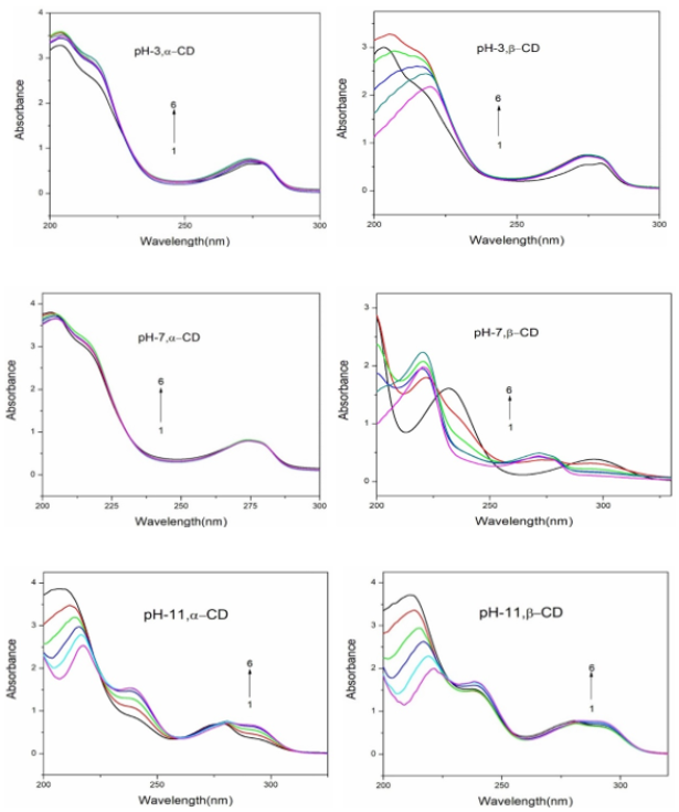
Figure 1. Absorption spectra of 25DMP in different α-CD and β-CD concentrations (M): (1) 0, (2) 0.002, (3) 0.004, (4) 0.006, (5) 0.008 and (6) 0.01.
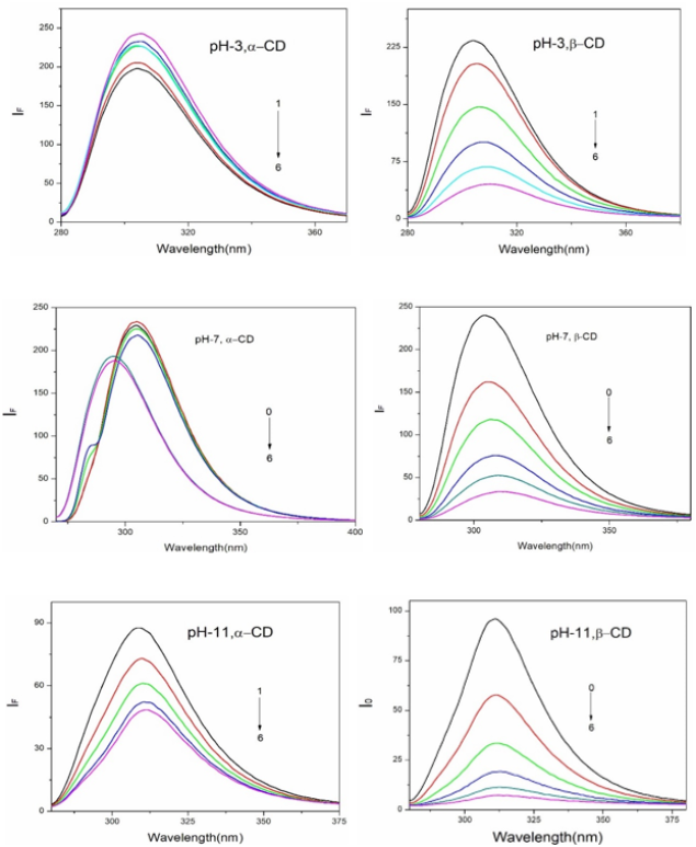
Figure 2. Fluorescence spectra of 25DMP in different α-CD and β-CD concentrations (M): (1) 0, (2) 0.002, (3) 0.004, (4) 0.006, (5) 0.008 and (6) 0.01.
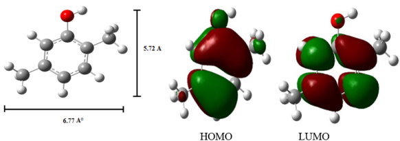
Figure 3. PM3 optimized structures of (a, b) 25DMP, (c, d) HOMO, LUMO of 25DMP.
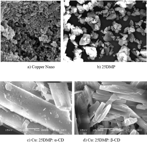
Figure 4. SEM images of (a) 25DMP, (b) Cu: 25DMP: α-CD, (b) Cu: 25DMP: β-CD nano.
Information

