
 ,
Saul Jalpilla,
Carolina Herrera,
Ingrid Cabrera,
Ivonne Lopez,
Fernanda Solares,
Vanessa Jimenez
Antecedents: Objective: Present the electrocardiographic changes secondary to methamphetamine use in an adolescent. Given that this is a pioneer case in the national literature, we consider its dissemination appropriate. Case Report: A 17-year-old obese female adolescent, smoker and methamphetamine (crystal) user, presented with ST-segment elevation (STE) 12 hours after a binge, without accompanying anginal equivalents. With the application of anti-ischemic and antithrombotic measures, improvement of the STE was observed on the follow-up electrocardiogram (ECG) 24 hours later. The patient did not develop cardiogenic shock or arrhythmias. Thrombolysis was not performed because she did not develop pathological Q waves. Total creatine phosphokinase (CPK) and creatine phosphokinase- MB (CPK-MB) levels were elevated, due to rhabdomyolysis. Conclusions: ST-segment elevation in methamphetamine users has been termed benign coronary spasm by some authors; however, due to the risk of thromboinflammation at the glycocalyx level, the problem could out-come obstructive. There is no consensus regarding treatment in pediatric patients. Treatment in adults prioritizes vasodilation and antithrombotic measures, especially in presence of cardiovascular risk factors such as obesity and smoking.
| Published in | Cardiology and Cardiovascular Research (Volume 10, Issue 4) |
| DOI | 10.11648/j.ccr.20261004.11 |
| Page(s) | 106-111 |
| Creative Commons |
This is an Open Access article, distributed under the terms of the Creative Commons Attribution 4.0 International License (http://creativecommons.org/licenses/by/4.0/), which permits unrestricted use, distribution and reproduction in any medium or format, provided the original work is properly cited. |
| Copyright |
Copyright © The Author(s), 2026. Published by Science Publishing Group |
ST-segment Elevation, Methamphetamines, Electrocardiogram, Thromboinflammation
STE | ST-segment Elevation |
ECG | Electrocardiogram |
CPK | Creatine Phosphokinase |
CPK-MB | Creatine Phosphokinase-MB |
SISVEA | Epidemiological Surveillance System for Addictions |
TAAR1 | Trace Amine-associated Receptor |
LSD | Lysergic Acid Diethylamide |
BMI | Body Mass Index |
BP | Blood Pressure |
HR | Heart Rate |
RR | Respiratory Rate |
SpO2 | Oxygen Saturation |
| [1] |
Secretaría de Salud, Direccion General de Epidemiología. (2026). Informe SISVEA 2024: Sistema de Vigilancia Epidemiologica de las Adicciones. Gobierno de Mexico.
https://www.gob.mx/cms/uploads/attachment/file/1053432/Informe_SISVEA2024.pdf |
| [2] | Uhlmann S, DeBeck K, Simo A, Kerr T, Montaner JS, Wood E. Crystal methamphetamine initiation among street-involved youth. J Psychoactive Drugs. 2014; 46(3): 198-207. |
| [3] | Paratz E, Cunningham N, MacIsaac A The Cardiac Complications of Methamphetamines Heart, Lung and Circulation, 2015; 25, 325-332Miranda CH, Schmidt A, Filho AP, Borges. |
| [4] | Kevil CG, Goeders NE, Woolard MD, Bhuiyan MS, Dominic P, Kolluru GK, et al. Methamphetamine use and cardiovascular disease. Arterioscler Thromb Vasc Biol. 2019; 39(9): 1739-46. |
| [5] | Chen J. P. (2007). Methamphetamine-associated acute myocardial infarction and cardiogenic shock with normal coronary arteries: refractory global coronary microvascular spasm. The Journal of invasive cardiology, 19(4), E89-E92. |
| [6] | Turnipseed, S. D., Richards, J. R., Kirk, J. D., Diercks, D. B., & Amsterdam, E. A. (2003). Frequency of acute coronary syndrome in patients presenting to the emergency department with chest pain after methamphetamine use. The Journal of emergency medicine, 24(4), 369-373. |
| [7] | Fuenzaiida A, Valdebenito M, Fajuri A. Acute Myocardial infarction associated with cocaine use: a different entity? Chilean Journal of Cardiology. 2014; 33(2): 123-126. |
| [8] | Schussler JM, Tomson C, Dresselhouse MP. Extreme Hyperthermia From Methamphetamine Toxicity Presenting as ST-Elevation Myocardial Infarction on Electrocardiogram: A Case Report Written With ChatGPT Assistance. Cureus. 2023 Mar 13; 15(3): e36101. |
| [9] | Miranda, C. H., Schmidt, A., Filho, A. P., & Borges, M. C. (2015). Endothelial glycocalyx damage in patients with acute coronary syndrome. Critical Care, 19(Suppl 2), P4. |
| [10] | Paratz ED, Zhao J, Sherwen AK, Scarlato RM, MacIsaac AI. Is an abnormal ECG just the tip of the ICE-berg? Examining the utility of electrocardiography in detecting methamphetamine-induced cardiac pathology. Heart Lung Circ. 2017 Jul; 26(7): 684-9. |
| [11] | Quintero-Aguirre, E., Balbuena-Robles, L. H., Barrera-Villegas, G., & Serna, D. (2015). Dolor precordial en los adolescentes y diagnostico diferencial de la onda T. Revista Mexicana de Pediatría, 82(1), 14-17. |
| [12] | El Menyar A. A. (2006). Drug-induced myocardial infarction secondary to coronary artery spasm in teenagers and young adults. Journal of postgraduate medicine, 52(1), 51-56. |
| [13] | Towbin J, Bricker J, Garson A Electrocardiographic criteria for diagnosis of acute myocardial infarction in childhood American Journal of Cardiology, 69, 1545-1548. |
| [14] |
Machado Villarroel, L., & Olmos Granados, H. M. (2022). Marcadores Biologicos en el diagnostico del infarto agudo al miocardio. Expresiones Medicas, 9(3), 7-13. Recuperado a partir de.
https://erevistas.uacj.mx/ojs/index.php/expemed/article/view/5368 |
| [15] | Maruyama, M., Kobayashi, Y., Kodani, E., Hirayama, Y., Atarashi, H., Katoh, T., & Takano, T. (2004). Osborn waves: History and significance. Japanese Heart Journal, 45(4), 505-511. |
| [16] | Iba T, Maier CL, Helms J, Ferrer R, Thachil J, Levy JH. Managing sepsis and septic shock in an endothelial glycocalyx-friendly way: from the viewpoint of surviving sepsis campaign guidelines. Ann Intensive Care. 2024; 14: 64. |
| [17] | Nehus, E., & Mitsnefes, M. (2019). Childhood Obesity and the Metabolic Syndrome. Pediatric clinics of North America, 66(1), 31-43. |
| [18] | Franks, P. W., Hanson, R. L., Knowler, W. C., Sievers, M. L., Bennett, P. H., & Looker, H. C. (2010). Childhood obesity, other cardiovascular risk factors, and premature death. The New England journal of medicine, 362(6), 485-493. |
| [19] | Suryawanshi, S. P., Das, B., & Patnaik, A. N. (2011). Myocardial infarction in children: Two interesting cases. Annals of pediatric cardiology, 4(1), 81-83. |
| [20] | Akhter MW, Aronow WS, Rashid M, Fredrick T, Campbell M. Amphetamine-related acute myocardial infarction requiring cardiac transplant. Int J Clin Pract. 2003 Jan; 57(1): 9-1. |
| [21] | Yeo KK, Mooney J, Wyrick K, Amsterdam EA. Methamphetamine-associated acute coronary syndrome: spasm versus thrombosis. Int J Cardiol. 2007 Apr 4; 121(1): e3-e4. |
| [22] | Khaheshi I, Mahjoob MP, Esmaeeli S, Eslami V, Haybar H. Simultaneous Thrombosis of the Left Anterior Descending Artery and the Right Coronary Artery in a 34-Year-Old Crystal Methamphetamine Abuser. Korean Circ J. 2015 Mar; 45(2): 158-160. |
| [23] | Westover, A. N., Nakonezny, P. A., & Haley, R. W. (2008). Acute myocardial infarction in young adults who abuse amphetamines. Drug and alcohol dependence, 96(1-2), 49-56. |
| [24] | Khattab, E., & Shujaa, A. (2013). Amphetamine abuse and acute thrombosis of left circumflex coronary artery. International Journal of Case Reports and Images, 4(12), 698-701. |
| [25] | Abdullah, C. S., Aishwarya, R., Alam, S., Morshed, M., Remex, N. S., Nitu, S., Kolluru, G. K., Traylor, J., Miriyala, S., Panchatcharam, M., Hartman, B., King, J., Bhuiyan, M. A. N., Chandran, S., Woolard, M. D., Yu, X., Goeders, N. E., Dominic, P., Arnold, C. L., Stokes, K.,. Bhuiyan, M. S. (2020). Methamphetamine induces cardiomyopathy by Sigmar1 inhibition-dependent impairment of mitochondrial dynamics and function. Communications biology, 3(1), 682. |
APA Style
Quintero, E., Jalpilla, S., Herrera, C., Cabrera, I., Lopez, I., et al. (2026). ST-Segment Elevation in Methamphetamine-Users Adolescents: An Emerging Treatment Paradigm, Case Report. Cardiology and Cardiovascular Research, 10(4), 106-111. https://doi.org/10.11648/j.ccr.20261004.11
ACS Style
Quintero, E.; Jalpilla, S.; Herrera, C.; Cabrera, I.; Lopez, I., et al. ST-Segment Elevation in Methamphetamine-Users Adolescents: An Emerging Treatment Paradigm, Case Report. Cardiol. Cardiovasc. Res. 2026, 10(4), 106-111. doi: 10.11648/j.ccr.20261004.11
@article{10.11648/j.ccr.20261004.11,
  author = {Eduardo Quintero and Saul Jalpilla and Carolina Herrera and Ingrid Cabrera and Ivonne Lopez and Fernanda Solares and Vanessa Jimenez},
  title = {ST-Segment Elevation in Methamphetamine-Users Adolescents: An Emerging Treatment Paradigm, Case Report},
  journal = {Cardiology and Cardiovascular Research},
  volume = {10},
  number = {4},
  pages = {106-111},
  doi = {10.11648/j.ccr.20261004.11},
  url = {https://doi.org/10.11648/j.ccr.20261004.11},
  eprint = {https://article.sciencepublishinggroup.com/pdf/10.11648.j.ccr.20261004.11},
  abstract = {Antecedents: Objective: Present the electrocardiographic changes secondary to methamphetamine use in an adolescent. Given that this is a pioneer case in the national literature, we consider its dissemination appropriate. Case Report: A 17-year-old obese female adolescent, smoker and methamphetamine (crystal) user, presented with ST-segment elevation (STE) 12 hours after a binge, without accompanying anginal equivalents. With the application of anti-ischemic and antithrombotic measures, improvement of the STE was observed on the follow-up electrocardiogram (ECG) 24 hours later. The patient did not develop cardiogenic shock or arrhythmias. Thrombolysis was not performed because she did not develop pathological Q waves. Total creatine phosphokinase (CPK) and creatine phosphokinase- MB (CPK-MB) levels were elevated, due to rhabdomyolysis. Conclusions: ST-segment elevation in methamphetamine users has been termed benign coronary spasm by some authors; however, due to the risk of thromboinflammation at the glycocalyx level, the problem could out-come obstructive. There is no consensus regarding treatment in pediatric patients. Treatment in adults prioritizes vasodilation and antithrombotic measures, especially in presence of cardiovascular risk factors such as obesity and smoking.},
 year = {2026}
}
TY  - JOUR
T1  - ST-Segment Elevation in Methamphetamine-Users Adolescents: An Emerging Treatment Paradigm, Case Report
AU  - Eduardo Quintero
AU  - Saul Jalpilla
AU  - Carolina Herrera
AU  - Ingrid Cabrera
AU  - Ivonne Lopez
AU  - Fernanda Solares
AU  - Vanessa Jimenez
Y1  - 2026/10/20
PY  - 2026
N1  - https://doi.org/10.11648/j.ccr.20261004.11
DO  - 10.11648/j.ccr.20261004.11
T2  - Cardiology and Cardiovascular Research
JF  - Cardiology and Cardiovascular Research
JO  - Cardiology and Cardiovascular Research
SP  - 106
EP  - 111
PB  - Science Publishing Group
SN  - 2578-8914
UR  - https://doi.org/10.11648/j.ccr.20261004.11
AB  - Antecedents: Objective: Present the electrocardiographic changes secondary to methamphetamine use in an adolescent. Given that this is a pioneer case in the national literature, we consider its dissemination appropriate. Case Report: A 17-year-old obese female adolescent, smoker and methamphetamine (crystal) user, presented with ST-segment elevation (STE) 12 hours after a binge, without accompanying anginal equivalents. With the application of anti-ischemic and antithrombotic measures, improvement of the STE was observed on the follow-up electrocardiogram (ECG) 24 hours later. The patient did not develop cardiogenic shock or arrhythmias. Thrombolysis was not performed because she did not develop pathological Q waves. Total creatine phosphokinase (CPK) and creatine phosphokinase- MB (CPK-MB) levels were elevated, due to rhabdomyolysis. Conclusions: ST-segment elevation in methamphetamine users has been termed benign coronary spasm by some authors; however, due to the risk of thromboinflammation at the glycocalyx level, the problem could out-come obstructive. There is no consensus regarding treatment in pediatric patients. Treatment in adults prioritizes vasodilation and antithrombotic measures, especially in presence of cardiovascular risk factors such as obesity and smoking.
VL  - 10
IS  - 4
ER  -
Pediatric Emergency Department, Xochimilco Maternal and Pediatric Hospital, Mexican Social Security Institute, Mexico City, Mexico
Pediatric Emergency Department, General Hospital of Zone 47, Mexican Social Security Institute, Mexico City, Mexico
Pediatric Emergency Department, General Hospital of Zone 47, Mexican Social Security Institute, Mexico City, Mexico
Faculty of Higher Studies Zaragoza, National Autonomous University of Mexico (UNAM), Mexico City, Mexico
Pediatric Emergency Department, Xochimilco Maternal and Pediatric Hospital, Mexican Social Security Institute, Mexico City, Mexico
Faculty of Medicine, National Autonomous University of Mexico (UNAM), Mexico City
Faculty of Medicine, National Autonomous University of Mexico (UNAM), Mexico City
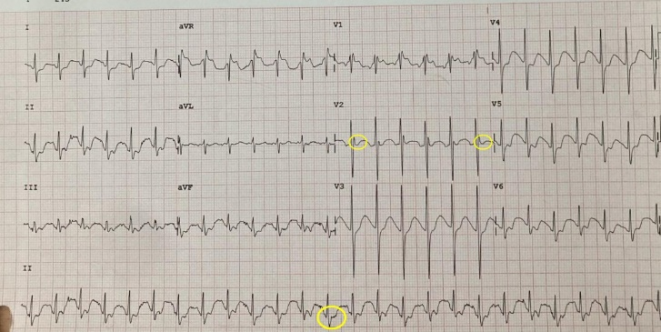
Figure 1. First electrocardiogram (June 7, 2023). Significant ST-segment elevation in the inferior leads (I, II, III, and aVF) and in leads V1-V6, with no Q waves. Sinus rhythm with an average ventricular rate of 125 beats/min and a prolonged QTc interval (0.578 seconds). An Osborn J wave is observed in lead II.
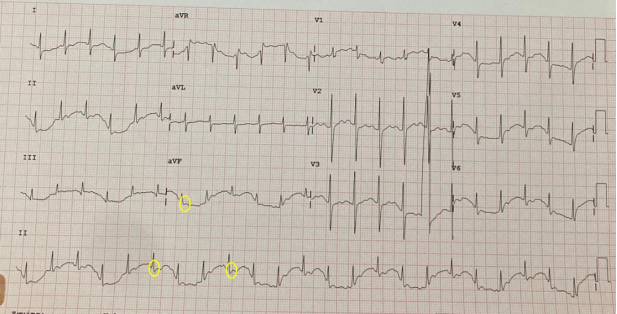
Figure 2. Follow-up electrocardiogram at 24 hours. A decrease in ST-segment elevation in DI and inferior leads were observed, with persistent QTc prolongation and no Q waves.

Information


 Share This Article
 Twitter
 Linked In
 Facebook

 Copy
 Download