Review Article | | Peer-Reviewed

Role of Chemistry in Determining Time Since Death

Received: 12 September 2026     Accepted: 4 October 2026     Published: 26 October 2026
Views:       Downloads:
Abstract

Accurate post‑mortem interval (PMI) estimation is fundamental to forensic science but traditional macroscopic indicators rapidly lose reliability. This review explains how chemical and molecular techniques are redefining PMI estimation. Early work focused on measuring electrolytes in vitreous humor (VH); the linear increase of potassium following cellular membrane breakdown remains a robust marker. Other metabolites such as creatinine, urea, glucose and hypoxanthine in VH, blood and synovial fluid extend the estimation window. Post‑mortem biochemical processes are also summarised, autolysis and putrefaction degrade macromolecules, produce volatile organic compounds and gases, and alter pH and redox potential, forming biochemical clocks. Biological matrices including VH, blood, cerebrospinal fluid, urine, muscle, liver and brain each provide different advantages and challenges. The review surveys tissues like muscle, liver and brain and describes how electrolyte leakage, metabolite accumulation and protein degradation offer complementary time markers. Key markers encompass electrolytes, metabolites, enzyme activities, protein degradation patterns, microRNAs and volatile organic compounds. Analytical tools such as gas/liquid chromatography, mass spectrometry, NMR spectroscopy and ion‑selective electrodes enable sensitive measurement. Emerging multi‑omics and spectroscopic techniques (ATR‑FTIR, Raman) integrated with machine‑learning models enhance resolution and show promise for multi‑organ metabolomics and proteomics. The review also notes limitations: environmental variability, inter‑individual differences, drugs and ethical constraints necessitate multidisciplinary approaches. Future research directions stress multi‑marker models, entomology integration, portable devices and population‑specific databases.

Published in Science Journal of Analytical Chemistry (Volume 14, Issue 3)
DOI 10.11648/j.sjac.20261403.12
Page(s) 45-60
Creative Commons

This is an Open Access article, distributed under the terms of the Creative Commons Attribution 4.0 International License (http://creativecommons.org/licenses/by/4.0/), which permits unrestricted use, distribution and reproduction in any medium or format, provided the original work is properly cited.

Copyright

Copyright © The Author(s), 2026. Published by Science Publishing Group

Keywords

Post Mortem Interval, Algor Mortis, Rigor Mortis, Livor Mortis, Chemistry in Death

1. Introduction
Determining the post‑mortem interval (PMI), the time between death and discovery, is central to forensic investigations. Accurate estimates guide investigations, aid identification and reconstruct events. Historically they relied on macroscopic observations. Cooling of the body (algor mortis), muscle stiffening (rigor mortis) and gravitational pooling of blood (livor mortis) were the main tools; however, they are heavily affected by ambient temperature, humidity, clothing and individual variation. Their usefulness is confined to the first 24-72 hours, after which the precision declines markedly, motivating the search for more objective methods .
This review recounts the shift from physical indicators to biochemical and chemical markers. Early studies measured electrolytes in vitreous humor; after death, cell membranes become permeable and potassium leaks into the vitreous fluid; the resulting linear increase provides a robust marker for PMI. Other metabolites such as creatinine, urea and glucose in vitreous humor and synovial fluid further reduce subjectivity. Metabolomics, machine‑learning and multivariate techniques are now being applied to tissues and fluids to process complex data and improve precision. Spectroscopic methods like attenuated total reflectance Fourier‑transform infrared (ATR‑FTIR) and Raman spectroscopy show promise for estimating PMI from skeletal remains .
The chemistry of decomposition, autolysis and putrefaction induce macromolecular breakdown, releasing sugars, free fatty acids, amino acids, volatile organic compounds and gases, while altering pH and redox potential is also summarized. These changes create biochemical clocks in tissues and fluids. Such chemical approaches are now central to forensic practice. By emphasising the role of chemistry, it was shown that how modern instrumentation such as mass spectrometry and nuclear magnetic resonance can translate biochemical changes into forensic timelines. The review outlines how different matrices, including vitreous humor, blood, cerebrospinal fluid, urine, muscles, liver and brain, offer distinct advantages and limitations for chemical PMI estimation. It sets out to survey key markers, analytical methods, limitations and future directions, demonstrating that forensic chemistry provides the objective backbone for modern PMI estimation and opens avenues for more reproducible timelines.
2. Historical Evolution of PMI Determination
The history of Postmortem Interval (PMI) determination has shifted its focus to earlier non-chemical measurements to advanced chemical and molecular techniques that have been able to improve the accuracy and validity of the forensic investigation considerably . Previously, PMI estimation was based on macroscopic physical alterations of the body, namely, rigor mortis, and livor mortis . The framework of predictability in a body temperature decline after death was involved in the use of algor mortis which means the cooling of a body . Nevertheless, its accuracy is highly compromised by multiple physical conditions, including environmental temperature, humidity, and the existence of clothing, may cause significant change in the rate of cooling and create large error margins . As an example, a basic cooling equation does not take into consideration complicated environmental conditions or internal temperature gradients in the body so that a basic temperature measurement will not give accurate PMI estimates.
The inflexibility of muscle is known as rigor mortis, which is the result of the biochemical alterations taking place inside muscle fibers . Although it has a general pattern, its initiation, course and lifespan depend on factors like the age of the person, physical status, cause of death, and conditions of the environment among others, which causes a lot of variation. Livor mortis, which is the gravitational accumulation of blood in the dependent body parts, is used to give information about the position of the body after death . However, as influenced in its development and fixation by the external and individual factors, as its general degree of reliability is limited, and it is difficult to distinguish between the blanchable and fixed lividity during the initial stages. These conventional approaches, as well as fundamental, are usually regarded to be effective only during the first 24 to 72 hours after death and their precision decreases significantly as the process of decomposition progresses .
A breakthrough in PMI determination involved the development of biochemical and chemical markers, which provided a more objective and repeatable analysis, due to the introduction of chemical methods . Initial studies were done on the fluctuations in electrolyte levels, especially the concentration of potassium in the vitreous humor. Vitreous humor is a quite valuable matrix because of its relative isolation and more predictable a postmortem alteration than blood. Once dead, cells membranes become permeable and the cell loses its ions and other molecules such as potassium into the fluid that surrounds the cell such as the vitreous humor. This rising trend in potassium concentration during the early postmortem period has an extremely linear association with time that elapsed after death, which implies that PMI can be estimated by simple computation. These analyses are not the only ones that have been investigated to be useful in early PMI estimation; other chemical analyses were also studied, including creatinine, urea, glucose in vitreous humor among others . These are the chemical methods that decrease the subjectivity of the physical observations and enhance the reproducibility of the estimations of PMI by furnishing quantifiable data . The study of other body fluids such as synovial fluid has also widened the scope of research to examine different types of biochemical changes to determine early PMI . Combining these biochemical indicators with the newer forensic techniques enables a deeper appreciation of the postmortem alterations and aids in explaining a portion of the variability observed with other non-chemical techniques .
New methods have also been introduced, such as application of attenuated total reflectance fourier transform infrared (ATR-FTIR) spectroscopy to monitor chemical changes in textile contaminated with emollients, but this is measurement of fire danger, not direct measurement of PMI . Nevertheless, PMI estimation on the basis of puparia of sarcosaprophagous flies has also been done using ATR-FTIR, in which chemical changes with time are analyzed . In the general context, such methods as Raman spectroscopy and multivariate data analysis show some potential in estimating PMI using skeletal remains, but both internal and external factors continue to affect the outcome . The rising use of machine learning algorithms and metabolomics that works to study the biochemical alterations of organs and tissues after death is a major advance toward more measurable and precise PMI prediction and not limited to the limitations of older methods .
3. Post-Mortem Chemical Changes in the Human Body
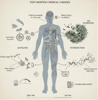
When the life of a human being ceases, the body constantly experiences a complex and irreversible process of post-mortem chemical transformations, starting with the immediate halting of metabolism and oxygen provision, and causing a breakdown of cellular homeostasis and the irreversible transition to equilibrium . It starts with autolysis, which is the self-digestion of the cells enabled by the extrusion of endogenous hydrolytic enzymes, mostly lysosomal, that breaks down the components of the cells . At the same time, putrefaction begins, which is associated with the multiplication and metabolism of microorganisms, and primarily, gut microbiota, which further enhances tissue decomposition by breaking down organic matter . All these enzymatic and microbial processes make tissues extremely chemically unstable after death, having a major effect on the further decomposition process .
The chemical pathway of decomposition consists of degradation of the basic macromolecules. Theological carbohydrates are broken into simpler sugars, lipids are broken down and oxidized, and this leads to the release of free fatty acids and other VOCs . Proteins are broken down into peptides and amino acids that are further deaminated to create ammonia and other nitrogenous products . Such metabolic changes in both intrinsic enzymes and microbial communities produce a variety of gases, such as methane, hydrogen sulfide, and carbon dioxide, which cause the typical bloating and smell of decomposition . Moreover, the development of acidic products of anaerobic activity and microbial activity results in the appearance of substantial changes in the tissue pH, whereas the redox potential of the internal environment is changed as the result of oxidative and reductive reactions . These biochemical changes are important because they can give quantifiable values of the PMI, which is the time interval that passed since death . As an illustration, the postmortem alterations of metabolites in blood and muscle tissue have been examined to approximate PMI, particularly when addressing problematic situations such as submersion . The dynamics of molecular damage of different molecules, including lncRNAs, in the brain tissue have also demonstrated the possibility of predicting the early PMI . Relative isolation of the vitreous humor makes it especially useful in measuring biochemical parameters of the body that are stable over time, such as potassium and glucose concentrations . PMI can also be estimated with greater accuracy, depending on the environmental temperature that causes a significant effect on the rate of decomposition and drugs or toxins that can modify microbial profiles and decomposing interactions . Volatile compounds measurement, or, in other words the volaboloma mortis, is a new physiological fingerprinting method of the decomposition process to estimate PMI . Newer post-mortem changes, such as metabolomics and proteomics, are being developed with the help of emerging omics technologies, which offer comprehensive biochemical adaptations, allowing the discovery of new biomarkers and enhancing the predictiveness of PMI . As an example, the degradation of skeletal muscle tissue proteins has been suggested as a promising molecular intervention, and a number of different proteinsensuously degrade over time . Moreover, research on skeletal muscle tissue stable nitrogen isotope ratio is investigating how it can be used to estimate PMI when putrefaction occurs . This is essential in investigating murder cases, as the methods of determining the time of death can be more accurate than the traditional ones that are environmentally delicate due to their reliance on time and place .
Figure 1. Post-mortem chemical changes in the human body.
After death, loss of oxygen and metabolism triggers autolysis (enzymatic self-digestion) and putrefaction (microbial decomposition). These processes lead to degradation of carbohydrates, lipids, and proteins into simple sugars, fatty acids, VOCs, peptides, amino acids, and nitrogenous compounds, accompanied by gas production, pH shifts, and redox changes. Tissue- and fluid-specific biomarkers, such as potassium and glucose in vitreous humor, brain lncRNAs, skeletal muscle proteins, and volatile compounds (volaboloma mortis), change predictably from early to late post-mortem intervals and can be used to estimate the PMI.
4. Biological Fluids and Tissues Used in Chemical PMI Estimation
The determination of the PMI, which is the duration by which the body has been dead but has not been found or analyzed, is a difficult and important role of forensic science, and modern fields such as thanatochemistry and the “omics” sciences have offered promising solutions to better estimate this time . The postmortem biochemical changes in various biological fluids and tissues are studied and provide different benefits and drawbacks to identify the PMI .
VH often has found favor in the estimation of PMI because it is anatomically isolated, and is largely resistant to postmortem contamination and degradation compared to other biological fluids . This is a stable fluid that is mostly avascular, and whose microbial activity and fast postmortem degradation are less susceptible, which makes it a good matrix of forensic examination . Once cells become damaged after death, ions and molecules that were contained within cells start leaking into fluids around them such as VH . Potassium (K+) and hypoxanthine (Hx) are among the most consistently studied VH markers to predict PMI . The increase in potassium during VH postmortem is remarkably linear, with the intracellular potassium leakage of the membrane occurring because of cellular breakdown. This consistent change is what renders K+ a highly applicable biomarker to determine the early PMI . Another potential indicator of PMI is hypoxanthine which is also present in VH due to adenosine triphosphate (ATP) breakdown . Beta-hydroxybutyrate (BHB) is also an example of other biochemical markers that have been examined in VH particularly concerning ketoacidosis . VH also serves in toxicological studies, in situations where blood or urine samples are lost or destroyed, or in postmortem studies of redistribution effects on concentrations of drugs . Research indicates its applicability to both measuring drugs such as quetiapine and its metabolites and in gamma-hydroxybutyrate (GHB), which may have postmortem increases in other matrices . The studies are still ongoing to identify new portable devices that can be used to conduct VH analysis to enable quick estimation of PMI on-site .
Although readily available, blood and serum pose some specific difficulties in terms of PMI estimation, in the first place, because of the phenomenon of postmortem redistribution (PMR) . PMR is a process that causes the passivity of drugs and endogenous substances in the body when the circulation process stops, resulting in artificial increases or decreases in concentration in various sites of the sample, e.g., central, versus peripheral blood. High volume of distribution lipophilic and basic drugs are especially vulnerable to PMR, which accumulates in the tissues during life, and then releases into the blood vessels near the body after death . In spite of these limitations, blood and serum are still valuable sources of information. As an example, postmortem unknown alterations in human serum metabolomic profiles have been examined in order to find promising biomarkers that can be used to monitor PMI . Also, such markers as creatine kinase (CK-Total, CK-MB), urea, creatinine, and uric acid in serum are correlated with PMI . Bloodstain analysis to determine the age of a bloodstain is also problematic as the anticoagulants added to samples as reference specimen may inhibit the clotting effect of the blood which is an unnatural inhibition of coagulation that may be used in forensic examination .
In addition to cerebrospinal fluid (CSF), urine is also used in PMI estimation, which can provide other information about the changes that occurred after death . CSF, like VH, is comparatively resistant to speedy postmortem decomposition and contamination, which offer a more constant environment to some biomarkers than blood . The biomarkers of brain injury such as glial fibrillary acidic protein (GFAP), neuron-specific enolase (NSE), and S100B have been studied in both CSF and VH as a predictor of PMI, with its use still being debated but promising . Urine is an excretory product, which may indicate the status of the metabolic processes before death and is not usually subject to redistribution that occurs after death as much as blood. Exoglycosidase activity (e.g. α-mannosidase, 2-galactosidase) in CSF and urine has been investigated as possible indicators in patients who had died of ethyl alcohol poisoning .
Tissue-specific chemical alterations in solid tissues like muscle, liver, and brain are actively studied and their application in new methods of analysis to estimate PMI is developing . These tissues go through very intricate processes of degradation after death, such as protein degradation and alterations in mRNA expression pattern . Protein degradation of skeletal muscles has become a promising biochemical biomarker of PMI, and extensive reviews indicate that proteomic technology can be used to identify reliable biomarkers . Postmortem production of ethanol and alcohol congeners have been investigated in the liver and muscle tissues and they can change considerably with time . Multi-organ samples are also being studied using metabolomics alongside machine learning algorithms to get more accurate estimations of PMI across different ambient temperatures such as liver and muscle samples . There are also postmortem changes in the brain tissue, and research has been conducted to determine the mRNA expression and patterns of long non-coding RNA (lncRNA) degradation to estimate early PMI especially in rat models . As an example, miR-122, miR-133a, and miR-206 have been denoted as possible miRNA biomarkers to estimate PMI in the brain tissue . Gastrocnemius muscle was applied in the context of metabolomics to estimate the duration of postmortem submersion, and it can be utilized in the absence of blood samples in late stages of decomposition, which is an alternative . They have also checked the myoglobin level in blood, liver, and parotid glands of albino rats to establish its association with PMI . These tissues are also affected by the dynamicity of drug distribution after death, as is observed in the postmortem distribution of substances, e.g., ropivacaine and its metabolites, in different body fluids and solid tissues of humans .
5. Key Chemical Markers Used in PMI Estimation
The correct determination of the PMI is an important but difficult task of forensic investigations, which influences the reconstruction of the events and the court process . The classical techniques that are based on macroscopic alterations such as rigor mortis, livor mortis, and algor mortis can be deemed as useful within the first 48-72 hours after death and are extremely sensitive to the environmental factors . Therefore, more sophisticated thanatochemistry and molecular methods have become potential solutions to improve PMI estimation through biochemical modification in different tissues and bodily fluids .
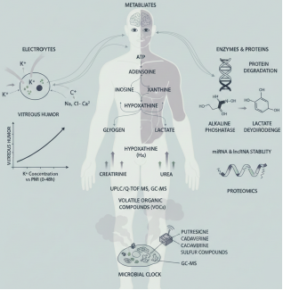
Electrolytes, especially K+, are a long-established gold standard in PMI estimation because they can be used to predict post-mortem diffusion . Once the cell becomes dead, the termination of active cellular transport aids in cell membrane degradation, and potassium, which is relatively abundant intracellularly, is forced to leave the cell and enter extracellular fluids, in particular, VH . This leakage causes an exceptionally linear rise in K+ concentration in the VH throughout the initial post-mortem duration, and it can be easily calculated to approximate PMI . Post-mortem changes occur in other electrolytes too, such as sodium, chloride, and calcium, but their applicability in PMI estimation is regarded as less reliable compared to potassium . The fact that VH is anatomically isolated and has strong properties reduces the chances of contamination and external interference, which makes it particularly helpful in this analysis .
Another important type of chemical markers that can be used to estimate PMI is metabolites, since their accumulation dynamics can be used to represent the current biochemical processes taking place after death . Adequately anaerobic to be degraded, Hx, a product of ATP breakdown, is accumulated over time postmortem, and therefore becomes a promising marker across a wide range of tissues, such as muscle and VH . It has been demonstrated to have a great potential in measuring PMI, commonly together with other purine bases, such as adenosine, inosine, and xanthine . Another important metabolite Lactate rapidly accumulates during the early post-mortem or early anaerobic glycolysis in tissues, particularly muscle . This initial accumulation is then succeeded by a more intricate kinetic profile though it is a good indicator of early PMI . Creatinine and urea, which are usually markers of renal performance in vivo also show post-mortem changes that can be related to PMI. Their concentrations are more likely to increase when renal clearance is stopped, which is a part of the wider metabolic phenotype of decomposition . Metabolomics, which applies the techniques such as ultra-high-performance liquid chromatography-quadrupole time-of-flight mass spectrometry (UPLC/Q-TOF MS), and gas chromatography-mass spectrometry (GC-MS) can completely profile these and thousands of other molecules, enabling more precise PMI estimates, even when conditions become difficult, such as high temperatures or submersion .
When enzymes and proteins began to degrade after death, it is possible to use this pattern to estimate PMI . Time-dependent variations are seen in the loss of enzyme activity (e.g., alkaline phosphatase and lactate dehydrogenase) and protein denaturation (depending on the factors, including temperature and pH), which also adds to the post-mortem biochemical landscape . These protein degradation patterns are increasingly investigated using proteomic methods, such as mass spectrometry, to give information about PMI in even highly advanced decomposition . An example is the work that examines the degradation of skeletal muscle proteins when subjected to controlled conditions to identify the reliable biomarkers . miRNAs and lncRNAs stability have also been explored as possible PMI biomarkers because the molecules are comparatively stable after death despite changes in cells . However, their degradation is affected by environmental factors and differences among individuals, which restricts their use in forensics and requires experimental designs and tracking methods .
VOCs are an important chemical indicator of decomposition, which is a result of microbial activity and biochemical processes of nature . These are the putrescine, cadaverine and other sulfurous compounds, known as the volaboloma mortis, and these give chemical fingerprint that as the decomposition continues undergoes changes . Non-invasive PMI estimation is provided by the analysis of VOCs by such methods as GC-MS, which is also essential to identify cadavers . The environmental factors play an important role in the formation and emission of VOCs, and studies are aimed at knowing how these profiles change over time under different conditions, including indoor settings . Microbial succession that occurs throughout the decomposition process, commonly called the microbial clock, is closely correlated with the production of VOC and has been identified as a promising and potentially more accurate measure of PMI, especially in later stages of decomposition . Microbial profiles may also be affected by the presence of drugs or toxins and cause the patterns of VOC emissions to change, further complicating but potentially informative of PMI determination . Combining these various chemical indicators with new analytical technologies, including multi-omics and machine learning models, constitutes the modern state of the art of redefining the PMI estimation so that it is more accurate and reliable in a forensic practice .
Figure 2. Key chemical markers used in post-mortem interval (PMI) estimation.
Overview of major biochemical indicators applied in PMI assessment, including electrolyte shifts (notably potassium in vitreous humor), post-mortem metabolite changes from ATP degradation (adenosine, inosine, hypoxanthine, xanthine), anaerobic glycolysis products (lactate), renal markers (creatinine, urea), enzyme and protein degradation patterns, RNA stability (miRNA and lncRNA), and volatile organic compounds (VOCs). These markers, analyzed using metabolomics, proteomics, and GC-MS-based approaches, reflect time-dependent biochemical and microbial processes (“microbial clock”) from early to late PMI.
6. Analytical Methods of Chemical PMI Determination
The accurate determination of the PMI is an important part of the forensic examination, and it is based on a wide array of different methods of analysis, including classical biochemist methods as well as advanced instrumental methods, and supported by strict quality control and validation . Although basic, classical chemical and biochemical techniques of PMI determination usually give initial or supportive data. Examples include colorimetric assays, where specific analytes are measured by a visual or spectrophotometric change and the concentration of a substance such as potassium or ammonia in a biological sample can be measured, which can change predictably after death. Ion-selective electrodes provide direct and selective detection of ionic species, i.e. potassium in VH, an index that is known to have post-mortem changes . Spectrophotometry can measure light absorbance/transmittance to determine biochemical markers that are associated with decomposition . These traditional methods tend to be economical and simple and might not be as specific and sensitive as may be needed in complicated forensic cases. To illustrate, a new non-enzymatic colorimetric sensor has been created on the assay of ephedrine in urine samples, and this shows the further development of the techniques .
Sophisticated instrumental methods have played an important role in improving the accuracy, sensitivity and specificity of chemical PMI analysis. The popular separation methods that are commonly used with MS, GC, and LC . GC is especially applicable to volatile and semi-volatile substances, and LC is applicable to non-volatile and thermolabile analytes . As an example, GC-MS is widely applicable in forensic toxicology to establish drug presence, detect specific substances such as synthetic cathinones and phenethylamines in hair, and determine different chemical profiles . It is also used to examine amphetamine chemical profiles through Bayesian predictive model to form connections in drug distribution networks . It is additionally possible to optimize the method to work with problematic analytes, including the nerve agents, by defying the strong retention hindrances on the silica sorbents . Equally, LC-MS, and its ultra-high-performance variants (UHPLC-MS/MS) offers unmatched sensitivity and specificity to complex mixtures to allow the determination of pesticides in food samples, antipsychotic drug residues in wastewater, and a variety of organic pollutants in environmental matrices . LC-MS/MS is viewed as the superior method of quantifying compounds, frequently utilizing stable isotope-labeled internal standards to increase the precision. Recently, a new and convenient method of LC-MS/MS quantification is standardization through infusion with a post column which is especially effective in the case of unstable isotope-label internal standards that are absent or too costly. In addition, the combination of LC with high-resolution MALDI-TOF MS and NMR spectroscopy using automatic full-contact splitless spotting interfaces further simplifies the process of studying in complex matrices of unknowns psychoactive substances . MS, itself is a potent instrument in analytical chemistry, and it allows identifying the molecules and providing structural data to be used in different applications, such as medical diagnostics, food analysis, and monitoring pollutants . MALDI MS, particularly can be utilized in the forensics area because of high specificity and capacity to determine different substances in complex samples without a long-term preparation . The high sensitivity of MeV SIMS is also vital in detecting the synthetic organic colorants with close molecular structures because the molecular structure is of high sensitivity . Another modern method is NMR spectroscopy, which is a non-destructive method of studying the structures and dynamics of molecules, detecting the presence of metabolic changes and products of decomposition correlating with PMI . One example of a system is NMR-based metabolomics used to investigate the chemical diversity of fungal extracts . Moreover, the LC-MS and 1H NMR have been used together to profile the metabolites of botanicals such as pomegranate .
Stringent quality control and validation measures are critical in determining the reliability and admissibility of methods of PMI determination in the field of forensic science. To achieve the consistency and comparability of outcomes between various laboratories and cases, the methods should be standardized . This also involves streamlining of methodologies, e.g., QuEChERS extraction combined with GC-MS to identify pesticides in grape, must and wine samples, which proved to be very linear and had a high recovery rate . The most important one is reproducibility, where the forensic evidence should be subject to intense examination during legal procedures . The approaches that are used in the analysis should be strictly validated in order to prove their accuracy, precision, sensitivity, and specificity. As an illustration, a wider multi-year investigation of pesticides in apples highlights the significance of the validated tool of assessing food quality and risk to human health . Moreover, external quality assessment systems, including the systems that were introduced in Italy to assess classical and new psychoactive drugs in oral fluid, are crucial in monitoring laboratory performance and reliability of the results . The effect of lost electronic fetal monitoring signals on perinatal asphyxia underscores the greater significance of data quality and sound measurements on medical and forensic matters . All in all, dynamic evolution and usage of these complex methods of analysis, as well as their strict validation, is vital in the further evolution of the sphere of chemical PMI determination and gives solid evidence to legal and scientific use.
7. Emerging and Modern Chemical Approaches
The metabolomics and proteomics fields have been greatly enhanced by emergent and modern chemical techniques which allow chemical profiling in large scale and also enhances PMI resolution especially when they are combined with spectroscopic and sensor-based methods and artificial intelligence . The chemical profiling of a large number of small-molecule metabolites and proteins in biological systems is also a characteristic of the modern day metabolomics and proteomics . This method offers a dynamic picture of physiological conditions and is essential to the detection of key biomarkers, the course of disease, and the clarification of treatment procedures . As one example, untargeted metabolomics, frequently using methods such as ultra-high-performance liquid chromatography-quadrupole time-of-flight mass spectrometry (UHPLC-qTOF-MS) can monitor thousands of molecules, providing information on biochemical alterations after death that are necessary to determine PMI correctly . On the same note, proteomic studies examine the degeneration of proteins to determine biomarkers that can be good clues of different forensic variables, particularly in problematic situations such as late PMI determination where soft tissues have fully tore . The dynamic and dynamic character of the proteins-metabolite interactions (PMIs) require high-resolution detection methods and chemoproteomics turned out to be an effective approach to capture and characterize the interactions with a resolution of proteome-wide nature .
The better PMI resolution of these enhanced methods is essential to medico-legal investigation to overcome the shortcomings of the conventional techniques, which are often unsuitable, particularly at long distance or when exposed to diverse environmental conditions . Metabolomic methods, including UHPLC-qTOF-MS, have been utilized to examine alterations of postmortem rat tissues and have demonstrated thousands of molecules, which can be used to estimate PMI with a high level of accuracy within the initial 4 days of death . Multi-organ metabolomics have also been studied to perform more accurate PMI estimation in various ambient temperatures, and it has been proven that machine-learning algorithms can deliver more accurate results compared to the use of a concrete change of substances . The ability of proteomic techniques to detect the important proteins and their degradative profiles can estimate late PMIs, even in human bones more than 15 years old, where pilot studies combining proteomics and machine learning have demonstrated this . It is noted that the systematic review of the sciences of omics indicates their capacity to give a comprehensive perspective of postmortem biochemical alterations, thus being able to defeat the flaws of the conventional approaches, especially when dealing with advanced stages of decomposition .
In this development, spectroscopic and sensor-based techniques are important. PMI estimation with machine learning and Infrared (IR) spectroscopy, specifically Attenuated Total Reflectance fourier Transform Infrared (ATR-FTIR) spectroscopy, has been applied to human skin, which presents a new, easy, precise, and dependable approach to address the issues in forensic pathology, even in the later stages of decomposition . As a label-free and non-destructive chemical analysis method, Raman spectroscopy is essential in studying the molecular compositions and can be accompanied by the sophisticated computational software such as RamanSPy to analyze the data . The two techniques IR and Raman spectroscopy are complementary, which means that they investigate molecular vibrations and also give chemical fingerprints. These methods produce large datasets, which need advanced analytical tools . Also, biosensors are designed to be used in quick and selective analysis, including new glutathione (GSH)-activable theranostic probes selectively to image and treat tumor cells . The quantitative determination of extracellular hydrogen peroxide (H2O2) released by single cells at a high spatial resolution has been performed with scanning electrochemical microscopy (SECM). These sensor-based methods provide high-specificity and sensitivity real-time data and can be invaluable in numerous biological and forensic studies . Moreover, in combination with machine learning and feature selection, near-infrared (NIR) hyperspectral imaging can be applied to identify adulteration in extra virgin olive oil, which proves its ability to identify complex chemicals. Near-infrared spectroscopy combined with chemometrics has been also found to be useful in the determination of rice mildew by simple and quick assays.
The introduction of artificial intelligence (AI) is a revolutionary feature of such new solutions, in particular, machine learning (ML) and chemometric models . AI is more effective in detecting, segmenting, and resolving data, and convolutional neural networks (CNNs) are effective at analyzing complex data . AI and ML play a central role in metabolomics to process large volumes of data in order to analyze it further to identify the peaks, a disease, annotate the metabolites, and integrate multi-omics . To enhance PMI estimation, machine learning is more often applied using metabolomics data on different types of tissues to enhance accuracy and objectivity . The machine learning framework to be used in real-time estimation of PMI combines the multimodal sensor data to address the limitation of conventional methods, particularly in field situations. To analyze enormous volumes of data produced by spectroscopic methods such as NIR spectroscopy, chemometric models are needed, based on the application of statistical and mathematical procedures to chemical data . The models, which are augmented with AI, make the data analysis more accurate and efficient. As an example, the AI models have been trained to forecast antioxidant activity in gentian root via FT-IR spectroscopy and chemometrics, which means that it would have been time-consuming with traditional techniques . PMI estimation with pathology images and deep learning is also developed with AI models which are further refined under different temperatures . The AI combined with the analytical methods have a tremendous potential to revolutionize the course of decisions and lead to innovative development in multiple industries . It is the synergy of metabolomics and proteomics with more advanced methods of spectroscopy and AI that is a significant paradigm shift in scientific research and forensic analysis and practice to become more precise, objective and efficient .
8. Limitations and Challenges of Chemical PMI Estimation
The calculation of the PMI with chemical markers poses a great challenge to forensic investigation because of a combination of factors, most of them being environmental, inter-individual differences, drug, trauma, and disease confounding, and inherent ethical and sampling constraints. Such complexities require that a strong multidisciplinary strategy is employed in order to enhance accuracy .
Biochemical postmortem alterations and their rate are greatly affected by environmental conditions, especially temperature and humidity, which affects the accuracy of chemical PMI estimations. As an example, increased temperatures can speed up the process of decomposition, enzymatic, and microbial activity, which is followed by faster degradation and conversion of chemical traces. On the other hand, these processes can be considerably slowed down by lower temperatures, which prolongs the conservation of some biomarkers . Humidity influences the process of tissue desiccation, bacterial growth, and it additionally adjusts decomposition dynamics and chemical indicator stability . The chemical and physical changes that occur between a dead body and the environment can provide interaction with the environment such that the skeleton is directly affected and thus the estimation of PMI is very tough, especially when skeletal remains are involved .
Another significant impediment is the inter-individual variability, which indicates that variables including age, sex, body mass index, underlying conditions, as well as genetic predispositions may change the baselines and the rate at which the chemical markers change postmortem . To illustrate, time-dependent information, in particular, biomechanical characteristics of brain tissue, which can be estimated in time of death, depends on several factors and their usefulness in practice can be restricted without factoring in such variability . Without considering these individual variations, the precision of decomposition-based regression models to estimate PMI is low when they are based on small samples and lack of decomposition scores . To reduce these errors, larger data sets and more complex methods, including artificial intelligence, are being considered .
Disease, trauma, and other drugs and toxins may produce a tremendous effect on metabolic pathways and the stability of various chemical markers, which introduce additional variability in PMI estimation . GFAP and NSE and S100B are the biomarkers of traumatic brain injury (TBI) identified as a possible source of PMI estimation, though their use is rather controversial because of the following factors . Drugs and toxins also have the ability to directly modify microbial profiles and succession patterns when decomposing a corpse, which subsequently influences the speed and character of chemical changes in a corpse. It requires the determination of postmortem levels of different poisonous elements and taking into account their influence on the nature of decomposition during the estimation of PMI .
Ethical issues and sampling are also very evident especially in regard to obtaining and manipulating human biological tissues in research. Human tissue samples that are essential in validating and developing new chemical markers to estimate PMI are usually limited by legal and ethical restrictions. The non-uniformity of the decomposition process in different people and different environments also complicates the development of standard sampling and analytical procedures . The challenge of getting adequate and representative samples particularly in later stages of decomposition may limit the creation of universal and broadly applicable chemical PMI models.
Considering these drawbacks, a greater agreement nowadays on the necessity of mixed techniques that can combine chemical markers and other forensic techniques is clear . Such multidisciplinary approach may include entomological, microbiological, physical and high-resolution imaging methods . As an example, luminol chemiluminescence is becoming a cost-effective and time-efficient screening method of skeletal remains capable of being also introduced into the overall forensic examination . Moreover, novel technologies, such as handheld hyperspectral imaging (HSI), are offered to examine changes occurring in skeletal remains after death through examining both chemical and physical alterations in the interrelation with the environments . There are also opportunities of using artificial intelligence and machine learning models such as those created on the geoFOR collaborative forensic taphonomy database that can enhance the predictions of PMI by using various data sources and considering the surrounding environment . Proteomic methods are also increasingly popular in the analysis of postmortem biological samples, and pattern of protein degradation is an excellent biomarker of PMI that can be used to introduce biomarkers that can be included in a holistic biochemical overview . Finally, a synthesized body of postmortem change knowledge, based on other scientific fields, is needed to improve the accuracy and reliability of PMI estimations in forensics .
9. Future Perspectives and Research Directions
The future of forensic science is somehow geared towards the creation of multi-marker models, which are necessary in improving the precision and dependability of investigations through using a variety of biological, chemical, and molecular information. The detailed models provide a more reliable method of estimating the PMI and species identification in comparison to single-molecule systems. As an example, by applying metabolomics (i.e. the large-scale study of metabolites) with machine learning algorithms, the issue of estimating PMI using inherent changes in metabolites globally, instead of focusing on particular substance changes, can be accurately estimated. With environmental data, such an integrated approach offers a potent, quantitative estimate of the lowest time a body has been in contact with insects.
One of the most important directions of development is the chemical combination with entomology and molecular biology. Chemical examinations and especially the examination of cuticular hydrocarbons (CHCs) and VOCs is beneficial to forensic entomology, which applies insect activity on decomposing remains. CHCs on insect exoskeletons can differ according to species, age, and environmental conditions and can be used to identify insect species along with estimating age of insect forensic insects (blowflies). As an example, the exact mixture of alkanes in CHCs is a chemical language, which can be used to monitor ecology. Moreover, molecular methods, including DNA barcoding, offer a clear-cut identification of species by sequencing standard parts of the DNA, in this case, the Cytochrome c Oxidase subunit I (COI) gene. This is an important requirement since various species do not need the same thermal requirements, and confusion may cause erroneous PMI estimations. Molecular fingerprints can also be obtained by high-resolution mass spectrometry to identify them.
There is also an urgent requirement in the development of portable forensic devices that will allow performing analyses on-site and minimize the time and resources needed to perform investigations in the laboratory. Machine learning-based portable FTIR spectroscopy, e.g., has shown a high level of accuracy in sex determination of third-instar larvae of Chrysomya rufifacies, with a potential of application in the field. Likewise, molecular biology also provides ways of quick identifications, including DNA barcode with PCR-HRM technology of necrophilous fly species; MALDI-TOF MS of arthropods, using the exuviae spectra as an identifier. Such technologies are especially useful in the distant environment or when it is necessary to analyze the situation immediately.
Lastly, population-specific reference data cannot be overestimated. Genetic and phenotypic differences between insect populations are also highly capable of affecting forensic research and, as such, localized data on development might be invaluable in estimating PMI, especially. As an example, the rates of development of the species of Lucilia sericata may be regionally different, and the casework requires population-specific data. By creating extensive and population-specific databases, including the HOFE forensic entomological database, it is better to manage the accuracy of forensic interpretations and provide global applicability and fairness in the field of forensic science associated with geographical variations. Such a fastidious nature of data collection and analysis, as well as high-tech integration, will further propel the field further and make forensic research more precise and efficient.
10. Conclusion
Chemistry has become central to post‑mortem interval estimation. Traditional indicators (algor, rigor and livor mortis) lose reliability over time, while chemical markers provide objective data across broader intervals. Electrolyte leakage, metabolite accumulation, enzyme activity and protein degradation in fluids and tissues form measurable timelines, and advanced techniques such as mass spectrometry, NMR, ATR‑FTIR and Raman spectroscopy enhance sensitivity. However, environmental factors, individual variability, drugs and trauma can confound these markers, and ethical and sampling constraints complicate validation. The review contends that robust PMI estimation requires multidisciplinary strategies combining chemical markers with entomological, microbiological and imaging data, aided by machine learning. Future research should prioritise multi‑marker and multi‑omics approaches, portable on‑site devices, entomology‑chemistry integration and population‑specific databases. Such innovations will help forensic scientists reconstruct timelines with greater precision and deliver fairer judicial outcomes.
Abbreviations

ATP

Adenosine Triphosphate

ATR-FTIR

Attenuated Total Reflectance-Fourier Transform Infrared

BHB

Beta-Hydroxybutyrate

CHCs

Cuticular Hydrocarbons

CK

Creatine Kinase

CK-MB

Creatine Kinase-MB / Creatine Kinase-Myocardial Band

CNNs

Convolutional Neural Networks

COI

Cytochrome C Oxidase Subunit I

CSF

Cerebrospinal Fluid

FTIR

Fourier Transform Infrared

GC

Gas Chromatography

GC-MS

Gas Chromatography-Mass Spectrometry

GHB

Gamma-Hydroxybutyrate

GFAP

Glial Fibrillary Acidic Protein

GSH

Glutathione

H2O2

Hydrogen Peroxide

HOFE

HOFE Forensic Entomological Database

HSI

Hyperspectral Imaging

Hx

Hypoxanthine

IR

Infrared

K⁺

Potassium Ion

LC-MS/MS

Liquid Chromatography-Tandem Mass Spectrometry

lncRNA

Long Non-Coding RNA

MALDI-TOF MS

Time-of-Flight Mass Spectrometry

MeV SIMS

Medium-Energy Secondary Ion Mass Spectrometry

miRNA

microRNA

ML

Machine Learning

NIR

Near-Infrared

NMR

Nuclear Magnetic Resonance

NSE

Neuron-Specific Enolase

PCR-HRM

Polymerase Chain Reaction-High-Resolution Melting

PMI

Post-Mortem Interval

PMR

Post-Mortem Redistribution

Q-TOF

Quadrupole Time-of-Flight

RNA

Ribonucleic Acid

SECM

Scanning Electrochemical Microscopy

S100B

S100 Calcium-Binding Protein B

TBI

Traumatic Brain Injury

UHPLC

Ultra-High-Performance Liquid Chromatography

VOC

Volatile Organic Compound

VH

Vitreous Humor

Author Contributions
Muhammad Zohaib Niaz: Conceptualization, Formal Analysis, Investigation, Methodology, Writing – original draft
Muhammad Farhan Rashid: Data curation, Investigation, Validation, Writing – review & editing
Hania Ali: Data curation, Investigation, Visualization, Writing – review & editing
Saad Imran: Formal Analysis, Validation, Writing – review & editing
Mubeen Ali Niaz: Project administration, Supervision, Validation, Writing – review & editing
Conflicts of Interest
The authors declare no conflict of interests.
References
[1] Kori, A., et al., Postmortem interval estimation through skeletal muscle protein degradation: a comprehensive review. International Journal of Legal Medicine, 2026: p. 1-13.
[2] Ngamloetphochit, S. and V. Vachirawongsakorn, Estimation of Post-mortem Interval Based on Livor Mortis using a Colorimeter in Thai Populations. Biomedical Sciences and Clinical Medicine, 2024. 63(2).
[3] Deng, M., et al., A novel perspective of ATR-FTIR spectroscopy combined with multiple machine learning methods for postmortem interval (PMI) human skin. Vibrational Spectroscopy, 2025. 138: p. 103800.
[4] Li, L., et al., New application of ATR-FTIR spectroscopy for postmortem interval estimation based on puparia of the sarcosaprophagous fly Chrysomya megacephala (Diptera: Calliphoridae). Forensic Chemistry, 2023. 33: p. 100484.
[5] Strete, G., et al., Current understanding and future research direction for estimating the postmortem interval: a systematic review. Diagnostics, 2025. 15(15): p. 1954.
[6] Teixeira, M. J., et al., Redefining postmortem interval estimation: the need for evidence-based research to bridge science and justice. Frontiers in Microbiology, 2025. 16: p. 1646907.
[7] Zapico, S. C. and J. Adserias-Garriga, Postmortem interval estimation: new approaches by the analysis of human tissues and microbial communities’ changes. Forensic Sciences, 2022. 2(1): p. 163-174.
[8] Aljeaid, R., Retracted: Application of Metabolomics and Machine Learning for the Prediction of Postmortem Interval. Cureus, 2024. 16(11).
[9] Yavuz-Kilicaslan, D., E. Emiral, and N. L. Satiroglu-Tufan, Cell free DNA and MiRNA analysis by quantitative Real-Time polymerase chain reaction in postmortem interval determination. International Journal of Legal Medicine, 2026. 140(1): p. 67-75.
[10] Singh, J., et al., Advancements in estimating post-mortem interval in medico-legal practice: A comprehensive review. Legal Medicine, 2025. 75: p. 102627.
[11] Pasaribu, R. S., et al., A small RNA, microRNA as a potential biomolecular marker to estimate post mortem interval in forensic science: a systematic review. International Journal of Legal Medicine, 2023. 137(5): p. 1313-1325.
[12] Singh, P., et al., Post-mortem interval estimation using miRNAs of road traffic accident cases: A forensic molecular approach. Science & Justice, 2023. 63(4): p. 485-492.
[13] Stigter, H., T. Krap, and W. Duijst, Estimation of the post-mortem interval; added value of mechanical excitability of human skeletal muscle. Journal of Forensic and Legal Medicine, 2024. 103: p. 102664.
[14] Salerno, M., et al., New trends in immunohistochemical methods to estimate the time since death: a review. Diagnostics, 2022. 12(9): p. 2114.
[15] Vieira, R. B., et al., Biochemical analysis of vitreous humor and synovial fluid in the estimation of early postmortem interval: A meta-analytical approach. Journal of forensic and legal medicine, 2024. 108: p. 102782.
[16] Hall, S. W., et al., Assessing the potential fire risk of laundered fabrics after contamination with emollients using ATR-FTIR spectroscopy and chemometrics. Science & Justice, 2021. 61(6): p. 779-788.
[17] Falgayrac, G., et al., Critical aspects of Raman spectroscopy as a tool for postmortem interval estimation. Talanta, 2022. 249: p. 123589.
[18] AlJuhani, A. A., et al., Advances in postmortem interval estimation: A systematic review of machine learning and metabolomics across various tissue types. Forensic Science, Medicine and Pathology, 2025. 21(3): p. 1428-1446.
[19] Fang, S., et al., A pilot study investigating early postmortem interval of rats based on ambient temperature and postmortem interval-related metabolites in blood. Forensic Science, Medicine and Pathology, 2024. 20(2): p. 560-568.
[20] Fu, Y., et al., Untargeted and targeted metabolomics analysis of CO poisoning and mechanical asphyxia postmortem interval biomarkers in rat and human plasma by GCMS. Journal of pharmaceutical and biomedical analysis, 2024. 251: p. 116443.
[21] Javan, G. T., et al., Complexity of human death: its physiological, transcriptomic, and microbiological implications. Frontiers in Microbiology, 2024. 14: p. 1345633.
[22] Nioi, M. and E. d’Aloja, Death as Rising Entropy: A Theory of Everything for Postmortem Interval Estimation. Forensic Sciences, 2025. 5(4): p. 76.
[23] Ho, J., et al., A holistic approach to understanding biochemical degradation of human tissues using high resolution MALDI MS. Analyst, 2025. 150(17): p. 3825-3837.
[24] Secco, L., et al., “Omics” and postmortem interval estimation: a systematic review. International journal of molecular sciences, 2025. 26(3): p. 1034.
[25] Huang, X., et al., 16S rRNA, metagenomics and 2bRAD-M sequencing to decode human thanatomicrobiome. Scientific Data, 2024. 11(1): p. 736.
[26] Strickland, M. S. and L. Lynch, Decomposer communities are universal in death: Environmental microbiology. Nature microbiology, 2024. 9(3): p. 585-586.
[27] Furuta, K., et al., Volatile organic compounds produced during postmortem processes can be linked via chromatographic profiles to individual postmortem bacterial species. Journal of Chromatography A, 2024. 1728: p. 465017.
[28] Mazzatenta, A., et al., Volabolomic fingerprinting for post-mortem interval estimation: a novel physiological approach. Biomolecules, 2024. 14(3): p. 286.
[29] Chhikara, A., et al., Protein degradation patterns as biomarkers for post-mortem interval estimation: a comprehensive review of proteomic approaches in forensic science. Journal of Proteomics, 2025. 310: p. 105326.
[30] Holzer, E., et al., Protein degradation in human skeletal muscle: advancing postmortem interval estimation under natural outdoor conditions. International Journal of Legal Medicine, 2026. 140(2): p. 1211-1225.
[31] McIntyre, D. B., et al., A review of multi-disciplinary decomposition research and key drivers of variation in decay. International journal of legal medicine, 2024. 138(5): p. 2181-2192.
[32] Steuer, A. E., et al., Postmortem metabolomics: influence of time since death on the level of endogenous compounds in human femoral blood. Necessary to be considered in metabolome study planning? AE Steuer et al. Metabolomics, 2024. 20(3): p. 51.
[33] Gadepally, S. S., et al., Multimodal forensic biomarkers of death: gene expression, enzyme kinetics, and histology for PMI estimation. Australian Journal of Forensic Sciences, 2025: p. 1-16.
[34] Mustafina, G. R., et al., Innovative strategies for estimating the postmortem interval in forensic practice: Multiomics, artificial intelligence, and hybrid models (a review). Russian Journal of Forensic Medicine, 2025. 11(3): p. 276-288.
[35] Zhang, F.-Y., et al., A fundamental study on postmortem submersion interval estimation by metabolomics analyzing of gastrocnemius muscle from submersed rat models in freshwater. International journal of legal medicine, 2024. 138(5): p. 2037-2047.
[36] Gao, H., et al., An experimental study to estimate the early postmortem interval based on the degradation of lncRNAs in rat brain tissue. Scientific Reports, 2024. 14(1): p. 19586.
[37] Palić, M., et al., A biochemical and histological assessment of postmortem changes to the eyes of domestic pigs: a preliminary study. Animals, 2024. 14(8): p. 1190.
[38] Saleh, A. R. and M. G. Makhloph, Postmortem period estimation through measuring potassium and glucose in synovial fluid and vitreous humor. Zagazig Journal of Forensic Medicine and Toxicology, 2024. 22(1): p. 147-160.
[39] Ushida, H., et al., Decrease in electrolyte after vitrectomy surgery may affect the results of forensic investigations using vitreous humor. BMC ophthalmology, 2024. 24(1): p. 219.
[40] Lanzinger, N., et al., Factors influencing the progression of post-mortem changes between scene and autopsy. Scientific Reports, 2026. 16(1): p. 1950.
[41] Li, S., et al., Influence of drugs and toxins on decomposition dynamics: Forensic implications. Molecules, 2024. 29(22): p. 5221.
[42] Stephenson, L., et al., Difficulties associated with the interpretation of postmortem toxicology. Journal of analytical toxicology, 2024. 48(6): p. 405-412.
[43] Beasley, M., et al., Using stable nitrogen isotope ratios from human skeletal muscle tissue for postmortem interval (PMI) estimation (part 1). Forensic Anthropology, 2025. 8(2): p. 131.
[44] Mróz, M., et al., As precisely as possible! Molecular methods of postmortem interval prediction-current prospects and limitations. Journal of forensic and legal medicine, 2025: p. 102946.
[45] Choi, S., et al., Postmortem changes in mRNA expression and tissue morphology in brain and femoral muscle tissues of rat. International Journal of Molecular Sciences, 2025. 26(15): p. 7059.
[46] Tukrul, M. V. and M. Gurav, Screening biochemical markers from muscles as PMI indicator.
[47] Kaehler, A., et al., Variance in intraindividual stability of vitreous humor for thanatochemistry. International Journal of Legal Medicine, 2025. 139(5): p. 2165-2175.
[48] Rab, E., et al., Simultaneous screening and quantitation of drugs and their metabolites in postmortem vitreous humor by liquid chromatography-high resolution mass spectrometry. Journal of Analytical Toxicology, 2025. 49(9): p. 645-654.
[49] Zelentsova, E. A., et al., Post-mortem changes in metabolomic profiles of human serum, aqueous humor and vitreous humor. Metabolomics, 2020. 16(7): p. 80.
[50] Notarstefano, V., et al., A new approach to assess post-mortem interval: A machine learning-assisted label-free ATR-FTIR analysis of human vitreous humor. Spectrochimica Acta Part A: Molecular and Biomolecular Spectroscopy, 2025. 327: p. 125326.
[51] Almas, F., et al., Postmortem interval estimation in cats using vitreous potassium: development of a regression model. 2025.
[52] Cardinale, A. N., et al., Thanatochemistry and the role of hypoxanthine in the post-mortem interval estimation: a systematic literature review. International Journal of Legal Medicine, 2025. 139(4): p. 1743-1780.
[53] Devika, S., Estimation of postmortem interval using vitreous potassium levels in dogs. Journal of Veterinary and Animal Sciences, 2024. 55(1): p. 156-159.
[54] Scendoni, R., et al., Transitioning from conventional to digital methods for estimating time since death: a multi-parameter forensic software. Journal of Forensic and Legal Medicine, 2025: p. 103009.
[55] Bauduin, A., et al., In vitro β-hydroxybutyrate stability evaluation in femoral blood and vitreous humor for integration into forensic toxicology practices. Journal of Forensic and Legal Medicine, 2024. 107: p. 102764.
[56] Moschovakou, D., et al., Vitreous humor in the forensic toxicology of quetiapine and its metabolites. Forensic Toxicology, 2024. 42(2): p. 212-220.
[57] Baudriller, A., et al., The interest of using vitreous humor for γ-hydroxybutyrate (GHB) quantification in related fatalities: Stability evaluation, case report and literature review. Journal of Forensic and Legal Medicine, 2024. 101: p. 102641.
[58] Bergmann, T., C. Leberecht, and D. Labudde, Analysis of the influence of EDTA-treated reference samples on forensic bloodstain age estimation. Forensic science international, 2021. 325: p. 110876.
[59] Defraia, B., et al., Postmortem redistribution of oxycodone. Drug Testing and Analysis, 2025. 17(9): p. 1614-1622.
[60] Kavin, G., et al., Correlation of vitreous and serum biochemical markers with postmortem interval and cold storage duration: A prospective observational study. Journal of Forensic and Legal Medicine, 2025: p. 102932.
[61] Ptaszyńska-Sarosiek, I., et al., Activity of exoglycosidases in blood, urine, cerebrospinal fluid, and vitreous humor in individuals who died from ethyl alcohol poisoning. Scientific Reports, 2024. 14(1): p. 22739.
[62] Cecchi, R., et al., Postmortem biochemistry of GFAP, NSE and S100B in cerebrospinal fluid and in vitreous humor for estimation of postmortem interval: a pilot study. Forensic Science, Medicine and Pathology, 2025. 21(2): p. 589-598.
[63] Nagappan, R., et al., Post-mortem utility of neuron specific enolase (NSE) and calcium binding protein B (S100B) for differentiating traumatic brain injury from other causes of death. International Journal of Legal Medicine, 2025. 139(1): p. 95-103.
[64] Trella, S., et al., The polychromatism of postmortem cerebrospinal fluid. Forensic Science, Medicine and Pathology, 2025. 21(2): p. 580-588.
[65] Fan, W., et al., Estimation of postmortem interval under different ambient temperatures based on multi-organ metabolomics and machine learning algorithm. International journal of legal medicine, 2025. 139(5): p. 2561-2575.
[66] Gao, Q., et al., A primary study of ethanol production in postmortem liver and muscle tissue of rats. Journal of Forensic and Legal Medicine, 2024. 102: p. 102653.
[67] Lee, E. J., et al., MiR-122, miR-133a, and miR-206 as potential biomarkers for post-mortem interval estimation. Genes & Genomics, 2024. 46(10): p. 1175-1182.
[68] Nasir, R., et al., Estimation of postmorterm interval by using myoglobin concentration in blood, liver and parotid glands of albino rats. Pakistan Armed Forces Med. J, 2024. 74: p. S247-S51.
[69] Zhang, X., et al., Postmortem distribution of ropivacaine and its metabolite in human body fluids and solid tissues by GC-MS/MS using standard addition method. Forensic Toxicology, 2025. 43(1): p. 163-171.
[70] Smith, D. H., et al., Modeling human decomposition: A Bayesian approach. Forensic science international, 2025. 367: p. 112309.
[71] Kalanjali, Y. and A. R. Isukapatla, Postmortem microbiome dynamics: Review of forensic microbial clock. Journal of forensic and legal medicine, 2025: p. 103024.
[72] Chen, Y., et al., UPLC/Q-TOF MS-based femoral muscle metabolomic analysis under high-temperature: a proof of concept for postmortem interval estimation. Frontiers in Medicine, 2025. 12: p. 1661063.
[73] Albloshi, A. M., M. F. El-Refaei, and E. A. Abdallah, Postmortem interval estimation based on protein analysis and marker studies in different organs in vivo. Forensic Science, Medicine and Pathology, 2025. 21(4): p. 1709-1719.
[74] Chighine, A., et al., Translating metabolomic evidence gathered from an animal model to a real human scenario: the post-mortem interval issue. Metabolomics, 2025. 21(5): p. 125.
[75] Dinges, C., et al., ACEF score and lactate: lifeline predictors in endocarditis valve procedures: insights from a single-center study. Clinical Research in Cardiology, 2024: p. 1-21.
[76] M. Løber, I. M., et al., Untangling the postmortem metabolome: A machine learning approach for accurate PMI estimation. Analytical Chemistry, 2025. 97(30): p. 16123-16132.
[77] Tersaruolo, C., O. Howe, and J. Frias, Estimating post-mortem interval (PMI) through purine analysis in muscle tissue using extreme gradient boosting. Forensic Chemistry, 2025: p. 100711.
[78] Arora, S., et al., Proteomic signatures: a review on forensic bone proteomics and estimation of post-mortem interval. Microchemical Journal, 2024. 205: p. 111317.
[79] Martínez-Jiménez, D., et al., Postmortem study of adrenomedullin and cortisol in femoral serum and pericardial fluid related to acute pulmonary edema. International Journal of Legal Medicine, 2025. 139(1): p. 353-359.
[80] Lv, Y., et al., Postmortem mitochondrial membrane potential dynamics as a temperature-independent biomarker for early postmortem interval estimation. Legal Medicine, 2025. 75: p. 102626.
[81] Hu, S., et al., Multimodal approaches based on microbial data for accurate postmortem interval estimation. Microorganisms, 2024. 12(11): p. 2193.
[82] Obafunwa, J. O., A. Roe, and L. Higley, Estimating the postmortem interval: applications of forensic acarology, palynology, and taphonomy. Forensic Science, Medicine and Pathology, 2025. 21(2): p. 940-951.
[83] Jangid, C., J. DAlAl, and K. KuMARI, Deciphering the microbial signature of death: advances in post-mortem microbial analysis. Problems of Forensic Sciences, 2023(134).
[84] Schieweck, A., et al., Catch me if you can—emission patterns of human bodies in relation to postmortem changes. International journal of legal medicine, 2024. 138(4): p. 1603-1620.
[85] Hamid, E., et al., Post-mortem intervals estimation by determination of insect diversity. Australian Journal of Forensic Sciences, 2025. 57(3): p. 373-385.
[86] Manandhar, S., et al., Determination of benzoate in cranberry and lingonberry by using a solid-contact benzoate-selective electrode. Talanta, 2024. 274: p. 125996.
[87] Akhoundian, M. and T. Alizadeh, An ultra-selective and non-enzymatic colorimetric sensor based on imprinted polymer for ephedrine assay in urine samples. Materials Today Communications, 2024. 39: p. 109193.
[88] Chaisri, N., et al., Rapid Apta-Chromogenic Detection Method for Nitrofuran Metabolite Determination. Molecules, 2024. 29(8): p. 1720.
[89] Korpinsalo, T., et al., Forensic comparison of amphetamine chemical profiles by bayesian predictive modelling. Journal of Chemometrics, 2024. 38(12): p. e3630.
[90] Rossi, I., Chemical fingerprinting: advances in analytical techniques for environmental monitoring. Revista Latinoamericana de Química, 2025. 52(1): p. 10-12.
[91] Zhang, Z., et al., Recent progress in the application of chemical attribution in forensic profiles. Forensic Chemistry, 2024. 37: p. 100556.
[92] Gątarek, P., A. Rosiak, and J. Kałużna-Czaplińska, Chromatographic methods for the determination of organic pollution in urban water: a current mini review. Critical Reviews in Analytical Chemistry, 2025. 55(5): p. 840-857.
[93] Simão, A. Y., et al., Microextraction by packed sorbent for the determination of selected synthetic cathinones and 2C-P in hair. Advances in Sample Preparation, 2025. 14: p. 100175.
[94] Sykalia, D. L., et al., Optimization and Validation of Analytical Methodology for Determination of Pesticides in Grape, Must and Wine Samples with QuEChERS Extraction and Gas Chromatography-Mass Spectrometry. Beverages, 2024. 10(3): p. 53.
[95] Mishra, G., et al., Overcoming strong retention barriers of A-and V-series nerve agents on silica sorbents. Journal of Chromatography A, 2024. 1736: p. 465385.
[96] Alabbas, A. B., et al., Environmental pollution monitoring via capillary zone electrophoresis and UHPLC simultaneous quantification of some antipsychotic drug residues in industrial wastewater effluents. Chemosensors, 2024. 12(7): p. 123.
[97] Can, H., et al., Development and validation of a UHPLC-ESI-MS/MS method for the simultaneous determination of organic acids and phenolic compounds in Filipendula vulgaris, Polygonum divaricatum, Hypericum linarioides, and Rheum ribes. Microchemical Journal, 2024. 201: p. 110683.
[98] Habler, K., et al., Standardization via Post Column Infusion—A Novel and Convenient Quantification Approach for LC-MS/MS. Molecules, 2024. 29(16): p. 3829.
[99] Song, L., et al., Chemical fingerprinting of PM2. 5 via sequential speciation analysis using electrochemical mass spectrometry. Environmental Science & Technology, 2024. 58(43): p. 19362-19371.
[100] Truong, C.-M., et al., Streamlining regular liquid chromatography with MALDI-TOF MS and NMR spectroscopy using automatic full-contact splitless spotting interface and flash-tap fractioning collection. Analytica Chimica Acta, 2024. 1298: p. 342401.
[101] Ivanova, B., Special Issue with Research Topics on “Recent Analysis and Applications of Mass Spectra on Biochemistry”. 2024, MDPI. p. 1995.
[102] Tsakalof, A., et al., Current role and potential of triple quadrupole mass spectrometry in biomedical research and clinical applications. Molecules, 2024. 29(23): p. 5808.
[103] Amin, M. O. and E. Al-Hetlani, Matrix-and surface-assisted laser desorption/ionization-mass spectrometry analysis of fingermark components for forensic studies: current trends and future prospects: Matrix-and surface-assisted laser desorption/ionization-mass spectrometry analysis of fingermark components for forensic studies: current trends and future prospects. Analytical and Bioanalytical Chemistry, 2024. 416(16): p. 3751-3764.
[104] de Almeida, C. M., et al., Applications of MALDI mass spectrometry in forensic science: CM de Almeida et al. Analytical and Bioanalytical Chemistry, 2024. 416(24): p. 5255-5280.
[105] Raicu, T., et al., Beyond Artists’ Colors: A Spectral Reference Database for the Identification of β‑Naphthol and Triarylcarbonium Colorants by MeV SIMS. ACS omega, 2024. 9(38): p. 39573-39583.
[106] Polcaro, L. M., et al., Development of a new extraction method for pomegranate and metabolite profiling by a LC-MS and 1H NMR combined approach. Foods, 2024. 13(10): p. 1429.
[107] de Jesus, V. E. T., et al., Mycobolome of Phialomyces Macrosporus Across OSMAC‐Based Assorted Culture Media. Chemistry & Biodiversity, 2024. 21(11): p. e202401547.
[108] Milman, B. and I. Zhurkovich, New Trends in Chemical Identification Methodology. Journal of Analytical Chemistry, 2024. 79(2): p. 119-133.
[109] Karmakar, D., et al., Impact of missing electronic fetal monitoring signals on perinatal asphyxia: a multicohort analysis. npj Digital Medicine, 2025. 8(1): p. 233.
[110] Łozowicka, B., et al., Comprehensive toxicological multi-year study on pesticides in apples: Control, trends and dietary risk assessment. Food chemistry, 2025. 464: p. 141897.
[111] Marchei, E., et al., NPS-EQA PART I: four years’ experience in external quality assessment program in Italy for classical and new psychoactive substances analysis in oral fluid. Journal of Pharmaceutical and Biomedical Analysis, 2024. 243: p. 116084.
[112] Wan, N., et al., Emerging trends in chemoproteomics: Mapping the landscape of protein-metabolite interactions. Current Opinion in Chemical Biology, 2025. 89: p. 102631.
[113] Zar, M. S., A. Bibi, and M. S. Akhtar, Unravelling time after death: A comprehensive review of multi-omics approaches in postmortem interval estimation. Forensic Science International, 2025: p. 112779.
[114] Song, Y., et al., High-confidence structural identification of metabolites relying on tandem mass spectrometry through isomeric identification: A tutorial. TrAC Trends in Analytical Chemistry, 2023. 160: p. 116982.
[115] Prakash, C., P. Moran, and R. Mahar, Pharmacometabolomics: An emerging platform for understanding the pathophysiological processes and therapeutic interventions. International Journal of Pharmaceutics, 2025. 675: p. 125554.
[116] Sillé, F. and T. Hartung, Metabolomics in preclinical drug safety assessment: current status and future trends. Metabolites, 2024. 14(2): p. 98.
[117] Singh, V., Current Advancements in Untargeted Metabolomics Analysis and Testing Driven by Machine Learning: Prospects for Artificial Intelligence in Patient-Centric Healthcare Transformation. Applied Biochemistry and Biotechnology, 2026. 198(2): p. 709-751.
[118] Garcés-Parra, C., et al., Enhancing late postmortem interval prediction: a pilot study integrating proteomics and machine learning to distinguish human bone remains over 15 years. Biological research, 2024. 57(1): p. 75.
[119] Georgiev, D., et al., RamanSPy: An open-source Python package for integrative Raman spectroscopy data analysis. Analytical chemistry, 2024. 96(21): p. 8492.
[120] Massei, A., et al., Exploring NIR spectroscopy data: A practical chemometric tutorial for analyzing freeze-dried pharmaceutical formulations. Chemometrics and Intelligent Laboratory Systems, 2025. 257: p. 105291.
[121] Liu, C., et al., A novel GSH-activable theranostic probe containing kinase inhibitor for synergistic treatment and selective imaging of tumor cells. Talanta, 2023. 260: p. 124567.
[122] Zhao, X., et al., Profiling H2O2 from single COS-7 cells by means of scanning electrochemical microscopy. Biosensors and Bioelectronics, 2023. 227: p. 115123.
[123] Kumar, M., et al., AI-enhanced prediction tools and sensor integration in advanced analytical chemistry techniques. Current Analytical Chemistry, 2026. 22(5): p. 830-850.
[124] Chi, J., et al., Artificial intelligence in metabolomics: a current review. TrAC Trends in Analytical Chemistry, 2024. 178: p. 117852.
[125] Xu, Y., et al., Integrating machine learning in metabolomics: a path to enhanced diagnostics and data interpretation. Small methods, 2024. 8(12): p. 2400305.
[126] Banerjee, S., et al., NIR spectroscopy—CNN‐enabled chemometrics for multianalyte monitoring in microbial fermentation. Biotechnology and Bioengineering, 2024. 121(6): p. 1803-1819.
[127] Li, X., et al., ResD-Net: A model for rapid prediction of antioxidant activity in gentian root using FT-IR spectroscopy. Spectrochimica Acta Part A: Molecular and Biomolecular Spectroscopy, 2024. 310: p. 123848.
[128] Liang, X., et al., A novel approach for estimating postmortem intervals under varying temperature conditions using pathology images and artificial intelligence models. International Journal of Legal Medicine, 2025. 139(4): p. 1809-1819.
[129] Weisensee, K. E., et al., geoFOR: a collaborative forensic taphonomy database for estimating the postmortem interval. Forensic science international, 2024. 355: p. 111934.
[130] Zwirner, J., et al., The influence of cooling on biomechanical time since death estimations using ovine brain tissue. International Journal of Legal Medicine, 2024. 138(6): p. 2541-2549.
[131] Ermida, C., et al., Postmortem interval estimation of human skeletonized remains through luminol chemiluminescence. International Journal of Legal Medicine, 2025. 139(1): p. 429-440.
[132] Schmidt, V.-M., et al., Handheld hyperspectral imaging as a tool for the post-mortem interval estimation of human skeletal remains. Heliyon, 2024. 10(4).
[133] Nau, A. M., et al., Identifying factors that help improve existing decomposition‐based PMI estimation methods. Journal of Forensic Sciences, 2025. 70(4): p. 1249-1260.
[134] Guidetti, C., et al., A novel artificial intelligence-based methodology to predict non-specific response to treatment. Psychiatry Research, 2025. 348: p. 116506.
[135] Bohnert, S., et al., Neuroforensomics: metabolites as valuable biomarkers in cerebrospinal fluid of lethal traumatic brain injuries. Scientific Reports, 2024. 14(1): p. 13651.
[136] Song, Z., et al., Heart proteomic profiling discovers MYH6 and COX5B as biomarkers for sudden unexplained death. Forensic Science International, 2024. 361: p. 112121.
[137] Zhai, C., Visual analysis of postmortem interval estimation trends and collaborative networks: a 15-year study (2006-2020). Forensic Science, Medicine and Pathology, 2025. 21(2): p. 599-609.
Cite This Article
  • APA Style

    Niaz, M. Z., Rashid, M. F., Ali, H., Imran, S., Niaz, M. A. (2026). Role of Chemistry in Determining Time Since Death. Science Journal of Analytical Chemistry, 14(3), 45-60. https://doi.org/10.11648/j.sjac.20261403.12

    Copy | Download

    ACS Style

    Niaz, M. Z.; Rashid, M. F.; Ali, H.; Imran, S.; Niaz, M. A. Role of Chemistry in Determining Time Since Death. Sci. J. Anal. Chem. 2026, 14(3), 45-60. doi: 10.11648/j.sjac.20261403.12

    Copy | Download

    AMA Style

    Niaz MZ, Rashid MF, Ali H, Imran S, Niaz MA. Role of Chemistry in Determining Time Since Death. Sci J Anal Chem. 2026;14(3):45-60. doi: 10.11648/j.sjac.20261403.12

    Copy | Download

  • @article{10.11648/j.sjac.20261403.12,
      author = {Muhammad Zohaib Niaz and Muhammad Farhan Rashid and Hania Ali and Saad Imran and Mubeen Ali Niaz},
      title = {Role of Chemistry in Determining Time Since Death},
      journal = {Science Journal of Analytical Chemistry},
      volume = {14},
      number = {3},
      pages = {45-60},
      doi = {10.11648/j.sjac.20261403.12},
      url = {https://doi.org/10.11648/j.sjac.20261403.12},
      eprint = {https://article.sciencepublishinggroup.com/pdf/10.11648.j.sjac.20261403.12},
      abstract = {Accurate post‑mortem interval (PMI) estimation is fundamental to forensic science but traditional macroscopic indicators rapidly lose reliability. This review explains how chemical and molecular techniques are redefining PMI estimation. Early work focused on measuring electrolytes in vitreous humor (VH); the linear increase of potassium following cellular membrane breakdown remains a robust marker. Other metabolites such as creatinine, urea, glucose and hypoxanthine in VH, blood and synovial fluid extend the estimation window. Post‑mortem biochemical processes are also summarised, autolysis and putrefaction degrade macromolecules, produce volatile organic compounds and gases, and alter pH and redox potential, forming biochemical clocks. Biological matrices including VH, blood, cerebrospinal fluid, urine, muscle, liver and brain each provide different advantages and challenges. The review surveys tissues like muscle, liver and brain and describes how electrolyte leakage, metabolite accumulation and protein degradation offer complementary time markers. Key markers encompass electrolytes, metabolites, enzyme activities, protein degradation patterns, microRNAs and volatile organic compounds. Analytical tools such as gas/liquid chromatography, mass spectrometry, NMR spectroscopy and ion‑selective electrodes enable sensitive measurement. Emerging multi‑omics and spectroscopic techniques (ATR‑FTIR, Raman) integrated with machine‑learning models enhance resolution and show promise for multi‑organ metabolomics and proteomics. The review also notes limitations: environmental variability, inter‑individual differences, drugs and ethical constraints necessitate multidisciplinary approaches. Future research directions stress multi‑marker models, entomology integration, portable devices and population‑specific databases.},
     year = {2026}
    }
    

    Copy | Download

  • TY  - JOUR
    T1  - Role of Chemistry in Determining Time Since Death
    AU  - Muhammad Zohaib Niaz
    AU  - Muhammad Farhan Rashid
    AU  - Hania Ali
    AU  - Saad Imran
    AU  - Mubeen Ali Niaz
    Y1  - 2026/10/26
    PY  - 2026
    N1  - https://doi.org/10.11648/j.sjac.20261403.12
    DO  - 10.11648/j.sjac.20261403.12
    T2  - Science Journal of Analytical Chemistry
    JF  - Science Journal of Analytical Chemistry
    JO  - Science Journal of Analytical Chemistry
    SP  - 45
    EP  - 60
    PB  - Science Publishing Group
    SN  - 2376-8053
    UR  - https://doi.org/10.11648/j.sjac.20261403.12
    AB  - Accurate post‑mortem interval (PMI) estimation is fundamental to forensic science but traditional macroscopic indicators rapidly lose reliability. This review explains how chemical and molecular techniques are redefining PMI estimation. Early work focused on measuring electrolytes in vitreous humor (VH); the linear increase of potassium following cellular membrane breakdown remains a robust marker. Other metabolites such as creatinine, urea, glucose and hypoxanthine in VH, blood and synovial fluid extend the estimation window. Post‑mortem biochemical processes are also summarised, autolysis and putrefaction degrade macromolecules, produce volatile organic compounds and gases, and alter pH and redox potential, forming biochemical clocks. Biological matrices including VH, blood, cerebrospinal fluid, urine, muscle, liver and brain each provide different advantages and challenges. The review surveys tissues like muscle, liver and brain and describes how electrolyte leakage, metabolite accumulation and protein degradation offer complementary time markers. Key markers encompass electrolytes, metabolites, enzyme activities, protein degradation patterns, microRNAs and volatile organic compounds. Analytical tools such as gas/liquid chromatography, mass spectrometry, NMR spectroscopy and ion‑selective electrodes enable sensitive measurement. Emerging multi‑omics and spectroscopic techniques (ATR‑FTIR, Raman) integrated with machine‑learning models enhance resolution and show promise for multi‑organ metabolomics and proteomics. The review also notes limitations: environmental variability, inter‑individual differences, drugs and ethical constraints necessitate multidisciplinary approaches. Future research directions stress multi‑marker models, entomology integration, portable devices and population‑specific databases.
    VL  - 14
    IS  - 3
    ER  - 

    Copy | Download

Author Information