
Background: COVID-19 is characterized not only by active SARS-CoV-2 replication but also by systemic inflammation, oxidative stress, endothelial dysfunction, and coagulation abnormalities, which contribute to severe disease and multiorgan injury. Therefore, agents capable of simultaneously modulating viral replication, oxidative imbalance, inflammatory responses, and hemostatic disturbances may represent a promising approach to comprehensive correction of coronavirus infection. Objective: This study aimed to evaluate the antioxidant, anti-infective, and anticoagulant properties of a Ganoderma lucidum- and Alkhadaya-based intervention in an experimental model of SARS-CoV-2 infection. Methods: One hundred experimental animals were intranasally infected with SARS-CoV-2 and divided into four groups (n=25 each) receiving a Ganoderma lucidum-based intervention, remdesivir, molnupiravir, or baicalin. The 30-day study included assessment of viral load by RT-PCR, SARS-CoV-2-specific antibody responses, cytokine and chemokine profiles, biochemical and coagulation parameters, antioxidant enzyme activity, histopathological changes, body-weight dynamics, and survival. Statistical evaluation included the Mann–Whitney U test, Wilcoxon test, Pearson χ² test, ROC analysis, logistic regression, and Kaplan–Meier survival analysis. Results: Animals receiving the Ganoderma lucidum-based intervention demonstrated a substantially lower pulmonary viral RNA level on day 3 (3.5 log copies/mL) compared with the remdesivir, molnupiravir, and baicalin groups (6.65, 6.40, and 6.78 log copies/mL, respectively). No deaths were recorded in this group, corresponding to 100% survival, whereas mortality reached 23% in the remdesivir and molnupiravir groups and 100% in the baicalin group. Treatment was also associated with enhanced SARS-CoV-2-specific humoral responses; antibody levels against the spike protein and RBD were significantly higher than in the comparator groups (p=0.0043 and p<0.0001, respectively). The reported efficacy coefficient reached 92.7%. In addition, treatment was associated with modulation of oxidative-stress-related parameters, including increased glutathione peroxidase activity after prolonged administration. Conclusion: The experimental findings indicate that the Ganoderma lucidum- and Alkhadaya-based approach has potential multimodal activity against SARS-CoV-2 infection, combining antiviral, immunomodulatory, antioxidant, and hemostasis-related effects. Its association with reduced viral load, enhanced antibody responses, and improved survival supports further controlled experimental studies to clarify its mechanisms, safety, and therapeutic potential.
| Published in | Science Discovery Immunology (Volume 1, Issue 1) |
| DOI | 10.11648/j.sdimmunol.20260101.12 |
| Page(s) | 8-25 |
| Creative Commons |
This is an Open Access article, distributed under the terms of the Creative Commons Attribution 4.0 International License (http://creativecommons.org/licenses/by/4.0/), which permits unrestricted use, distribution and reproduction in any medium or format, provided the original work is properly cited. |
| Copyright |
Copyright © The Author(s), 2026. Published by Science Publishing Group |
СOVID-19, SARS-CoV-2, Coronavirus Infection, Ganoderma Lucidum (Reishi mushroom), Alkhadaya, Antioxidant Properties, Anticoagulant Properties, Antiviral Activity
Indicators | I | II | III | IV | p |
|---|---|---|---|---|---|
Prothrombin index (PTI) | 93.6±2.5 | 94.5± 4.8 | 92.7±1.2 | 90.8±3.2 | <0.05 |
International Normalized Ratio (INR) | 1.2±0.03 | 2.7±1.1 | 3.5±1.4 | 2.0±0.4 | < 0.05 |
Activated partial thromboplastin time (APTT) | 27.4±0.7 | 24.8±0.6 | 23.9±0.5 | 26.5±0.7 | |
D- dimer | 341.3±45.8 | 80.8±26.1 | 110.3±30.5 | 200.7±34.9 | < 0.05 |
Indicators. pg / ml | I | II | III | IV | Reference values |
|---|---|---|---|---|---|
IFN-α | 0.78 (95% CI: 0; 2.25) | 0.66 (95% CI: 0; 2.01 | 0.89 (95% CI 0; 2.12) | 0 | Up to 10 |
IFN-γ | 2.24 (95% CI: 0.71; 2.96) | 0.55 (95% CI: 0; 1.09) | 1.12 (95% CI: 0.12; 2.01) | 0.98 (95% CI: 0.22; 1.34) | Up to 50 |
IL-1 | 9.41 (95% CI: 3.99; 14.83) | 5.54 (95% CI: 1.52; 11.95) | 7.03 (95% CI: 1.96; 12.03) | 6.73 (95% CI: 2.31; 13.47) | Up to 5 |
IL-6* | 159.53 (95% CI: 94.14; 224.91) | 29.08 (95% CI: 10.65, 47.52) | 50.92 (95% CI: 30.84; 78.77) | 98.26 (95% CI: 42.45; 147.03) | Up to 7 |
IL-10 * | 74.27 (95% CI: 40.27; 108.26) | 65.83 (95% CI: 31.55; 97.84) | 0 | 4.26 (95% CI: 0.64; 7.89) | Up to 9.1 |
MIP-1α* | 103.64 (95% CI: 66.86; 140.43) | 3.71 (95% CI: 0.56; 6.85) | 69.44 (95% CI: 42.62; 97.33) | 45.13 (95% CI: 22.06; 77.83) | Up to 15.6 |
MIP-1β* | 131.12 (95% CI: 86.34; 175.91) | 55.28 (38.64; 101.72) | 7.53 (95% CI: 4.64; 10.43) | 97.61 (95% CI: 56.94; 154.37) | Up to 7.8 |
IP-10* | 176.21 (95% CI: 138.2; 214.22) | 55.67 (95% CI: 48.09, 63.25) | 90.44 (95% CI: 66.71; 138.49) | 122.86 (95% CI: 100.45; 188.63) | Up to 3.1 |
| [1] | Abilov PM, Makhkamova FT (2018) Clinical and functional evaluation of the effectiveness of treatment of chronic catarrhal gingivitis in children with the use of biologically active additives based on Ganoderma Lutsidum. Pediatrics, Scientific and Practical Journal, Tashkent 1: 108-111. |
| [2] | Abilov PM Efficienty of individual prophylaxis of Dental Caries using dental gel Ispring based on Ganoderma Lucidum in schoolchildren in Tashkent. Journal of Dentistry and Oral Health. 2018; 5: 1 -4. |
| [3] | Abilov PM, Iriskulov BU, Saydalikhodjaeva OZ, Seytkarimova GS, Norboeva SA, Musaev Kh.A. An experimental assessment of the influence of Ganoderma lucidum on the state of oxidative stress.International journal of scientific & technology research 2020; 9(3): 6645-6649. |
| [4] | Abdullah, F., Myers, J., Basu, D., Tintinger, G., Ueckermann, V., Mathebula, M., et al. (2022). Decreased severity of disease during the first global omicron variant covid-19 outbreak in a large hospital in tshwane, South Africa. Int. J. Infect. Dis. 116, 38–42. |
| [5] | Abu- Raddad, L. J., Chemaitelly, H., and Butt, A. A. National Study Group for COVID-19 Vaccination (2021). Effectiveness of the BNT162b2 Covid-19 vaccine against the B.1.1.7 and B.1.351 variants. N.Engl. J. Med. 385, 187–189. |
| [6] | Ackermann, M., Verleden, SE, Kuehnel, M., Haverich, A., Welte, T., Laenger, F., et al. (2020). Pulmonary vascular Endothelialitis, thrombosis, and angiogenesis in Covid-19. N.Engl. J. Med. 383, 120–128. |
| [7] | Afkhami, S., D'Agostino, M.R., Zhang, A., Stacey, H.D., Marzok, A., Kang, A., et al. (2022). Respiratory mucosal delivery of next-generation COVID-19 vaccine provides robust protection against both ancestral and variant strains of SARS-CoV-2. Cell 185, 896–915. |
| [8] | Alonso- Canovas, A., Kurtis, M.M., Gomez- Mayordomo, V., Macías-García, D., Gutiérrez Viedma, A., Mondragón Rezola, E., et al. (2023). Functional neurological disorders after COVID-19 and SARS-CoV-2 vaccines: a national multicentre observational study. J. Neurol. Neurosurg. Psychiatry 94, 776–777. |
| [9] | Alu, A., Chen, L., Lei, H., Wei, Y., Tian, X., and Wei, X. (2022). Intranasal COVID-19 vaccines: from bench to bed. EBioMedicine 76: 103841. |
| [10] | Andreano, E., Piccini, G., Licastro, D., Casalino, L., Johnson, N.V., Paciello, I., et al. (2021). SARS-CoV-2 escape from a highly neutralizing COVID-19 convalescent plasma. Proc. Natl. Acad. Sci. USA 118: 54118. |
| [11] | Ao, D., He, X., Liu, J., and Xu, L. (2023). Strategies for the development and approval of COVID-19 vaccines and therapeutics in the post-pandemic period. Signal Transduct. Target. Ther. 8: 466. |
| [12] | Arvin, A. M., Fink, K., Schmid, M. A., Cathcart, A., Spreafico, R., Havenar-Daughton, C., et al. (2020). A perspective on potential antibody-dependent enhancement of SARS-CoV-2. Nature 584, 353–363. |
| [13] | Baum, A., Fulton, B.O., Wloga, E., Copin, R., Pascal, K.E., Russo, V., et al. (2020). Antibody cocktail to SARS-CoV-2 spike protein prevents rapid mutational escape seen with individual antibodies. Science 369, 1014–1018. |
| [14] | Berardicurti, O., Ruscitti, P., Ursini, F., D'Andrea, S., Ciaffi, J., Meliconi, R., et al. (2020). Mortality in tocilizumab -treated patients with COVID-19: a systematic review and meta-analysis. Clin. Exp. Rheumatol. 38, 1247–1254. |
| [15] | Blanco- Melo, D., Nilsson- Payant, B.E., Liu, W.C., Uhl, S., Hoagland, D., Møller, R., et al. (2020). Imbalanced host response to SARS-CoV-2 drives development of COVID-19. Cell 181, 1036–1045. |
| [16] | Boehm, E., Kronig, I., Neher, R.A., Eckerle, I., Vetter, P., Kaiser, L., et al. (2021). Novel SARS-CoV-2 variants: the pandemics within the pandemic. Clin. Microbiol. Infect. 27, 1109–1117. |
| [17] | Borczuk, A. C. (2021). Pulmonary pathology of COVID-19: a review of autopsy studies. Curr. Opin. Pulm. Med. 27, 184–192. |
| [18] | Bourgonje, AR, Abdulle, AE, Timens, W., Hillebrands, JL, Navis, GJ, Gordijn, S. J., et al. (2020). Angiotensin-converting enzyme 2 (ACE2), SARS-CoV-2 and the pathophysiology of coronavirus disease 2019 (COVID-19). J. Pathol. 251, 228–248. |
| [19] | Bournazos, S., Gupta, A., and Ravetch, J. V. (2020). The role of IgG fc receptors in antibody-dependent enhancement. Nat. Rev. Immunol. 20, 633–643. |
| [20] | Bricker, T.L., Darling, T.L., Hassan, A.O., Harastani, H.H., Soung, A., Jiang, X., et al. (2020). A single intranasal or intramuscular immunization with chimpanzee adenovirus vectored SARS-CoV-2 vaccine protects against pneumonia in Hamsters. bioRxiv [Preprint]. |
| [21] | Bukhari K, Mulley G, Gulyaeva AA, et al. Description and initial characterization of metatranscriptomic nidovirus -like genomes from the proposed new family abyssoviridae, and from a sister group to the coronavirinae, the proposed genus alphaletovirus. Virology. 2018; 524: 160–171. |
| [22] | Butowt, R., Bilinska, K., and Von Bartheld, C. S. (2020). Chemosensory dysfunction in COVID-19: integration of genetic and epidemiological data points to D614G spike protein variant as a contributing factor. ACS Chem. Neurosci. 11, 3180–3184. |
| [23] | Cameroni, E., Bowen, J.E., Rosen, L.E., Saliba, C., Zepeda, S.K., Culap, K., et al. (2022). Broadly neutralizing overcome antibodies SARS-CoV-2 omicron antigenic shift. Nature 602, 664–670. |
| [24] | Campos, K. R., Sacchi, C. T., Abbud, A., and Caterino -de-Araujo, A. (2021). SARSCoV-2 variants in severely symptomatic and deceased persons who had been vaccinated against COVID-19 in São Paulo, Brazil. Rev. Panam. Salud Publica 45: e 126. |
| [25] | Cao, Z., Gao, W., Bao, H., Feng, H., Mei, S., Chen, P., et al. (2023). VV116 versus Nirmatrelvir -ritonavir for Oral treatment of Covid-19. N.Engl. J. Med. 388, 406–417. |
| [26] | Carvajal JJ, Garc´ıa -Castillo V, Cuellar SV, Campillay-Ve´liz CP, Salazar -Ardiles C, Avellaneda AM, Muñoz CA, Retamal-D´ıaz A, Bueno S.M, Gonza´lez PA, Kalergis AM and Lay MK (2024) New insights into the pathogenesis of SARS-CoV- 2 during and after the COVID-19 pandemic. Front. Immunol. 15: 1363572. |
| [27] | Chemaitelly, H., Yassine, H.M., Benslimane, F.M., al Khatib, H.A., Tang, P., Hasan, M.R., et al. (2021). mRNA-1273 COVID-19 vaccine effectiveness against the B.1.1.7 and B.1.351 variants and severe COVID-19 disease in Qatar. Nat. Med. 27, 1614–1621. |
| [28] | Chen, J., Wang, P., Yuan, L., Zhang, L., Zhang, L., Zhao, H., et al. (2022). A live attenuated virus-based intranasal COVID-19 vaccine provides rapid, prolonged, and broad protection against SARS-CoV-2. Sci. Bull. 67, 1372–1387. |
| [29] | Choudhary, O.P., Priyanka, M.T.A., Mohammed, T.A., and Singh, I. (2021). Intranasal COVID-19 vaccines: is it a boon or bane? Int. J. Surg. 94: 106119. |
| [30] | Chu, H., Chan, J. F., Wang, Y., Yuen, T. T., Chai, Y., Hou, Y., et al. (2020). Comparative replication and immune activation profiles of SARS-CoV-2 and SARS- CoV in human lungs: an ex vivo study with implications for the pathogenesis of COVID-19. Clin. Infect. Dis. 71, 1400–1409. |
| [31] | Colson, P., Bader, W., Fantini, J., Dudouet, P., Levasseur, A., Pontarotti, P., et al. (2023). From viral democratic genomes to viral wild bunch of quasispecies. J. Med. Virol. 95: e 29209. |
| [32] | Costa, R., Olea, B., Bracho, M.A., Albert, E., de Michelena, P., Martínez -Costa, C., et al. (2022). RNA viral loads of SARS-CoV-2 alpha and Delta variants in nasopharyngeal specimens at diagnosis stratified by age, clinical presentation and vaccination status. J. Infect. 84, 579–613. |
| [33] | Davies, N.G., Abbott, S., Barnard, R.C., Jarvis, C.I., Kucharski, A.J., Munday, J.D., et al. (2021). Estimated transmissibility and impact of SARS-CoV-2 lineage B.1.1.7 in England. Science 372: 3055. |
| [34] | de Melo, G.D., Lazarini, F., Levallois, S., Hautefort, C., Michel, V., Larrous, F., et al. (2021). COVID-19-related anosmia is associated with viral persistence and inflammation in human olfactory epithelium and brain infection in Hamsters. Sci. Transl. Med. 13: 8396. |
| [35] | de Wit, E., van Doremalen, N., Falzarano, D., and Munster, V. J. (2016). SARS and MERS: recent insights into emerging coronaviruses. Nat. Rev. Microbiol. 14, 523–534. |
| [36] | Dejnirattisai, W., Huo, J., Zhou, D., Zahradník, J., Supasa, P., Liu, C., et al. (2022). SARS-CoV-2 omicron -B.1.1.529 leads to widespread escape from neutralizing antibody responses. Cell 185, 467–484. |
| [37] | Delgado, S., Somovilla, P., Ferrer -Orta, C., Martínez -González, B., Vázquez- Monteagudo, S., Muñoz-Flores, J., et al. (2024). Incipient functional SARSCoV-2 diversification identified through neural network haplotype maps. Proc. Natl. Acad. Sci. USA 121: e 2317851121. |
| [38] | Domizio, J.D., Gulen, M.F., Saidoune, F., Thacker, V.V., Yatim, A., Sharma, K., et al. (2022). The cGAS -STING pathway drives type I IFN immunopathology in COVID-19. Nature 603, 145–151. |
| [39] | Du, L., He, Y., Zhou, Y., Liu, S., Zheng, B. J., and Jiang, S. (2009). The spike protein of SARS-CoV-2 is a target for vaccine and therapeutic development. Nat. Rev. Microbiol. 7, 226–236. |
| [40] | Ellul, M.A., Benjamin, L., Singh, B., Lant, S., Michael, B.D., Easton, A., et al. (2020). Neurological associations of COVID-19. Lancet Neurol. 19, 767–783. |
| [41] | Fan, Y., Li, X., Zhang, L., Wan, S., Zhang, L., and Zhou, F. (2022). SARS-CoV-2 omicron variant: recent progress and future perspectives. Signal Transduct. Target. Ther. 7: 141. |
| [42] | Faria, NR, Mellan, TA, Whittaker, C., Claro, IM, Candido, DDS, Mishra, S., et al. (2021). Genomics and epidemiology of the P.1 SARS-CoV-2 lineage in Manaus, Brazil. Science 372, 815–821. |
| [43] | Fernández-Castañeda, A., Lu, P., Geraghty, A.C., Song, E., Lee, M.H., Wood, J., et al. (2022). Mild respiratory COVID can cause multi-lineage neural cell and myelin dysregulation. Cell 185, 2452–2468. |
| [44] | Fiolet, T., Kherabi, Y., MacDonald, C. J., Ghosn, J., and Peiffer-Smadja, N. (2022). Comparing COVID-19 vaccines for their characteristics, efficacy and effectiveness against SARS-CoV-2 and variants of concern: a narrative review. Clin. Microbiol. Infect. 28, 202–221. |
| [45] | Fisman, D. N., and Tuite, A. R. (2021). Evaluation of the relative virulence of novel SARS-CoV-2 variants: a retrospective cohort study in Ontario, Canada. J Assoc Med Can 193, E1619–E1625. |
| [46] | Forchette, L., Sebastian, W., and Liu, T. (2021). A comprehensive review of COVID-19 virology, vaccines, variants, and therapeutics. Curr Med Sci. 41, 1037–1051. |
| [47] | Gao, Y.D., Ding, M., Dong, X., Zhang, J.J., Kursat Azkur, A., Azkur, D., et al. (2021). Risk factors for severe and critically ill COVID-19 patients: a review. Allergy 76,428–455. |
| [48] | Garcia-Beltran, W.F., Lam, E.C., St. Denis, K., Nitido, A.D., Garcia, Z.H., Hauser, B.M., et al. (2021). Multiple SARS-CoV-2 variants escape neutralization by vaccine-induced humoral immunity. Cell 184, 2372–2383. |
| [49] | Georg, P., Astaburuaga-García, R., Bonaguro, L., Brumhard, S., Michalick, L., Lippert, L.J., et al. (2022). Complement activation induces excessive T cell cytotoxicity in severe COVID-19. Cell 185, 493–512. |
| [50] | Georgieva, E.; Ananiev, J. et al. COVID-19 Complications: Oxidative Stress, Inflammation, and Mitochondrial and Endothelial Dysfunction. Int. J. Mol. Sci. 2023, 24, 14876. |
| [51] | GeurtsvanKessel, CH, Geers, D., Schmitz, KS, Mykytyn, AZ, Lamers, MM, Bogers, S., et al. (2022). Divergent SARS-CoV-2 omicron-reactive T and B cell responses in COVID-19 vaccine recipients. Sci Immunol. 7: 2202. |
| [52] | Greaney, AJ, Loes, AN, Crawford, KHD, Starr, TN, Malone, KD, Chu, HY, et al. (2021). Comprehensive mapping of mutations in the SARS-CoV-2 receptorbinding domain that affect recognition by polyclonal human plasma antibodies. Cell Host Microbe 29, 463–476. |
| [53] | Greene, C., Connolly, R., Brennan, D., Laffan, A., O'Keeffe, E., Zaporojan, L., et al. (2024). Blood-brain barrier disruption and sustained systemic inflammation in individuals with long COVID-associated cognitive impairment. Nat. Neurosci. 27, 421–432. |
| [54] | Gruell, H., Vanshylla, K., Tober -Lau, P., Hillus, D., Schommers, P., Lehmann, C., et al. (2022). mRNA booster immunization elicits potent neutralizing serum activity against the SARS-CoV-2 omicron variant. Nat. Med. 28, 477–480. |
| [55] | Gupta, A., Madhavan, M.V., Sehgal, K., Nair, N., Mahajan, S., Sehrawat, T.S., et al. (2020). Extrapulmonary manifestations of COVID-19. Nat. Med. 26, 1017–1032. |
| [56] | Hadj, H. I. (2022). Covid-19 vaccines and variants of concern: a review. Rev. Med. Virol. 32: e 2313. |
| [57] | Hadj Hassine, I., Ben M'hadheb, M., and Menéndez-Arias, L. (2022). Lethal mutagenesis of RNA viruses and approved drugs wit h antiviral mutagenic activity. Viruses 14: 841. |
| [58] | Hadjadj, J., Yatim, N., Barnabei, L., Corneau, A., Boussier, J., Smith, N., et al. (2020). Impaired type I interferon activity and inflammatory responses in severe COVID-19 patients. Science 369, 718–724. |
| [59] | Harvey, W.T., Carabelli, A.M., Jackson, B., Gupta, R.K., Thomson, E.C. Harrison, E.M., et al. (2021). SARS-CoV-2 variants, spike mutations and immune escape. Nat. Rev. Microbiol. 19, 409–424. |
| [60] | Hashimoto, T., Perlot, T., Rehman, A., Trichereau, J., Ishiguro, H., Paolino, M., et al. (2012). ACE2 links amino acid malnutrition to microbial ecology and intestinal inflammation. Nature 487, 477–481. |
| [61] | Hassan, AO, Kafai, NM, Dmitriev, IP, Fox, JM, Smith, BK, Harvey, IB, et al. (2020). A single-dose intranasal ChAd vaccine protects upper and lower respiratory tracts against SARS-CoV-2. Cell 183, 169–184. |
| [62] | Heurich, A., Hofmann-Winkler, H., Gierer, S., Liepold, T., Jahn, O., and Pöhlmann, S. (2014). TMPRSS2 and ADAM17 cleave ACE2 differentially and only proteolysis by TMPRSS2 augments entry driven by the severe acute respiratory syndrome coronavirus spike protein. J. Virol. 88, 1293–1307. |
| [63] | Ho, C.Y., Salimian, M., Hegert, J., O'Brien, J., Choi, S.G., Ames, H., et al. (2022). Postmortem assessment of olfactory tissue degeneration and microvasculopathy in patients with COVID-19. JAMA Neurol. 79, 544–553. |
| [64] | Hoffmann, M., Kleine -Weber, H., Schroeder, S., Krüger, N., Herrler, T., Erichsen, S., et al. (2020). SARS-CoV-2 cell entry depends on ACE2 and TMPRSS2 and is blocked by a clinically proven protease inhibitor. Cell 181, 271–280. |
| [65] | Holter, J.C., Pischke, S.E., de Boer, E., Lind, A., Jenum, S., Holten, A.R., et al. (2020). Systemic complement activation is associated with respiratory failure in COVID-19 hospitalized patients. Proc. Natl. Acad. Sci. USA 117, 25018–25025. |
| [66] | Huang, C., Wang, Y., Li, X., Ren, L., Zhao, J., Hu, Y., et al. (2020). Clinical features of patients infected with 2019 novel coronavirus in Wuhan. Lancet 395, 497–506. |
| [67] | Hue, S., Beldi-Ferchiou, A., Bendib, I., Surenaud, M., Fourati, S., Frapard, T., et al. (2020). Uncontrolled innate and impaired adaptive immune responses in patients with COVID-19 acute respiratory distress syndrome. Am. J. Respira. Crit. Care Med. 202, 1509–1519. |
| [68] | Hui, KPY, Ho, JCW, Cheung, MC, Ng, KC, Ching, RHH, Lai, KL, et al. (2022). SARS-CoV-2 omicron variant replication in human bronchus and l ung ex vivo. Nature 603, 715–720. |
| [69] | Comparative Pathogenesis of Severe Acute Respiratory Syndrome Coronaviruses. Annu. Rev. Pathol. Mech. Dis. 2024. 19: 423–51. |
| [70] | Li X, Mi Z, Liu Z and Rong P (2024) SARS-CoV-2: pathogenesis, therapeutics, variants, and vaccines. Front. Microbiol. 15: 1334152. |
| [71] | Mart M. Lamers, Bart L. Haagmans (2022) SARS-CoV-2 pathogenesis. Nature. |
| [72] | Mohammad Shah Alam, Daniel M. Czajkowsky (2021) SARS-CoV-2 infection and oxidative stress: Pathophysiological insight into thrombosis and therapeutic opportunities. Cytokine and Growth Factor Reviews 63 (2022) 44–57 |
| [73] | Natalie M. Kirk, Yuying Liang & Hinh Ly (2024) Pathogenesis and virulence of coronavirus disease: Comparative pathology of animal models for COVID-19, Virulence, 15: 1, 2316438, |
| [74] | Zhang, X., Wu, S., Wu, B., Yang, Q., Chen, A., Li, Y., et al. (2021). SARS-CoV-2 omicron strain exhibits potent capabilities for immune evasion and viral entrance. Signal Transduct. Target. Ther. 6: 430. |
APA Style
Melisovich, A. P. (2026). Antioxidant, Anti-infective and Anticoagulant Properties of Ganoderma Lucidum and Alkhadaya in the Comprehensive Correction of Coronavirus Infection COVID-19. Science Discovery Immunology, 1(1), 8-25. https://doi.org/10.11648/j.sdimmunol.20260101.12
ACS Style
Melisovich, A. P. Antioxidant, Anti-infective and Anticoagulant Properties of Ganoderma Lucidum and Alkhadaya in the Comprehensive Correction of Coronavirus Infection COVID-19. Sci. Discov. Immunol. 2026, 1(1), 8-25. doi: 10.11648/j.sdimmunol.20260101.12
@article{10.11648/j.sdimmunol.20260101.12,
  author = {Abilov Pulat Melisovich},
  title = {Antioxidant, Anti-infective and Anticoagulant Properties of Ganoderma Lucidum and Alkhadaya in the Comprehensive Correction of Coronavirus Infection COVID-19},
  journal = {Science Discovery Immunology},
  volume = {1},
  number = {1},
  pages = {8-25},
  doi = {10.11648/j.sdimmunol.20260101.12},
  url = {https://doi.org/10.11648/j.sdimmunol.20260101.12},
  eprint = {https://article.sciencepublishinggroup.com/pdf/10.11648.j.sdimmunol.20260101.12},
  abstract = {Background: COVID-19 is characterized not only by active SARS-CoV-2 replication but also by systemic inflammation, oxidative stress, endothelial dysfunction, and coagulation abnormalities, which contribute to severe disease and multiorgan injury. Therefore, agents capable of simultaneously modulating viral replication, oxidative imbalance, inflammatory responses, and hemostatic disturbances may represent a promising approach to comprehensive correction of coronavirus infection. Objective: This study aimed to evaluate the antioxidant, anti-infective, and anticoagulant properties of a Ganoderma lucidum- and Alkhadaya-based intervention in an experimental model of SARS-CoV-2 infection. Methods: One hundred experimental animals were intranasally infected with SARS-CoV-2 and divided into four groups (n=25 each) receiving a Ganoderma lucidum-based intervention, remdesivir, molnupiravir, or baicalin. The 30-day study included assessment of viral load by RT-PCR, SARS-CoV-2-specific antibody responses, cytokine and chemokine profiles, biochemical and coagulation parameters, antioxidant enzyme activity, histopathological changes, body-weight dynamics, and survival. Statistical evaluation included the Mann–Whitney U test, Wilcoxon test, Pearson χ² test, ROC analysis, logistic regression, and Kaplan–Meier survival analysis. Results: Animals receiving the Ganoderma lucidum-based intervention demonstrated a substantially lower pulmonary viral RNA level on day 3 (3.5 log copies/mL) compared with the remdesivir, molnupiravir, and baicalin groups (6.65, 6.40, and 6.78 log copies/mL, respectively). No deaths were recorded in this group, corresponding to 100% survival, whereas mortality reached 23% in the remdesivir and molnupiravir groups and 100% in the baicalin group. Treatment was also associated with enhanced SARS-CoV-2-specific humoral responses; antibody levels against the spike protein and RBD were significantly higher than in the comparator groups (p=0.0043 and pGanoderma lucidum- and Alkhadaya-based approach has potential multimodal activity against SARS-CoV-2 infection, combining antiviral, immunomodulatory, antioxidant, and hemostasis-related effects. Its association with reduced viral load, enhanced antibody responses, and improved survival supports further controlled experimental studies to clarify its mechanisms, safety, and therapeutic potential.},
 year = {2026}
}
TY  - JOUR
T1  - Antioxidant, Anti-infective and Anticoagulant Properties of Ganoderma Lucidum and Alkhadaya in the Comprehensive Correction of Coronavirus Infection COVID-19
AU  - Abilov Pulat Melisovich
Y1  - 2026/10/14
PY  - 2026
N1  - https://doi.org/10.11648/j.sdimmunol.20260101.12
DO  - 10.11648/j.sdimmunol.20260101.12
T2  - Science Discovery Immunology
JF  - Science Discovery Immunology
JO  - Science Discovery Immunology
SP  - 8
EP  - 25
PB  - Science Publishing Group
UR  - https://doi.org/10.11648/j.sdimmunol.20260101.12
AB  - Background: COVID-19 is characterized not only by active SARS-CoV-2 replication but also by systemic inflammation, oxidative stress, endothelial dysfunction, and coagulation abnormalities, which contribute to severe disease and multiorgan injury. Therefore, agents capable of simultaneously modulating viral replication, oxidative imbalance, inflammatory responses, and hemostatic disturbances may represent a promising approach to comprehensive correction of coronavirus infection. Objective: This study aimed to evaluate the antioxidant, anti-infective, and anticoagulant properties of a Ganoderma lucidum- and Alkhadaya-based intervention in an experimental model of SARS-CoV-2 infection. Methods: One hundred experimental animals were intranasally infected with SARS-CoV-2 and divided into four groups (n=25 each) receiving a Ganoderma lucidum-based intervention, remdesivir, molnupiravir, or baicalin. The 30-day study included assessment of viral load by RT-PCR, SARS-CoV-2-specific antibody responses, cytokine and chemokine profiles, biochemical and coagulation parameters, antioxidant enzyme activity, histopathological changes, body-weight dynamics, and survival. Statistical evaluation included the Mann–Whitney U test, Wilcoxon test, Pearson χ² test, ROC analysis, logistic regression, and Kaplan–Meier survival analysis. Results: Animals receiving the Ganoderma lucidum-based intervention demonstrated a substantially lower pulmonary viral RNA level on day 3 (3.5 log copies/mL) compared with the remdesivir, molnupiravir, and baicalin groups (6.65, 6.40, and 6.78 log copies/mL, respectively). No deaths were recorded in this group, corresponding to 100% survival, whereas mortality reached 23% in the remdesivir and molnupiravir groups and 100% in the baicalin group. Treatment was also associated with enhanced SARS-CoV-2-specific humoral responses; antibody levels against the spike protein and RBD were significantly higher than in the comparator groups (p=0.0043 and pGanoderma lucidum- and Alkhadaya-based approach has potential multimodal activity against SARS-CoV-2 infection, combining antiviral, immunomodulatory, antioxidant, and hemostasis-related effects. Its association with reduced viral load, enhanced antibody responses, and improved survival supports further controlled experimental studies to clarify its mechanisms, safety, and therapeutic potential.
VL  - 1
IS  - 1
ER  -
Department of Normal and Pathological Physiology, Tashkent State Medical University (TSMU), Tashkent, Uzbekistan
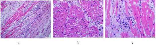
Figure 1. Lymphocytic myocarditis.
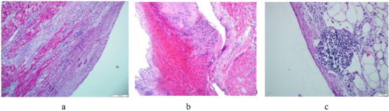
Figure 2. Endocardium and pericardium in the main group.
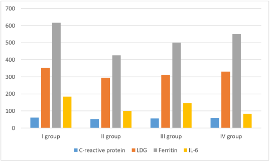
Figure 3. Laboratory parameters in the study groups.
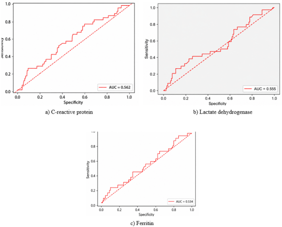
Figure 4. ROC analysis of the logistic regression model based on the dynamics of CRP, LDH and ferritin levels and the progression of COVID-19.
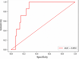
Figure 5. ROC analysis of the logistic regression model based on the D- dimer level and the presence of thrombosis.
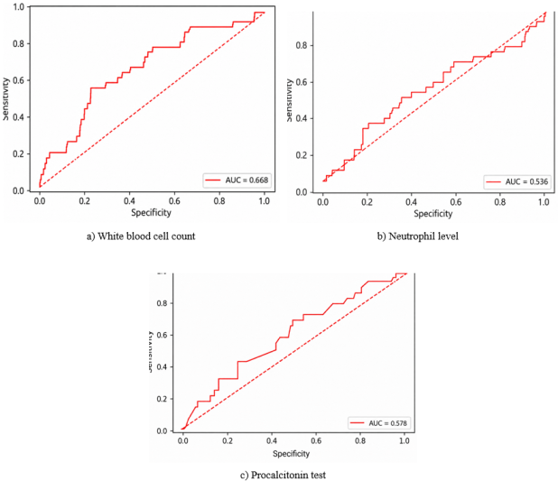
Figure 6. ROC analysis of the logistic regression model based on the dynamics of leukocyte levels, procalcitonin test, neutrophils and the presence of bacterial infection.
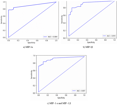
Figure 7. ROC analysis of logistic regression models based on levels MIP-1α and MIP-1β in blood serum as a marker of disease severity.
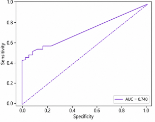
Figure 8. ROC analysis of logistic regression models based on serum IL-10 concentration and mortality.
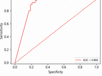
Figure 9. ROC analysis of the logistic regression model based on the dynamics of MIP-1α levels and mortality in experimental animals with COVID-19.
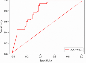
Figure 10. ROC analysis of the logistic regression model based on dynamics MIP-1β levels and mortality in experimental animals with COVID-19.
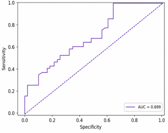
Figure 11. ROC analysis of the logistic regression model based on dynamics IP-10 levels and mortality in experimental animals with COVID-19.
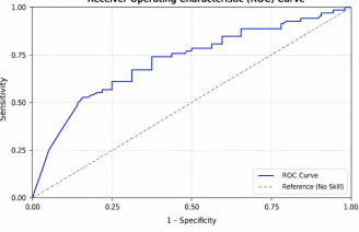
Figure 12. ROC curve characterizing the discriminatory ability of the antibody level in predicting the incidence of the new coronavirus infection.
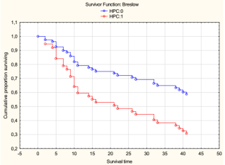
Figure 13. Kaplan-Meier curve in groups: 1-main group, 0-control group.
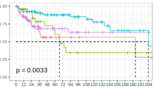
Figure 14. Kaplan-Meier curve for all 4 groups.
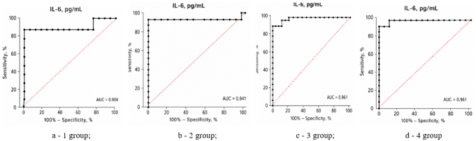
Figure 15. ROC curves characterizing the sensitivity and specificity of determining IL-6 in the blood of rats with coronavirus infection after correction.
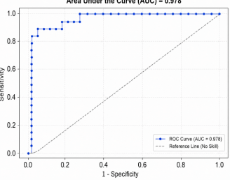
Figure 16. ROC curve of the logistic regression model for predicting cognitive impairment in rats with coronavirus infection.
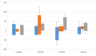
Figure 17. Study of the effect of the drugs used on the immunogenicity of SARS - CoV -2.
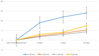
Figure 18. Comparative analysis of the immunogenicity of the SARS - CoV -2 S -protein in various drugs used.

Information



 Share This Article
 Twitter
 Linked In
 Facebook
 Copy
Copy Download