
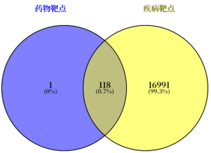
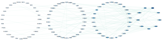
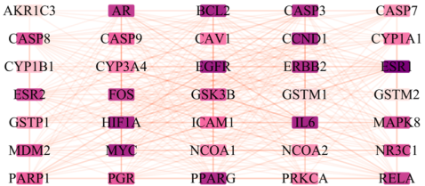
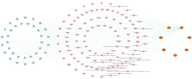
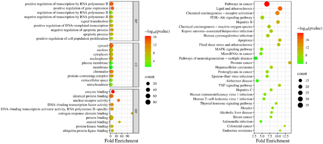
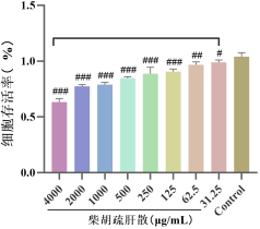
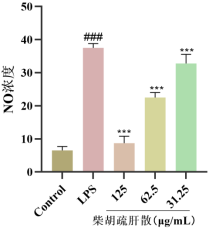
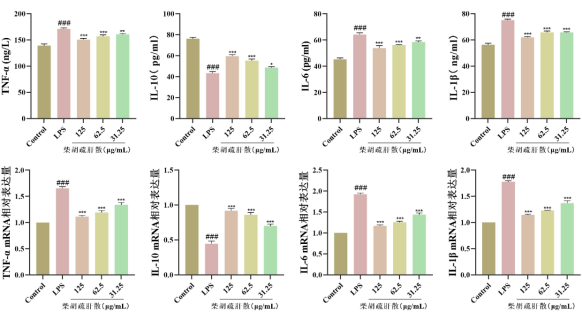
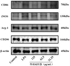
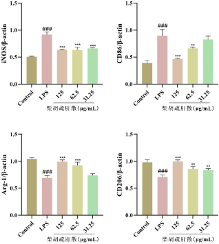
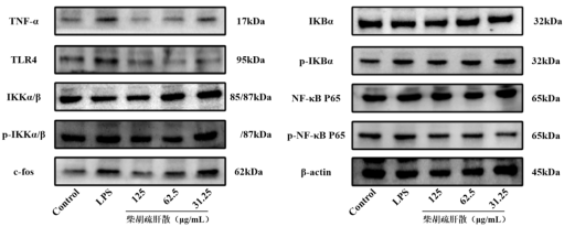
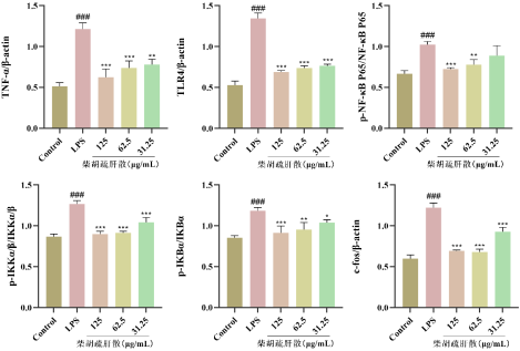
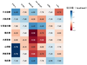
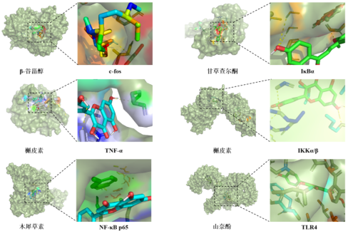
Objective To explore the mechanism by which Chaihu Shugan Powder inhibits LPS-induced inflammation in BV2 cells based on network pharmacology, molecular docking and in vitro experiments. Method The antidepressant mechanism of Chaihu Shugan Powder was predicted by network pharmacological analysis, and the key active ingredients were screened in combination with molecular docking technology. Further construct an LPS-induced inflammatory model of BV2 cells and provide intervention treatment with Chaihu Shugan Powder. The protein levels and mRNA expression levels of TNF-α, IL-1β, IL-6 and IL-10 in the cell supernatants and intracellular tissues of each group were detected by ELISA kits and qRT-PCR. The level of NO in the cell supernatant was detected by the Greiss method; The expressions of microglial polarization markers iNOS, CD86, CD206 and Arg-1, as well as key proteins of the TNF-α/NF-κB pathway in each group of cells were detected by Western blot. Result The results of network pharmacological analysis showed that a total of 118 intersection targets were obtained. Based on the degree value > 3 (median), 35 core targets were screened out, and the traceability analysis obtained 8 key components that played a role. The results of pathway enrichment indicated that the antidepressant effect of Bupleurum evacuation might be related to signaling pathways such as TNF, PI3K-Akt, Apoptosis and MAPK. The results of the CCK-8 experiment determined that the high, medium and low administration doses of Chaihu Shugan Powder were 125, 62.5 and 31.25μg/mL, respectively. The results of the Greiss method for detecting the NO level in the cell supernatant showed that different doses of Chaihu Shugan Powder could significantly reduce the release of NO induced by LPS (P<0.001). ELISA and PCR results showed that the Chaihu Shugan Powder administration group could significantly adjust back the abnormal changes in the levels of various inflammatory factors (P<0.001, P<0.01, P<0.05). The results of Western blot detection showed that compared with the Control group, the expressions of M1-type polarization markers iNOS and CD86 in microglia of the LPS group significantly increased (P<0.001), while the expressions of M2-type polarization markers CD206 and Arg-1 significantly decreased (P<0.001). The protein contents of TNF-α, TLR4, p-IKKα/β, p-IκBα, NF-κB p65 and c-fos proteins increased (P<0.001); After treatment with Chaihu Shugan Powder, the abnormal expression of the above proteins could be significantly improved in all cases (P<0.001,P<0.01,P<0.05). The findings from molecular docking indicated that all eight essential components demonstrated strong binding affinities with the proteins previously mentioned. Conclusion Chaihu Shugan Powder can exert an antidepressant effect by inhibiting the polarization of microglia to M1 type and regulating the TNF-α/NF-κB signaling pathway, thereby suppressing the expression of inflammatory factors.
| Published in | Science Research (Volume 14, Issue 5) |
| DOI | 10.11648/j.sr.20261405.15 |
| Page(s) | 296-307 |
| Creative Commons |
This is an Open Access article, distributed under the terms of the Creative Commons Attribution 4.0 International License (http://creativecommons.org/licenses/by/4.0/), which permits unrestricted use, distribution and reproduction in any medium or format, provided the original work is properly cited. |
| Copyright |
Copyright © The Author(s), 2026. Published by Science Publishing Group |
Chaihu Shugan Powder, Lipopolysaccharide, BV2 Cells, Depression
基因 | 引物序列(5,→3,) |
|---|---|
IL-6 | gccttcttgggactgatgct |
agcctccgacttgtgaagtg | |
TNF-α | atggcctccctctcatcagt |
aaggtacaacccatcggctg | |
IL-1β | gggctgcttccaaacctttg |
aagacacaggtagctgccac | |
IL-10 | cagagaagcatggcccagaa |
gctccactgccttgctctta | |
β-actin | tactgctctggctcctagca |
cggactcatcgtactcctgc |
| [1] | FERRARI A J, SOMERVILLE A J, BAXTER A J, et al. Global variation in the prevalence and incidence of major depressive disorder: a systematic review of the epidemiological literature [J]. Psychological Medicine, 2012, 43(3): 471-81. |
| [2] | TIAN W, YAN G, XIONG S, et al. Burden of depressive and anxiety disorders in China and its provinces, 1990-2021: findings from the Global Burden of Disease Study 2021 [J]. The British journal of psychiatry: the journal of mental science, 2025: 11-. |
| [3] | WANG J, ZHAO Z, YANG J, et al. The Association Between Depression and All-Cause, Cause-Specific Mortality in the Chinese Population - China, 2010-2022 [J]. China CDC weekly, 2024, 6(40): 1022-7. |
| [4] | 张慧. 浅论《内经》与抑郁症的病因 [J]. 四川中医, 2007, (10): 37-8. |
| [5] | 马柯, 张洪秀, 董振飞, 等. 百合地黄汤治疗抑郁症的研究进展 [J]. 中成药, 2019, 41(04): 874-8. |
| [6] | 张鸿. 柴胡疏肝散加减联合黛力新治疗乳腺癌相关性抑郁的临床观察 [D], 2022. |
| [7] | 王常瞵, 高冬梅, 高明周, 等. 柴胡疏肝散化学成分和药理作用研究进展及质量标志物的预测分析 [J]. 中华中医药学刊, 2022, 40(11): 124-31+271. |
| [8] | 张喆, 赵静洁, 王永志, 等. 柴胡疏肝散药理作用及机制研究进展 [J]. 中国中医药信息杂志, 2017, 24(09): 128-31. |
| [9] | 高雪松, 赵静洁. 柴胡疏肝散治疗抑郁症研究进展 [J]. 河南中医, 2022, 42(04): 629-33. |
| [10] | MONROE S M, HARKNESS K L. Major Depression and Its Recurrences: Life Course Matters [J]. Annual review of clinical psychology, 2022, 18: 329-57. |
| [11] | 林洁筠, 段阳, 龙淼青, 等. 柴胡疏肝散在抗抑郁治疗中的药效物质与作用机制研究进展 [J]. 中国实验方剂学杂志, 2024, 30(10): 246-57. |
| [12] | PHAN VAN T, HUYEN TON NU BAO T, LEYA M, et al. Amlexanox attenuates LPS-induced neuroinflammatory responses in microglial cells via inhibition of NF-κB and STAT3 signaling pathways [J]. Sci Rep, 2024, 14(1): 2744. |
| [13] | LIN X, KONG J, WU Q, et al. Effect of TLR4/MyD88 signaling pathway on expression of IL-1β and TNF-α in synovial fibroblasts from temporomandibular joint exposed to lipopolysaccharide [J]. Mediators of inflammation, 2015, 2015: 329405. |
| [14] | ZANNAS A S, JIA M, HAFNER K, et al. Epigenetic upregulation of FKBP5 by aging and stress contributes to NF-κB-driven inflammation and cardiovascular risk [J]. Proceedings of the National Academy of Sciences of the United States of America, 2019, 116(23): 11370-9. |
| [15] | VALLABHAPURAPU S, KARIN M. Regulation and function of NF-kappaB transcription factors in the immune system [J]. Annual review of immunology, 2009, 27: 693-733. |
| [16] | DUDEK K A, DION-ALBERT L, LEBEL M, et al. Molecular adaptations of the blood-brain barrier promote stress resilience vs. depression [J]. Proceedings of the National Academy of Sciences of the United States of America, 2020, 117(6): 3326-36. |
| [17] | MILLER A H, RAISON C L. The role of inflammation in depression: from evolutionary imperative to modern treatment target [J]. Nat Rev Immunol, 2016, 16(1): 22-34. |
| [18] | BRáS J P, BRAVO J, FREITAS J, et al. TNF-alpha-induced microglia activation requires miR-342: impact on NF-kB signaling and neurotoxicity [J]. Cell death & disease, 2020, 11(6): 415. |
| [19] | SI-LONG D, JIAN-GUO C, FANG W. Microglia: A Central Player in Depression [J]. Current medical science, 2020, 40(3): 391-400. |
| [20] | YIRMIYA R, RIMMERMAN N, RESHEF R. Depression as a Microglial Disease [J]. Trends in Neurosciences, 2015, 38(10): 637-58. |
| [21] | DEPARTMENT OF NEUROSCIENCE C F A N, FRIEDMAN BRAIN INSTITUTE I S O M A M S, NEW YORK., DEPARTMENT OF NEUROSCIENCE C F A N, et al. Central and Peripheral Inflammation Link Metabolic Syndrome and Major Depressive Disorder [J]. Physiology (Bethesda, Md), 2019, 34(2): 123-33. |
| [22] | TROUBAT R, BARONE P, LEMAN S, et al. Neuroinflammation and depression: A review [J]. Eur J Neurosci, 2021, (1). |
| [23] | EMORY UNIVERSITY SCHOOL OF MEDICINE W C I, ATLANTA, 30322 GEORGIA, USA., SCHOOL OF HUMAN ECOLOGY U O W-M, MADISON, 53706 WISCONSIN, USA. The role of inflammation in depression: from evolutionary imperative to modern treatment target [J]. Nature reviews Immunology, 2016, 16(1): 22-34. |
| [24] | JIANG B, WANG H, WANG J L, et al. Hippocampal Salt-Inducible Kinase 2 Plays a Role in Depression via the CREB-Regulated Transcription Coactivator 1-cAMP Response Element Binding-Brain-Derived Neurotrophic Factor Pathway [J]. Biol Psychiatry, 2019, 85(8): 650-66. |
| [25] | ZHONG Q, YU H, HUANG C, et al. FCPR16, a novel phosphodiesterase 4 inhibitor, produces an antidepressant-like effect in mice exposed to chronic unpredictable mild stress [J]. Progress in neuro-psychopharmacology & biological psychiatry, 2019, 90: 62-75. |
| [26] | ALBONI S, BENATTI C, COLLIVA C, et al. Vortioxetine Prevents Lipopolysaccharide-Induced Memory Impairment Without Inhibiting the Initial Inflammatory Cascade [J]. Front Pharmacol, 2020, 11: 603979. |
| [27] | OLEG B, P J M, S M C, et al. Identification of a unique TGF-β-dependent molecular and functional signature in microglia [J]. Nature neuroscience, 2014, 17(1): 131-43. |
| [28] | RUBEN O, A M C, JEAN H G. Microglial M1/M2 polarization and metabolic states [J]. British journal of pharmacology, 2016, 173(4): 649-65. |
| [29] | LI Q, XIE Y, LIN J, et al. Microglia Sing the Prelude of Neuroinflammation-Associated Depression [J]. Molecular neurobiology, 2025, 62(4): 5311-32. |
| [30] | WANG Y F, CHEN C Y, LEI L, et al. Regulation of the microglial polarization for alleviating neuroinflammation in the pathogenesis and therapeutics of major depressive disorder [J]. Life sciences, 2025, 362: 123373. |
| [31] | LI J, YANG Z, ZHAO J, et al. Nomilin Alleviates Neuroinflammation and Depressive-like Behaviors in Mice through the Upregulation of TAX1BP1-Mediated Modulation of Microglia Polarization [J]. ACS chemical neuroscience, 2025. |
| [32] | HAN C, PEI H, SHENG Y, et al. HIPK2 mediates M1 polarization of microglial cells via STAT3: A new mechanism of depression-related neuroinflammation [J]. J Cell Physiol, 2024, 239(3): e30994. |
| [33] | 王艳军, 戴国梁, 陈佩瑶, 等. 延胡索乙素通过作用于α7nAChR介导BV2小胶质细胞的炎症反应与极化调控 [J]. 中国中药杂志, 2025, 50(11): 3117-26. |
APA Style
Fajing, Q., Xiaoyun, X., Huimin, L. (2026). The Effect of Chaihu Shugan Powder on Neuroinflammation in Depression Was Explored Based on the LPS-induced BV2 Inflammatory Cell Model. Science Research, 14(5), 296-307. https://doi.org/10.11648/j.sr.20261405.15
ACS Style
Fajing, Q.; Xiaoyun, X.; Huimin, L. The Effect of Chaihu Shugan Powder on Neuroinflammation in Depression Was Explored Based on the LPS-induced BV2 Inflammatory Cell Model. Sci. Res. 2026, 14(5), 296-307. doi: 10.11648/j.sr.20261405.15
@article{10.11648/j.sr.20261405.15,
  author = {Qiang Fajing and Xu Xiaoyun and Li Huimin},
  title = {The Effect of Chaihu Shugan Powder on Neuroinflammation in Depression Was Explored Based on the LPS-induced BV2 Inflammatory Cell Model},
  journal = {Science Research},
  volume = {14},
  number = {5},
  pages = {296-307},
  doi = {10.11648/j.sr.20261405.15},
  url = {https://doi.org/10.11648/j.sr.20261405.15},
  eprint = {https://article.sciencepublishinggroup.com/pdf/10.11648.j.sr.20261405.15},
  abstract = {Objective To explore the mechanism by which Chaihu Shugan Powder inhibits LPS-induced inflammation in BV2 cells based on network pharmacology, molecular docking and in vitro experiments. Method The antidepressant mechanism of Chaihu Shugan Powder was predicted by network pharmacological analysis, and the key active ingredients were screened in combination with molecular docking technology. Further construct an LPS-induced inflammatory model of BV2 cells and provide intervention treatment with Chaihu Shugan Powder. The protein levels and mRNA expression levels of TNF-α, IL-1β, IL-6 and IL-10 in the cell supernatants and intracellular tissues of each group were detected by ELISA kits and qRT-PCR. The level of NO in the cell supernatant was detected by the Greiss method; The expressions of microglial polarization markers iNOS, CD86, CD206 and Arg-1, as well as key proteins of the TNF-α/NF-κB pathway in each group of cells were detected by Western blot. Result The results of network pharmacological analysis showed that a total of 118 intersection targets were obtained. Based on the degree value > 3 (median), 35 core targets were screened out, and the traceability analysis obtained 8 key components that played a role. The results of pathway enrichment indicated that the antidepressant effect of Bupleurum evacuation might be related to signaling pathways such as TNF, PI3K-Akt, Apoptosis and MAPK. The results of the CCK-8 experiment determined that the high, medium and low administration doses of Chaihu Shugan Powder were 125, 62.5 and 31.25μg/mL, respectively. The results of the Greiss method for detecting the NO level in the cell supernatant showed that different doses of Chaihu Shugan Powder could significantly reduce the release of NO induced by LPS (PPPPPPPPPPConclusion Chaihu Shugan Powder can exert an antidepressant effect by inhibiting the polarization of microglia to M1 type and regulating the TNF-α/NF-κB signaling pathway, thereby suppressing the expression of inflammatory factors.},
 year = {2026}
}
TY  - JOUR
T1  - The Effect of Chaihu Shugan Powder on Neuroinflammation in Depression Was Explored Based on the LPS-induced BV2 Inflammatory Cell Model
AU  - Qiang Fajing
AU  - Xu Xiaoyun
AU  - Li Huimin
Y1  - 2026/09/04
PY  - 2026
N1  - https://doi.org/10.11648/j.sr.20261405.15
DO  - 10.11648/j.sr.20261405.15
T2  - Science Research
JF  - Science Research
JO  - Science Research
SP  - 296
EP  - 307
PB  - Science Publishing Group
SN  - 2329-0927
UR  - https://doi.org/10.11648/j.sr.20261405.15
AB  - Objective To explore the mechanism by which Chaihu Shugan Powder inhibits LPS-induced inflammation in BV2 cells based on network pharmacology, molecular docking and in vitro experiments. Method The antidepressant mechanism of Chaihu Shugan Powder was predicted by network pharmacological analysis, and the key active ingredients were screened in combination with molecular docking technology. Further construct an LPS-induced inflammatory model of BV2 cells and provide intervention treatment with Chaihu Shugan Powder. The protein levels and mRNA expression levels of TNF-α, IL-1β, IL-6 and IL-10 in the cell supernatants and intracellular tissues of each group were detected by ELISA kits and qRT-PCR. The level of NO in the cell supernatant was detected by the Greiss method; The expressions of microglial polarization markers iNOS, CD86, CD206 and Arg-1, as well as key proteins of the TNF-α/NF-κB pathway in each group of cells were detected by Western blot. Result The results of network pharmacological analysis showed that a total of 118 intersection targets were obtained. Based on the degree value > 3 (median), 35 core targets were screened out, and the traceability analysis obtained 8 key components that played a role. The results of pathway enrichment indicated that the antidepressant effect of Bupleurum evacuation might be related to signaling pathways such as TNF, PI3K-Akt, Apoptosis and MAPK. The results of the CCK-8 experiment determined that the high, medium and low administration doses of Chaihu Shugan Powder were 125, 62.5 and 31.25μg/mL, respectively. The results of the Greiss method for detecting the NO level in the cell supernatant showed that different doses of Chaihu Shugan Powder could significantly reduce the release of NO induced by LPS (PPPPPPPPPPConclusion Chaihu Shugan Powder can exert an antidepressant effect by inhibiting the polarization of microglia to M1 type and regulating the TNF-α/NF-κB signaling pathway, thereby suppressing the expression of inflammatory factors.
VL  - 14
IS  - 5
ER  -
Clinical Pharmacy Department, Yulin Xingyuan Hospital, Yulin, China
Clinical Pharmacy Department, Yulin Xingyuan Hospital, Yulin, China
Clinical Pharmacy Department, Yulin Xingyuan Hospital, Yulin, China;Science and Education Department, Yulin Hospital, the First Affiliated Hospital of Xi'an Jiaotong University, Yulin, China

Information



 Share This Article
 Twitter
 Linked In
 Facebook
 Copy
Copy Download